Thursday, December 22, 2022

Iris Publishers-Open access Journal of Oceanography & Marine Biology | Status of Sabaki Tilapia Aquaculture in the Kingdom of Saudi Arabia


Authored by Benjamin C Young*,

Opinion

Since 1980s, shrimp farming has been increasingly prosperous in the Kingdom of Saudi Arabia (henceforth, Saudi Arabia) owing to economic development, market demand, and environmental factors. Initially, the primary species being cultured was the Indian prawn (Fenneropenaeus indicus). However, the white spot syndrome virus, which emerged in 2010, decimated this species and since then aquaculture industries in Saudi Arabia have started farming specific pathogen-free white shrimp (Litopenaeus vannamei) to cover the shortage of producing Indian prawns. Currently, shrimp farming is widespread in Saudi Arabia to meet global demand. Presently, the total aquaculture production of Saudi Arabia is ~83,000 tons, and shrimp farming accounts for ~70% of the total aquaculture production. The primary farmed fish species Sabaki tilapia (Oreochromis spilurus), Nile tilapia (Oreochromis niloticus), Asian sea bass (Lates calcarifer), and gilt-head bream (Sparus aurata) contributed 15,000 tons to the total production in 2019. Note that because of overproduction, the price of Asian sea bass has been decreasing. Moreover, the fingerling source of gilt-head bream depends on imports; therefore, tilapia’s culture is gaining popularity. Among the various tilapia species, the Sabaki tilapia is one of the unique and higher economic value species in the global tilapia aquaculture industry. Due to market demand and environmental factors, fisheries authority selected Sabaki tilapia to target the aquaculture development projects’ species, while the commercial culture of Sabaki tilapia had not been developing in Saudi Arabia. The project has been beginning since 2018. However, in the farming period, the Sabaki tilapia culture had confronted several difficulties, such as extreme salinity. On the coastline of Jeddah, West Saudi Arabia, seawater’s salinity is 42 to 45‰ during the producing period in 35 to 42℃ water temperature. Furthermore, the regulations strictly prohibit the installation of pumping wells, and freshwater only purchased through water companies, so salinity adjustment is also one of the critical costs. In addition, there is a lack of information on the commercial scale of Sabaki tilapia in Saudi Arabia. Despite Sabaki tilapia becoming more prevalent in aquaculture production in Saudi Arabia, there is a lack of information on the farming operations. Therefore, we conducted trial systems that focused on the types of Sabaki tilapia aquaculture practices in Saudi Arabia. We attempted several farming systems to understand the best result for the spawning and grow-out stage. The four farming systems are earth pond, net cage, raceway, and arena, respectively. The practice results, the raceway, and arena have superior fry production results than the net cage and earth pond. Also, the net cage required further management skills and costs. Furthermore, the reproductive performance of a female brood stock can spawn 120 to 550 fries. The hatching rate is more than 95%. The fry of Sabaki tilapia has strong cannibalism. Therefore, the grading is necessary. The nutritional requirements of Sabaki tilapia were crude protein 35 to 45% and crude fat 10 to 15%. The feed conversion ratio (FCR) of the grow-out stage was 1.2 to 1.4; the raceway and net cage have superior growth performance than the earth pond. Generally, the farming measure of Sabaki tilapia has not vast differences comparing other tilapia species. However, because of the unique farming environment in Saudi Arabia, the farming environment was under high salinity and temperature in the producing period. Notably, the bacterial infection continually emerges in the winter season. Moreover, our survey reported that the primary costs of Sabaki tilapia aquaculture production were feed (50.06%–69.33%), labor (14.51%–20.98%), and fry (5.29%–5.81%). Most aquaculture companies in Saudi Arabia are either industrial or large-scale businesses. In developing countries, this is important because the scale of farming operations makes a significant difference. Significantly, the industrial-scale producers spent less on feed as they could produce feed in their facilities. Because the major operating costs in Sabaki tilapia production were feed, labor, and fry, future aquaculture operations could emphasize cost control for producers and specific marketing and disease prevention strategies to enable the Sabaki tilapia industry’s sustainability in Saudi Arabia.

To read more about this article...Open access Journal of Oceanography & Marine Biology

Please follow the URL to access more information about this article

https://irispublishers.com/aomb/fulltext/status-of-sabaki-tilapia-aquaculture-in-the-kingdom-of-saudi-arabia.ID.000536.php

To know more about our Journals...Iris Publishers

To know about Open Access Publishers

Iris Publishers-Open access Journal of Rheumatology & Arthritis Research | Virtual Clinics: Descriptive Survey of A Sample of Models Used Within Orthopedics in The United Kingdom


Authored by Lindsay K Smith*,

Abstract

Objectives: To gain a snapshot of the models of virtual orthopedic clinics currently in use within the United Kingdom through data collected from non-medical healthcare professionals. Information collected included types of patients referred, staff members involved, facilities used, methods of follow up, benefits and challenges.

Method: A short online survey, collected quantitative data from non-medical healthcare professionals affiliated with two recognized professional bodies who were directly involved or had knowledge about virtual clinics.

Results: Responses were received from 11 virtual clinics. The majority of individuals involved were physiotherapists (73%) with 27% overseeing them. Most clinics were conducted Monday-Friday (45%) in an office space (73%), using a computer, desk, and telephone (91%), with referrals mainly coming from orthopedic services (54%) and Accident and Emergency (54%).

Conclusion: The results of this survey provide an insight into orthopedic virtual clinics in the UK prior to the COVID-19 pandemic. Although missing from previous research, there is evidence to show the involvement of physiotherapists in addition to other non-medical healthcare professionals. Equipment needed for set up is minimal and benefits include patient satisfaction, efficiency, and the ability to meet orthopedic guidelines.

Keywords: Virtual clinics; Orthopedics; Physiotherapy; Survey

Background

The National Health Service (NHS) is currently over stretched and over-burdened [1,2] with an ongoing challenge to match capacity to demand [3]. The number of outpatient clinic appointments is growing, leading to delays, dissatisfaction, and non-compliance with national guidelines [4]. With rising pressures on primary care, the NHS Long Term Plan (2019) supports the development of digitally enabled services to replace the unsustainable growth of current outpatient care, with the aim to reduce unnecessary faceto- face ((FTF) appointments [5]. Virtual clinics (VCs) are one way of achieving this goal, and have been deemed safe, cost-effective, and associated with high levels of patient satisfaction [3,6-8].

While the use of VCs in orthopedics has become increasingly popular over the past decade, literature surrounding the topic is scarce. Research has primarily focused on virtual fracture clinics (VFCs) replacing traditional FTF fracture pathways [7-9] with some mention of joint arthroplasty follow-ups being replaced by virtual orthopedic clinics (VOCs) [4,10-12]. King D, et al [13] report that VCs should be considered as one part of the musculoskeletal pathway but that the paradigm for delivery is still evolving.

Although guidelines and protocols provide a framework for professionals to follow and recommendations of how healthcare should be delivered [14], to-date, there are no standards pertaining Although guidelines and protocols provide a framework for professionals to follow and recommendations of how healthcare should be delivered [14], to-date, there are no standards pertaining

Method

This was a descriptive survey, collecting quantitative data from non-medical healthcare professionals (HCP) working within orthopedics, about the models of VCs currently in use across the UK. The online survey comprised of seven questions, and sought information relating to; types of patients/staff involved, facilities used, frequency of clinics, follow up methods as well as a chance to gain an insight into the benefits and challenges that VCs pose for HCP. The survey received ethical approval (University of the West of England, Bristol REC Reference No: HAS.20.01.107, 7th February 2020) and was disseminated nationally via email to 500 members of the Association of Trauma and Orthopedic Chartered Physiotherapists (ATOCP) and 61 members of the Arthroplasty Care Practitioner’s Association (ACPA ) [15]. The survey remained open for four weeks, with data analyzed thereafter.

Results

Responses were received from 11 clinics and data provided a snapshot of orthopedic VCs immediately prior to the COVID-19 pandemic. The clinics did not involve patient contact but were constructed to review medical records and imaging results plus other diagnostic tests to facilitate a decision about further treatment. Clinics were staffed by advanced practice physiotherapists and/or advanced practice nurses; other staff included orthopedic consultants (4 clinics) and administrators. Frequency of clinics varied from once a month to every weekday; dedicated office space was available for some VCs, but the majority used shared office space. The essential equipment included a desk, computer, and telephone. Referrals to the orthopedic VCs were from primary care, emergency departments, minor injury units and secondary care. Patients were excluded if their condition required surgery or admission to hospital; one response indicated that those with hearing problems were excluded. Follow-up information was offered to patients through a mixture of letter, email, telephone, and pre-printed leaflets. No patients were contacted by video-call. Benefits listed were speed and efficiency, avoidance of hospital visits, patient satisfaction, and the ability to meet targets. While challenges included the administrative burden, access to medical records, quality of radiographic images, staff training, appropriate referrals, and the time taken to implement decisions and contact patients after the VC.

Discussion

In our study, physiotherapists played a key role in the operation of VCs in eight cases (73%) and were overseen by physiotherapists in the remaining 27%. This is in contrast with the current body of literature whereby a majority of VCs are run by a consultant and a nurse specialist. The majority of participants in this study listed various minor soft tissue injuries and fractures within their inclusion criteria. One of the exclusion criteria stated by the participants was hearing problems. This emphasizes a need to adapt communication methods within VC’s to ensure care is provided to a broader patient group.

A strength of this survey was that it attempted to gather information lacking in current research such as the frequency of clinics. The study revealed that the majority of VC’s operate during weekdays (45%) with 27% operating 2-4 times a week. Most respondents of the survey used a shared or private office which implies that VC’s are well established within these trusts and that HCP’s are convening together to conduct these clinics. Assuming that space was not an issue, a VC would be a cost-effective addition to most trusts’ services as already well documented in the literature. Current literature suggests that patients are mainly followed up by a telephone call [9,16]. This is in contrast with our study whereby the majority of patients received a letter by post.

The findings of this study are similar to the findings of other research conducted into VCs in that the majority of referrals were reported as coming from the Emergency Department, orthopedic services, and the general practitioner. Some benefits of running a VC were reported as efficiency, patient satisfaction and patients being streamlined to the appropriate specialist. Challenges included contacting patients by telephone, administrative support, and the referral of inappropriate patients.

Limitations

The limited response rate of the survey decreases the generalizability and therefore reduces the external validity of the study. This may have been due to VC’s being less common in practice or the target community not being responsive to electronic survey’s and time-poor with winter pressures on service delivery. In addition, towards the end of data collection saw the outbreak of the Covid-19 pandemic which would have impacted on respondents’ capacity to complete the questionnaire.

Future research should consider the incorporation of multiple professions involved within orthopedics thus enabling a more reflective target population. In addition, the development of an evidence-based framework enabling the provision of consistent guidelines would ensure the continuity of high-quality care and improved patient outcomes.

Conclusion

In a culture where delivery of care by virtual methods is rapidly evolving, this survey has given some insight into orthopedic VCs in the UK prior to the COVID-19 pandemic, and the results provide evidence for the involvement of physiotherapists in addition to other non-medical health professionals, a detail missing from previous research. Equipment and facilities needed are minimal, equating to a low-cost set-up and the benefits of efficiency, patient satisfaction, and the ability to deliver care in accordance with orthopedic guidelines are consistent with existing literature.

To read more about this article...Open access Journal of Rheumatology & Arthritis Research

Please follow the URL to access more information about this article

To know more about our Journals...Iris Publishers

To know about Open Access Publishers

Wednesday, December 21, 2022

Iris Publishers-Open access Journal of Anaesthesia & Surgery | Ultrasound-guided Ilioinguinal-Iliohypogastric nerve block in inguinal hernia repair surgery under general anesthesia: a randomized controlled study


Authored by Nabil A Mageed*,

Abstract

The number of health workers infected with COVID-19 is increasing exponentially. Recent reports indicate that about 787 frontline health workers died of COVID-19 in the United States of America. In Ghana, not less than 779 health workers especially doctors, certified registered Anaesthetists and nurses have been infected with COVID-19, with nine death. Aerosol and fomite transmission of SARS-CoV-2 to individuals is plausible. Anaesthesia professionals who are in close contact as they help patients breathe through airway equipment are at maximum risk of exposure to COVID-19. This study aimed to assess the knowledge, attitudes, and practices of Anaesthetists towards the prevention of the spread of the COVID-19 infection. Certified Registered Anaesthetists were required to complete a self-administered structured questionnaire. A total of 356 respondents were recruited in this study. The data showed that 73.3 % of the respondents recruited for this study were males and 26.7 % of them were females. The mean age of the respondents was 37.1 ± 5.3 years old. We realized that 4.8% of them always had access to appropriate personal protective equipment, whereas 95.2 % of them had no access during work. The data also showed that 34.0 % of them always had access to a face mask and hand sanitizers, while 66.0 % of them had no access to a face mask and hand sanitizers during work. There is, the need for regular inservice training of Certified Registered Anaesthetists and other health workers on the right attitude and practices to prevent the spread of the virus among health workers. Policymakers should ensure the regular supply of basic PPE at the various hospitals.

Keywords: Ilioinguinal; Iliohypogastric; Block; Ultrasound; Inguinal hernia repair

Introduction

Abdominal wall hernias repair surgery is one of the most common surgical procedures. Inguinal hernias are undoubtedly the commonest hernia type (70% - 75%) with incidence rate 11 for each 10,000 in persons between 16 and 24 years of age, 200 for each 10,000 in persons more than 75 years of age [1]. The management of post-operative pain is oftentimes unsatisfactory. Opioids, non-steroidal anti-inflammatory drugs, and analgesics are routinely used to alleviate post-operative pain, but they are associated with several undesirable effects and do not seem to be completely effective on preventing and treating postoperative pain [2]. The estimated incidence rate of chronic pain after inguinal herniorrhaphy is about 10%. It represents a major problem that significantly affects daily activities. The severity of early postoperative pain correlates with the risk of developing chronic pain [3]. Ilioinguinal-iliohypogastric (II/IH) nerve blocks have been increasingly utilized in patients for perioperative analgesia. The use of ultrasound guidance was associated with improved perioperative analgesia compared to landmark based methods [4].

The ultrasound technology has been used for many types of peripheral nerve blocks in both adult and children. It has the advantages of increasing the success rate by easier identification of the site of the nerve, correct deposition of the local anesthetic around the nerve and decrease the incidence of complication associated with this block [5]. Bupivacaine is an amino amide highly potent local anesthetic that blocks the peripheral afferents nerve by acting on voltage dependent sodium channels so, it prevents the generation of action potential. Bupivacaine is being used for intraoperative anesthesia and postoperative analgesia [6].

In this study, the analgesic efficacy of adding II/IH nerve block to general anesthesia in inguinal hernia repair surgery was evaluated. We hypothesized that II/IH nerve block may reduce perioperative opioids requirements in such patients. The primary outcome measures were the total dose of postoperative fentanyl requirements and the postoperative pain score in the first 24 hours postoperatively. While the secondary outcomes were the hemodynamic parameters, time to first analgesic request and side effects.

Materials and Methods

This prospective, randomized, double blinded, controlled study was conducted from May 2017 to May 2018 after approval from the Institutional Research Board, Faculty of Medicine, Mansoura University given a code number (MS/17.04.46) and after obtaining an informed written consent from each patients prior to surgery. A total of 80 patients of either sex with age ranging from 20-60 years and ASA I and II scheduled for elective unilateral open inguinal hernia repair surgery were included in this study. The exclusion criteria included, patient refusal, coagulation disorders, local skin infection at site of the block, known allergy to the bupivacaine study drug, body mass index > 40 Kg m2-1 and bilateral inguinal hernia repair to be performed at the same procedure.

Patients were randomly into two groups, each group 35 patients by a computer-generated randomization table and closed envelope method. Control group: general anesthesia group and Study group (II/IH group): general anesthesia combined with ilioinguinal-iliohypogastric nerve block group. Patients in II/ IH group were injected with 20 ml of 0. 25% bupivacaine, while patients in the control group were injected with a placebo (20 ml of 0.9 saline). The anesthetist who preparing the injected drugs was not participated in the study and unaware of the patient group allocation. The surgeons, patients and ICU personnel’s who recorded the postoperative data were unaware of group allocation.

All patients were assessed preoperatively by history, physical examination, laboratory evaluation (complete blood picture, liver function, renal function tests) and ECG. The day before the surgery, the study protocol and II/IH nerve block procedure were explained to all patients. Patient was familiar with the use of 10-cm visual analogue scale score (VAS) identifying 0 as no pain and 10 as worst imaginable pain [7]. On arrival of the patient to the recovery room routine monitoring including electrocardiography, non-invasive blood pressure, and pulse oximetry was done. Peripheral intravenous cannula (20 G) was inserted, and Lactated Ringer’s started to be infused (500 mL). All patients were premedicated using midazolam 0.03 mg kg-1 IV. General anesthesia in both groups was induced using IV propofol at dose of 2-3 mg kg-1, fentanyl IV 1μ kg-1 and atracurium besylate 0.6 mg kg-1 to facilitate intubation. Patient was mechanically ventilated using a volume control mode with tidal volume 6-8 ml kg-1, respiratory rate 10-14 breath min- 1 and I: E ratio 1:2 to maintain end tidal CO2 around 35mmHg. Anesthesia was maintained using isoflurane 1, 2% and 60% air in oxygen mixture and top up dose of atracurium. Intravenous fluids were given per body weight and according to intraoperative loss. Patients in both groups received paracetamol infusion (10mg kg- 1) 15 minutes before extubation. Ketorolac amp were given to all patients every 8 hours. All patients were extubated at the end of surgery after neuromuscular reversal with administration of 0.05 mg kg-1 of neostigmine and 0.02 mg kg-1 of intravenous atropine.

The technique of II/IH nerve block was described by Wang Y, et al. [4]. Ilioinguinal-iliohypogastric nerve block was performed by ultrasound guided nerve blocks. In a supine position, the skin overlying the injection site was sterilized with complete antiseptic solution and the probe surface in contact with the skin should be covered with a sterile adhesive dressing. Place a high frequency linear probe (10 MHz or greater) medial and superior to the anterior superior iliac spine in an oblique manner at the line joining the umbilicus and the anterior superior iliac spine (ASIS). The ultrasound image revealed three muscle layers, separated by hyper echoic fascia: external oblique, the internal oblique, and the transversus abdominis muscles. The fascia transversalis is located below transversus abdominis muscle, just above the peritoneum and the abdominal cavity. Position the probe such that the bony shadow from the ASIS is visible on one side of the image on the screen. Identify the peritoneum, transversus abdominis muscle, and internal oblique muscle. The external oblique muscle may not be visible as a distinct muscle layer at this level. Sliding the probe in a cephalic direction up over the iliac crest, while maintaining the orientation of the probe along a line to the umbilicus, will bring all three muscles into view as three distinct layers. This may be useful if there is any doubt about the anatomy and the relevant planes. Always identify the deepest structures first (i.e. the peritoneum) and work toward the superficial structures to identify each layer. The ilioinguinal and Iliohypogastric nerves are seen in close proximity to one another as two small round hypo echoic structures with a hyper echoic border. They lie in the plane between the internal oblique muscle and the transversus abdominis muscle close to the ASIS. Insert the block needle in plane from medial to lateral and ensure that there is a good image of the needle tip at all times as the needle is advanced. 20 mL of 0. 25% bupivacaine was injected around the nerves in the transversus abdominis plane.

The primary outcome measures were total dose of fentanyl consumption in the first postoperative 24 hours, the time period for the first analgesic requirement and postoperative pain scores was assessed by 10 point visual analogue scale (VAS) [7]. When the patients experienced pain (VAS score >3), a bolus dose of IV fentanyl 0.5μ kg-1 was administered and can be repeated till visual analogue scale score ≤ 3 mm was attained. VAS was assessed at 30min after the end of the surgery, 2, 4, 8, 12 and 24 hours postoperatively.

The secondary outcome measures were heart rate (HR) and mean arterial pressure (MAP). These parameters were measured before induction of anesthesia (basal value), after skin incision (approximately 15 min after the block), 30, 45, 60 and 90 minutes intraoperatively. In the post-surgical care unit, the above parameters were recorded at 0, 2, 4, 8, 12, 18, and 24 hours post-operative. Other secondary measures including postoperative complications (nausea, vomiting and itching) and duration of surgery were also recorded.

Statistical Analysis

The determination of the sample size was based on a pilot study on 10 patients (5 in each group) depending on the total dose of fentanyl consumption in the first postoperative 24 hours (123±53 versus 88±31) as a primary end point. The total required sample size was 70 patients (35 in each group) to obtain a power of 95%, and type I α error of 0.05. The total number of patients was increased to 80 to avoid 10 % dropped out cases.

The statistical analysis of data was done by using Statistical Package for Social Science (SPSS) program version 22. To test the normality of data distribution, Shapiro-Wilk test was done. Unpaired student-t test was used for comparisons between the means of two groups for quantitative data. Mann-Whitney U test was used for nonparametric data (VAS). Chi square test was used for the analysis of categorical data. The description of data done in the form of mean (±SD) for quantitative data, frequency, and proportion for categorical data and median (range) for nonparametric data. Any difference or change showing probability (P) less than 0.05 were considered statistically significant at confidence interval 95.
irispublishers-openaccess-anaesthesia-surgery

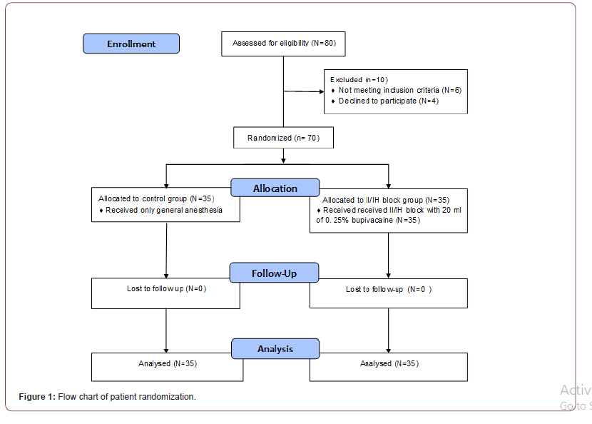
As shown in Figure 1, a total of 80 patients were included in this prospective, randomized, double blind controlled study and 10 patients were excluded or discontinued the intervention. A final of 70 patients were studied, 35 patients in the control group and 35 in the II/IH block group (Figure 1).

There were no statistically significant differences between the studied groups as regard: age, gender, BMI, ASA status, and duration of surgery (Table 1).

As regard the primary outcome parameters, VAS was significantly lower in the II/IH group at 0,2,4, 8 hours postoperative when compared to the control group with P value < 0.001 but no statistically significant differences were detected between the two studied groups at 12, 18 and 24 h postoperatively (Table 2). The total post-operative fentanyl consumption in the first 24h was statistically significantly lower in the II/IH group (78.00 ± 55.72 μg) than the control group (174.14 ± 27.32 μg) (Table 3). The time to the first request of analgesia showed statistically significant longer in the II/IH group (305.57 ± 12.22 min) than the control group (49.29 ± 19.45min) (Table 3).

Table 1:Demographic data of studied groups.

irispublishers-openaccess-anaesthesia-surgery

Table 2:Visual analogue score for visceral pain at rest during the first postoperative 24 hours.

irispublishers-openaccess-anaesthesia-surgery

Table 3:First request of analgesia, total post-operative fentanyl (μg) consumption in the first 24 h and complications.

irispublishers-openaccess-anaesthesia-surgery

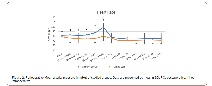
The heart rate and the mean arterial pressure showed statistically significant difference between the two studied groups intraoperatively at 15m, 30m, 45m, 1h, 1.5h. However, the heart rate and mean arterial pressure showed no statistically significant differences between the two studied groups at 0, 2h, 4h, 8h, 12h, 18h, 24h postoperative (Figure 2&3). The incidence of postoperative nausea and vomiting (PONV) was statistically significantly higher in control group than II/IH group (31.1% versus 5.7% respectively). However, there was no statistically significant difference between both studied groups as regard the Pruritus (Table 3).

Discussion

Acute postoperative pain following open inguinal hernia repair is maximum during the first 24 h period. Various modalities have been adopted to reduce this pain which includes parenteral opioids, non-steroidal anti-inflammatory drugs (NSAID), central neuroaxial analgesia and regional techniques. Among all these techniques, ultrasound guided ultrasound guided II/IH nerve block and transversus abdominis plane (TAP) block is effective and easy to perform with least complications [8].
irispublishers-openaccess-anaesthesia-surgery

This study was done to evaluate the efficacy of adding ultrasound guided II/IH nerve block to general anesthesia in cases of unilateral inguinal hernia repair surgery. It was found that adding the block was effective in prolonging the time of first analgesic request, decreasing the total dose of fentanyl analgesics required in the first 24 hours postoperative and providing better postoperative pain scores (VAS) and lower complications like PONV.

The present study demonstrated that the total analgesic consumption of fentanyl during 24 h postoperative was statistically significantly decreased in the study group (78.00 ± 55.72 μg) when compared to the control group (174.14 ± 27.32μg). Also, there was statistically significant prolongation of the time of first analgesic request in the study group (5.09 ± 0.2h) in comparison to the control group (0.82 ± 0.32h). This coincides with the study of Toivonen J, et al. [9] who evaluated the effect of pre-incisional ilioinguinal and iliohypogastric nerve block using 10 ml of 0.5% plain bupivacaine on postoperative analgesic requirement in patients undergoing herniorrhaphy under spinal anesthesia [9]. It demonstrated that the mean number and the total dose of supplementary analgesics was smaller in II/IH nerve block group (15 ± 25 μg) than the other group (50 ± 59 μg). Also, the average time latency was higher in block group 8.1 (0.9-54.7) h than the other group 4.3 (0.4-20.6) h. Also, our results are parallel with the study of Al Dehayat and Al Momany [10] who examined if there was difference in morphine requirements and pain scores in patients undergoing caesarean section, with and without ilioinguinal - iliohypogastric nerve block. They concluded that the quantity of morphine administered in 24 hours was 10mg (range 10-45mg) in bilateral IL and IH nerve block group and 30mg (range 0-25mg) in no block group [10].

However, these findings were in contrast with a previous study by Ding and White, 2010 who evaluated the effect of an ilioinguinalhypogastric nerve block with either saline or bupivacaine on the postoperative analgesic requirement in patients undergoing inguinal herniorrhaphy and found that a similar percentage of patients in the two groups required parenteral opioid analgesic medication in the early postoperative period and also the total dose of the fentanyl showed no statistical difference between the two groups [11]. This may be attributed to their blind technique increasing block failure rate and small sample size of only thirty patients.

In this study pain scores were statistically significantly decreased in the II/IH block group than the control group at 0, 2, 4 and 8 h. This findings are coincides with the study of Al Dehayat and Al Momany [10] who examined morphine requirements and pain scores in patients undergoing caesarean section, with and without ilioinguinal-iliohypogastric nerve block and found that Mean (SD) pain scores for block group were lesser than no block group (p<0.05) at 1, 4, 8 and 24 h [10]. The longer duration of this study may be attributed to the higher bupivacaine concentration 0.5%.

Moreover, Radhakrishnan and Kumar, 2017 who evaluated postoperative pain analgesia with ilioinguinal and iliohypogastric nerve block with 10 mL of 0.75% ropivacaine following repair of inguinal hernia surgery and time of discharge with spinal anesthesia and demonstrated that VAS at rest during different post-operative periods at 3 h, 6 h, and 12 h was assessed and found that they were significant statistically which is parallel to this current study [2].

The result of the current study revealed that heart rate and mean arterial blood pressure in II/IH block group were significantly lesser than control group intraoperatively, which may be attributed to the more effective analgesia produced by the ilioinguinal- iliohypogastric nerve block leading to more abolishing of the stress response to pain result in reducing both heart rate and blood pressure [12]. While the post-operative heart rate and mean arterial blood, pressure showed no significant statistical difference in the two groups mostly due to lower pain intensity and adding of rescue analgesia on patient demand.

The study done by Fekry DM, et al. [13] who compared Ultrasound-guided ilioinguinal- iliohypogastric, and genitofemoral nerve block with 25 mL of 0.5 % bupivacaine versus spinal subarachnoid blockade for inguinal hernia repair [13]. Both the heart rate and mean arterial blood pressure were the same without variations intra and post-operative that is parallel to our study. While the spinal anesthesia group showed decreased blood pressure at 5, 15, 30, and 45 min after spinal anesthesia.

The study done by Fekry DM, et al. [13] who compared Ultrasound-guided ilioinguinal- iliohypogastric, and genitofemoral nerve block with 25 mL of 0.5 % bupivacaine versus spinal subarachnoid blockade for inguinal hernia repair [13]. Both the heart rate and mean arterial blood pressure were the same without variations intra and post-operative that is parallel to our study. While the spinal anesthesia group showed decreased blood pressure at 5, 15, 30, and 45 min after spinal anesthesia.

Conclusion

This study proved that ultrasound guided Iliohypogastric/ ilioinguinal nerve block in open unilateral inguinal hernia repair under general anesthesia is effective in providing better quality of analgesia with prolonged time of first analgesic request, decreasing the total dose of fentanyl analgesic consumption, improving postoperative VAS values and decreasing incidence of postoperative complications as nausea and vomiting.

To know about Open Access Publishers

Iris Publishers-Open access Journal of Cardiology Research & Reports | Prognostic Value of Serum Uric Acid Levels to the Cardiovascular Events in Hypertensives


Authored by A Karamanou*,

Abstract

Aim: The aim of the study was to investigate the association of serum uric acid (SUA) levels with the major cardiovascular events (MACE) in hypertensive patients.

Methods and Materials: This is a prospective analysis including 652 hypertensive patients (52,6% female) of mean age at the entry: 57,2±13,1 years, mean office systolic/diastolic blood pressure (S/DBPo): 152,3±19,1/94,4±11,83 mmHg, mean office heart rate (HRo): 76,0±12 beats/min, treated or newly diagnosed untreated. The median follow-up period was 9 years. SUA were measured at the entry of the study. Major cardiovascular events (MACE) (myocardial infarction, unstable angina, transient ischemic attack or stroke, peripheral vascular intervention, heart failure events, cardiovascular death) were registered. We estimated the prognostic value of SUA to the MACE. Cox proportional hazard model was employed to determine the prognostic value of uric acid.

Result:The median follow-up period was 9 years. There were 264 (40,5%) MACE at the end of the study. Cox regression statistical analysis revealed that SUA was a strong predictor of MACE (HR: 1,14, 95%CI: 1,06 – 1,2 p-value=0,000)

Conclusion: SUA levels have statistically significant prognostic value to MACE in hypertensive patients.

Introduction

Uric acid is a product of the activity of xanthine oxidase, an enzyme increasingly implicated as a mechanistic participant in oxidant stress and cardiovascular disease. Contributing factors responsible for hyperuricemia are alcoholic and high-purine foods consumption, low water consumption and poorly exercising. In adults with essential hypertension an association with hyperuricemia is very common. Prevalence of hyperuricemia is 30% in untreated hypertensives and >75% in malignant hypertension. Uric acid is thought to play a pathogenic role in hypertension [1-3] mediated by several mechanisms such as inflammation, vascular smooth muscle cell proliferation in renal microcirculation, endothelial dysfunction, and activation of the renin – angiotensin – aldosterone system [4-7]. Animal models have shown that acute elevations of serum urate, by inhibition of uricase, induce a prompt rise in blood pressure and that chronic urate elevation maintains the rise in pressure and induces irreversible vascular damage and glomerular changes, and results in a form of salt-sensitive hypertension [8, 9]. Furthermore, studies have shown that in overweight and obese subjects, hyperinsulinemia secondary to insulin resistance may enhance their absorption of uric acid and thus contribute to the association of hyperuricemia with hypertension [10].

Also, numerous studies have noted an association of elevated serum uric acid (SUA) levels also with heart failure [11], coronary artery disease [12] and stroke [13]. Several pathophysiological mechanisms have been postulated including multiple proatherogenic processes, increased oxidative stress [14, 15], vascular smooth muscle cell proliferation [16], leukocyte activation [17], platelet adhesiveness and aggregation [18] and crystal formation within coronary atherosclerotic plaques [19]. Xanthine oxidase activity is increased during ischemia and heart failure, and treatment with xanthine oxidase inhibitors has favorable effects on myocardial oxygen consumption and endothelium- dependent vascular function [20, 21]. Purpose of this study was to evaluate the predictive role of SUA levels with respect MACE (myocardial infarction, stroke, cardiovascular death) in a large Greek cohort of hypertensive men and women, in whom the SUA levels were routinely obtained at baseline.

Patient and Method

Patient Population

We studied 652 consecutive hypertensives (52,6% female) of mean age 57,2±13,1 years, treated or newly diagnosed nevertreated with anti-hypertensive drugs who were self-referred to our outpatient cardiology hypertensive clinic for BP evaluation. The median follow-up period was 9(5-14) years. Patients were excluded from the study, if they suffered from any cardiovascular disease, secondary hypertension, and any other clinically significant concurrent medical condition such as thyroidal, psychiatric, neuromuscular, chronic kidney disease, respiratory, hepatic or gastrointestinal illness, or systemic disease. None of the participants had any history or clinical/laboratory evidence of recent infection, inflammation or underwent any medical treatment (including antiinflammatory treatment and hormone replacement therapy) the last month before entry into the study. Patients under treatment for hyperuricemia were also excluded from the study. The primary endpoint of the trial was combined events of myocardial infarction or unstable angina, transient ischemic attack or stroke, peripheral vascular intervention, heart failure events or cardiovascular death during the study period. The study was approved by our hospital’s ethics committee and conformed to the 1964 Declaration of Helsinki. All subjects gave their written informed consent at the baseline of our study.

Baseline Measurements

At baseline, all participants were individually interviewed and information on gender, age, weight, height, waist circumference, hip circumference, smoking status, physical exercise status and diet was recorded. Body mass index (BMI) and waist to hip ratio (WHR) determined. Venous blood samples were drawn from all participants after an overnight fast (8-12 h) for the determination of SUA, fasting glucose (glo), lipids and serum creatinine (sCr). At baseline, resting sitting office BP was measured twice, using Cuff’s of a size appropriated to the arm circumference, with at least 5 min intervals using an automatic sphygmomanometer. If the difference between the first and second measurement was >10 mm Hg, then repeated measurements were performed. The average of the last two measurements was used for screening. Moreover, a bilateral measurement was performed to define the arm subjected to the relatively higher hemodynamic load and accordingly was used for all the following measurements.

Outcomes Measure

Follow-up data, based on interviews, health care facility medical records, and death certificates, were collected during the follow up period. Cardiovascular death was ascertained by either certificate, a proxy interview, or both. In the former case, the underlying cause of death was coded according to the International Classification of Disease, Ninth-tenth version (ICD-9 and ICD-10). Years of follow-up for each patient were calculated from baseline to the date of death for decedents and to the date of follow-up for those still alive.

Statistical Analysis

Continuous variables are presented as either mean (±standard deviation) or medians (interquartile range), and categorical variables as percentages. The significance of differences in baseline characteristics of the participants according to SUA were compared using Pearson’s Chi-square test or Student’s t-tests, as appropriate. Correlation analyses were performed using Pearson’s correlation coefficient. Cox proportional hazard regression models were used to estimate the hazard ratio (HR) and 95% confidence interval (CI) for the effect of baseline SUA. To assess independent associations of SUA with outcomes, covariates were considered confounders and were entered into the summary model. Statistical significance was set at p value<0,05. Results were analyzed with SPSS for Windows software.

Result

Clinical and laboratory characteristics of the study population are shown in (Table 1). The mean age of the study population at baseline was 57,2 year old (52.6% females) and the mean BMI was 28,0±4,3 kg/m2, median W/H: 0,88 (0,84-0,95). Moreover 57,8 % patients were newly diagnosed hypertensives. The mean serum glo was 104,3±31,6 mg/dl and the mean sCr was 0,95±0,4 mg/dl. Moreover, mean SUA level was 5,1 ±1,7 mg/dl (Table 1). During a median 9(5-14) years of follow-up, 270 (41,4%) persons of the study population developed MACE events. A total of 85 patients, (13 %) of the study population, developed myocardial infarction (MI), or unstable angina, 102 patients (15,6 %) developed transient ischemic attack (TIA) or stroke ,13 patients (1,99%) had hospitalization for heart failure, 12 patients (1,84%) underwent peripheral vascular intervention and 58 patients (8,89%) died from cardiovascular causes (Table 2) The median age at the time of occurring MACE was 67 (59-75) years old. Males had statisticaly significant increase risk of developing MACE compared to females Moreover, treated patients at baseline did not differ as far as it concern occurring MACE, compared to newly diagnosed nevertreated with anti-hypertensive drugs patients at baseline.

According to chi-square test hypertensive patients on treatment had statistically significant more cardiovascular events compared to hypertensive patients without treatment (p<0,001). Additionally, male hypertensive patients had significantly more cardiovascular events compared to female (p<0,001) (Table 3). At baseline, higher serum uric acid levels were associated with age (r=0,085, p=0,029), BMI (r=0,100, p<0,011), waist (r=0,247, p<0,001), waist to hip (r=0,288, p<0,001) and creatinine clearance (r=0,354, p<0,001) while SBP, DBP and heart rate were not significantly associated with serum uric acid (Table 4).

Table 1: Demographic and clinical characteristics of the study population at baseline (n=652).

irispublishers-openaccess-cardiovascular-research

Table 2: Incidence of MACE during follow-up.

irispublishers-openaccess-cardiovascular-research

Table 3: Comparison between MACE and No MACE outcome.

irispublishers-openaccess-cardiovascular-research

Table 4: Correlations of serum uric acid in overall study population (n=751).

irispublishers-openaccess-cardiovascular-research

Table 5: Cox regression analysis.

irispublishers-openaccess-cardiovascular-research

Table 6: Hazard ratio for uric acid in entire cohort.

irispublishers-openaccess-cardiovascular-research

Cox regression analysis showed that serum uric acid, age, male gender and serum fasting glucose were independent predictors for MACE (Table 5). Finally, we found that, in entire cohort, serum uric acid was an independent predictor for MACE (HR:1,13, 95%CI: 1,004-1,2, p=0,038) (Table 6) (Figure 1).

irispublishers-openaccess-cardiovascular-research

Discussion

The main finding of our study is that elevated serum uric acid is a strong predictor of combined endpoint of myocardial infarction, stroke hospitalization for HF vascular intervention and cardiovascular death in essential hypertensive patients in a long term follow-up. Our findings are in accordance with several other studies and meta-analysis, which have shown that elevated uric acid levels predict an increased risk of cardiovascular events and cardiovascular and all cause mortality [22-27]. Moreover, a systematic review and meta-analysis found that hyperuricaemia may modestly increase the risk of CHD events independently of traditional CHD risk factors. Women were found to have a more pronounced increase in risk for CHD mortality than for men.28 A similar meta-analysis was performed for hyperuricaemia and stroke showing that hyperuricaemia modestly increased the risk of stroke incidence and mortality, independent of known risk factors, but without gender difference [29-31].

In contrary, some studies have failed to show uric acid as an independent predictor of death but they were underpowered, included too few events or did not adjust for known confounders [32-34]. Moreover, recent studies of losartan and atorvastatin showed that uric acid reduction contributes to attenuation of cardiovascular risk [35-36]. In a small randomized clinical trial, [37] allopurinol treatment in newly-diagnosed, hypertensive adolescents was associated with significant reductions in casual and 24-hour ambulatory blood pressure compared to placebo. Interestingly, a cohort study of hypouricemic patients enrolled in Veterans Affairs medical centers in the Pacific Northwest reported that the use of allopurinol was associated with a 23% lower allcause mortality rate [38].

The major strength of our study was that we used a large sample size and excluded for the presence of secondary hypertension, cardiovascular disease, chronic kidney disease, malignancy, and any other medical treatment. However, when interpreting our results, some limitations should be considered. First, bias from follow-up loss may have affected our results. Loss to follow-up is expected, especially in those who are in poor health. However, loss to followup of high-risk people would probably lead to a conservative bias and subsequent underestimation of risk. Furthermore, we were not able to include some important confounders in this study, such as the presence of or use of medication to treat gout and any dietary habits.

Conclusion

our findings, which were obtained from large cohort of Greek hypertensives, indicated that serum UA may be a predictor for the development of MACE in a large follow-up period, and this association was significant after adjustment for baseline covariates.

To read more about this article...Open access Journal of Cardiology Research & Reports

Please follow the URL to access more information about this article

To know more about our Journals...Iris Publishers

To know about Open Access Publishers

Iris Publishers-Open access Journal of Anaesthesia & Surgery | Managing Difficult Airway for Emergency Liver Transplant: More Than One Challenge to Manage

  Authored by  Wasan Sarhan*, Introduction Results of orthotopic liver transplantation (OLT) are consistently improving. Patients who were n...