Monday, February 28, 2022

Iris Publishers-Open access Journal of Gynecology & Womens Health | Atwal Striae Gravidarum Score for Prediction of Perineal Tears During Vaginal Delivery: A Cross- Sectional Study


Authored by  Ahmed M Abbas*

Abstract

Objective: To evaluate the value of Atwal striae gravidarum (SG) score for prediction of occurrence of perineal tears (PT) during vaginal delivery.

Methods: A cross-sectional study conducted between October 2016 and April 2018 included all multiparous women presented in the active phase of labor. Assessment of SG score was done using Atwal score in the abdomen, hips, breasts and buttocks. According to the total striae score (TSS) women were classified into two groups; group (I): mild SG (TSS ≤12) and group (II) moderate/severe SG (TSS >12). The primary study outcome was the rate of PT in both groups.

Results: The study included 421 women; 188 in group I and 233 in group II. The rate of PT was significantly higher in group II than group I (56.2% vs. 3.2% respectively, p<0.001). Additionally, para-urethral, vaginal and cervical tears were more common in group II (p=0.029, 0.025 and 0.047 respectively). Additionally, we found significantly higher TSS among women with PT versus those without PT (17.07±3.56 vs. 7.93±6.94, p <0.001). Multivariate regression analysis revealed that the presence of abdominal striae and the TSS >12 were associated with increased risk of PT.

Conclusion: Assessment of Atwal SG score could precisely predict the occurrence of PT in multiparous women during vaginal delivery.

Keywords: Striae gravidarum; Perineal tears; Vaginal delivery; Atwal score

Introduction

Perineal trauma is common during vaginal delivery varied from minor superficial mucosal lacerations to major tears involving the musculature of the perineum and rectum [1]. It can lead to a numerous complication as hemorrhage, dyspareunia, perineal abscess with subsequent rectovaginal fistula and incontinence which have a major negative effect on physical, psychological aspects and women’s quality of life [2].

During vaginal delivery, the fetal head exerts considerable pressure on the vaginal and perineal tissues. This could lead to tissue tears even with a small baby and apparently easy delivery [3]. Others can deliver with intact perineum in spite of large babies or malpresentation.

Striae gravidarum (SG) are stretch marks occurred during pregnancy that may be an indicator of poor skin elasticity [4]. Women who does not have SG may have better skin elasticity and less liable to have vaginal or perineal tears during delivery [5]. SG is caused by changes in the structural connective tissue as a result of a hormonal elect on the alignment and reduced elastin and fibrillin in the dermis [6]. SG usually occurs on the hips, buttocks, abdomen, breasts, and thighs and usually appears after the 24th week of gestation [7]. The incidence of SG is variable ranges between 43% to 88% [8].

Scoring of SG severity was published using the numerical scoring system of Atwal [9]. This score provides a rank based on observation of four common areas of SG (abdomen, hips, buttocks, and breast). The final score for each body area ranges from 0 to 6 according to the number and color of SG. The total striae score (TSS) ranges from 0 to 24. Women having TSS score ≤12 indicated mild striae, TSS score (13-18) indicated moderate striae and TSS >18 indicated severe striae [9].

In a previous study by Halperin et al., 2010 reported a significant association between the degree of PT and severity of SG. Additionally, SG especially on the hips and breast predicted the occurrence of first- and second-degree PT [5].

Therefore, the aim of the present study is to determine the value of assessment of Atwal SG score in laboring women for prediction of the occurrence of PT during vaginal delivery.

Patients and Methods

The current study was a cross-sectional study conducted in a central hospital between October 2016 and April 2018. The Institutional ethical review board approved the study protocol and informed written consent was obtained from all participants after discussing the nature of the study.

Eligible participants

All multiparous women attended the reception unit of the hospital in the active phase of labor were invited to participate in the study if they met our inclusion criteria. We included women aged 18-40 years, has spontaneous onset of labor, singleton pregnancy, fetus with cephalic presentation at gestational age 37-40 weeks and estimated fetal weight by ultrasound (2500-4000gm). We excluded women with multiple pregnancy, scarred uterus, malpresentation, suspected fetal macrosomia, preterm labor, medical disorders as diabetes or hypertension and those refused to participate in the study.

Recruitment

A detailed history was taken from all women included age, parity, gestational age, residency, educational level, previous miscarriages, previous episiotomy, gestational weight gain and body mass index (BMI) was calculated for each participant.

Intervention

Inspection of the four body areas described in the Atwal SG score (abdomen, hips, buttocks, and breast) was done and the TSS was calculated for each woman [9]. The score includes the following criteria (a) the number of SG at each body site (0=no striae signs, 1=1-4 striae, 2=5-10 striae, 3=more than 10 striae) and (b) the color of the SG which ranges from pale to purple (0=no redness, 1=pink, 2=dark red, 3=purple). The final score for each body site, relating to number and color, ranges from 0 to 6. Accordingly, the TSS ranges from 0 to 24. Women having TSS score up to 12 were considered to be having mild SG, TSS score (13-18) indicated moderate SG and TSS more than 18were considered to be having severe SG.

According to the TSS, women were classified into two groups:

Group (I): women with no or mild SG (TSS ≤12)

Group (II): women with moderate or severe SG (TSS >12)

All study participants were followed until the second stage of labor using the portogram. All women were delivered by the same obstetrician through slow assisted delivery of fetal head with perineal support. Examination of the birth canal after delivery of the placenta with the active method was done. Any perineal tear was documented as regards the degree, location, number and length. The length of perineal tear was measured by a metal graduated and sterilized ruler. Presence of any other vaginal or cervical tears was recorded. Additionally, the neonatal data (Apgar score, weight, admission to pediatric intensive care unit) was recorded.

Study outcomes

The primary outcome was the difference in the rate of PT between both groups. Secondary outcomes included the rate of perineal tears according to each degree, number and length of perineal tears, the rate of para-urethral, vaginal and cervical tears in both groups.

Statistical analysis

Data were collected, tabulated, statistically analyzed by computer using SPSS version 22 (SPSS Inc., Chicago, IL), two types of statistics were done: Quantitative data were expressed as the mean, and standard deviation (SD). Qualitative data were expressed as frequencies and percentage. Normality of the quantitative variables was assessed using the Kolmogorov-Smirnov test. Chisquare (x2) and independent t-test were used to compare both groups if the data were normally distributed. Otherwise, Mann- Whitney and Fisher’s exact tests were used. A multivariate logistic regression model was performed for the predictors of occurrence of PT including, presence of SG at each body site, parity >3, BMI>30 Kg/m2, duration of second stage of labor >5 minutes, and TSS >12. P-value <0.05 was considered statistically significant.

Receiver operating characteristics (ROC) curve was performed to detect the best cut-off value for Atwal score for prediction of PT generally, and 3rd, 4th degree PT specifically. The best cut-off on the ROC curve has the highest true positive rate together with the lowest false positive rate.

Results

Five hundred twenty-three women were approached to participate in the study. We excluded 41 cases as they did not meet the inclusion criteria. The remaining 482 women were classified according to TSS into two groups: group (I) included 217 women with TSS ≤12 and group (II) included 265 women with TSS >12. Twenty-nine women in group (I) and 32 women in group (II) were excluded from the final analysis as they did not deliver vaginally. Therefore, 188 women in group (I) and 233 women in group (II) were included in the final analysis (Figure 1).

The mean age of the study participants was 26.26±4.6 years. The mean parity was 2.84±1.7 and the mean gestational age at inclusion was 38.31±1.52 weeks. SG were present in 88.1% of the study participants. The mean TSS of the whole participants was 10.91±7.41. Of the included 421 women, 50 women (11.9%) had no striae, while 138 (32.6%), 159 (37.8%) and 74 (17.7%) women had mild, moderate and severe striae, respectively. The most common site of SG was the abdomen (85.7%); other sites were hips (63.9%), buttocks (57.7%) and breasts (49.2%).

No statistically significant difference between the baseline characteristics of both study groups (Table 1). The duration of second stage was quite similar in both study groups (5.05±1.24 vs. 5.21±1.14 min, p=0.175). No difference between both study groups regarding the neonatal birth weight (p=0.945), Apgar score at 5 minutes (p=0.072), and the need for pediatric intensive care unit admission (p=0.307) (Table 1).

Regarding the study outcomes, the overall incidence of PT was significantly higher in group II than group I (56.2% vs. 3.2% respectively, p<0.001). Similarly, the incidence of the four degrees of PT was significantly higher in group II. Additionally, paraurethral, vaginal and cervical tears were more common in group II with statistically significant difference (p=0.029, 0.025 and 0.047 respectively). PT occurred in group II were lengthier than group I (p=0.016). Multiple tears were more common in group II (41 cases vs. none) in group I (p<0.001) (Table 2).

Perineal tears occurred in 137 (32.5%) women of the whole participants. We found significantly higher TSS among women with PT as compared with women without PT (17.07±3.56 vs. 7.93±6.94, p <0.001). Figure 2 compares SG scores at each body site as well as for the TSS among women with and without PT.

Table 3 illustrates the results of multivariate logistic model of predictors of PT. According to the results of multivariate regression analysis, presence of abdominal striae and the TSS >12 were associated with increased risk of spontaneous PT during vaginal delivery. Therefore, they are considered as significant predictors of PT. Pregnant women with abdominal striae had 10.3 times risk of PT in comparison to those without abdominal striae (RR=10.315, 95%CI: 1.33 – 79.968; p= 0.026). Other sites of striae were not associated with increased risk of PT. Pregnant women with TSS >12 had 12.9 times risk of PT in comparison to those with TSS ≤12 (RR=12.924, 95%CI: 3.183- 52.476; p= 0.000). The duration of the second stage was not associated with increased risk of development of PT.

On ROC analysis (Figure 3), the area under the curve (AUC) for prediction of PT at vaginal delivery based on the TSS (AUC= 0.857, P=0.000, 95%CI: 0.822-0.892). The ROC curve shows that the best cut off value using the TSS was ≥14 with 92% sensitivity (95%CI: 84.8-96.5) and 71.5% specificity (95%CI: 64.8-77.5) for prediction of PT with an overall accuracy of 78.2% (95%CI: 73.1-82.7).On ROC analysis (Figure 4), the AUC for prediction of 3rd&4th degree PT at vaginal delivery based on the TSS (AUC=0.728, P=0.000, 95%CI: 0.647-0.808). The ROC curve shows that the best cut off value using the TSS was ≥15 with 89.7% sensitivity (95%CI: 82.3-94.8) and 59 % specificity (95%CI: 48.7-68.7) for prediction of 3rd & 4th degree PT with an overall accuracy of 74.9% (95CI: 68.4-80.6).

Discussion

In the present study, assessment of SG by Atwal score could significantly predict the occurrence of PT during vaginal delivery in multiparous women.

The prediction of PT at vaginal delivery has been studied several times in the Literature. Multiple risk factors have been reported including primiparity, advanced age, higher BMI, African ethnicity, gestational age ≥40 weeks, longer second stage, nonsmoking, instrumental delivery, higher neonatal birth weight [10-13]. All of these variables may serve as predisposing risk factors, but none of them are pathophysiological similar to PT. Rather than using such parameters as risk factors for PT, similar connective tissue disorders such as SG may serve as a promising risk factor for the prediction of PT. Women with SG should be informed about their risk of developing PT.

The prevalence of SG in the present study (88.1%) was higher than previous studies as Wahman et al. [14] study who reported a rate of 53.6% among 168 American women, and Lerdpienpitayakul et al. [15] who reported a rate of 68.6% among 272 Thailandian women. However, it was nearly similar to other studies which found rates of 82.8% among a cohort of 587 Iranian women [16]. These data reflected that racial differences could affect the presence of SG. The high prevalence of SG in our study could be attributed to the characteristics of the study participants as all of them were multipara and young age [6,17]. The mean age of our study participants was 26.26±4.6 years. Halperin et al. reported that younger maternal age was significantly associated with SG [5]. Women with SG were younger than women without SG (26.74±3.57 vs. 29.29±4.40 years respectively, p=0.005). This could be attributed to the pregnancy hormones induced age dependent responses of the maternal skin. Younger women connective tissue has more collagen with less cross-linking of collagen; therefore, it is more liable to partial tearing that occurs due to the stretch associated with SG formation [18].

In order to evaluate the severity of SG, two main scoring systems were reported in the literatures, Atwal’s score system [9], and Davey’s score [19]. In Davey’s score, SG was evaluated only in the abdomen by calculating the number of striae in the four quadrants [19]. On the other hand, Atwal’s score depends on both the number and color of striae at four body sites; abdomen, breasts, hips and buttocks. Therefore, we used Atwal’s score for our study because we supposed that it is more accurate in evaluation of SG.

The incidence of PT in the present study was 32.5%. This was similar to Halperin et al. study (34%) [5], but higher than Sereshti and her colleagues’ study (14.5%) [16]. We found a significantly higher TSS among women with PT versus those without PT (17.07±3.56 vs. 7.93±6.94, p<0.001). This finding coincides with Halperin et al. study in which TSS was 3.60±0.39 among women with tears versus 2.31±0.23 among those without tears (p=0.003) [5]. Our results show significantly higher rate of moderate/severe SG among women with PT versus no tears (95.6% vs. 35.9%, respectively, p<0.001). Similarly, Kapadia et al. reported a higher rate of moderate/severe striae among same group versus those with no tears (70% vs. 40.7%, respectively, p=0.001) [3].

To read more about this article.....Open access Journal of Gynecology & Womens Health

Please follow the URL to access more information about this article

https://irispublishers.com/wjgwh/fulltext/atwal-striae-gravidarum-score-for-prediction-of-perineal-tears-during-vaginal-delivery.ID.000587.php

To know more about our Journals...Iris Publishers

To know about Open Access Publishers

Friday, February 25, 2022

Iris Publishers-Open access Journal of Current Trends in Clinical & Medical Sciences | Turning Back the Clock: The Effect of the COVID-19 Response in Sub-Saharan Africa and Other Low- Income Countries


Authored by  Sanghvi Reema*

Introduction

The COVID-19 pandemic has affected everyone across the world, either through the disease itself or with our response to the disease as healthcare professionals, for the better part of a year. In the matter of a few months, it managed to spread from its little corner of the world to a true pandemic. In response to this global pandemic many affluent nations have instituted lockdown procedures to protect their population, a practice that has been adopted by many low to low- middle income countries (LMICs) as well.

Unfortunately, this pandemic does not take place in a vacuum, and individuals can have more than one condition with resultant needs outside their homes. This becomes readily apparent when considering someone receiving medical treatment for a disease such as HIV or TB, who may struggle keeping their treatment appointments. It also can restrict patients who rely on public transportation to reach hospitals or clinics, for example, pregnant women attempting to see their obstetrician or when going into labor. A lockdown of this magnitude and duration, while bothersome in wealthy countries, can be catastrophic in LMICs. In addition to access to medical care, this lockdown can also have an effect on a family’s financial stability. Many work in the informal economy even a day without work could lead to an inability to place food on the table [1].

Many individuals in these countries live in overcrowded spaces, leading to local outbreaks within a community. Enforcement of the lockdown has also seen an increase in violence towards those violating the order. In this review we will examine the literature, with a few firsthand accounts as well, of how the pandemic is currently being handled. We will examine the multitude of effects these policies are having on the local populace, particularly in Sub-Saharan Africa, the socioeconomic impact it is having, and hopefully elicit agreed upon recommendations for how national health agencies could create an individualized approach to their own COVID-19 response. We will also highlight areas that would benefit from additional research in the coming months to years.

Background

As of August 2020, there had been over 19 million confirmed cases of COVID-19 and 716 thousand deaths worldwide. By the end of 2020 this jumped to 81.9 million cases and 1.8 million deaths [2]. Through 2020 a little less than half of these cases occurred in the Americas, the United States leading with 19.6 million cases and 341 thousand deaths [3]. By contrast, all of Africa has 1.9 million cases with 42 thousand deaths. This all translates to 86.6 deaths per 100,000 in the Americas and only 4.2 per 100,000 in Africa. It is also important to point out that, for example, in the country of Malawi the average age is 17 with only 6.6% of the population over the age of 60 [4]. In comparison, in the United States the median age is 38.5 with 16.9% greater than the age of 65, in the United Kingdom the median age is 40.6 with 18.5% greater than the age of 65, and in Japan the median age is 48.6 with 29.2% over the age of 65 [5]. This should be an important consideration seeing that advanced age is a significant risk factor for morbidity and mortality due to this disease. The response to this pandemic is constantly changing based on recommendations from the various international health organizations. As areas are hit harder by the disease, restrictions there may increase and vice versa. Nations have adopted their own policies in how to combat this pandemic from quarantining the sick to full on lockdown. It has been speculated and even shown that minimizing interactions within a population, along with better hygiene practices, can slow the spread of disease, the so-called flattening of the curve. This is of course a good thing for health care systems, and the more well-developed nations have benefited from this practice. The problem lies when considering how this could affect the developing world [6].

Many LMICs have also adopted these lockdown policies while their health care systems are significantly lacking compared to wealthier countries. There is evidence that non-COVID-19 deaths (such as cancer deaths, measles, women dying in labor) are increasing due to disruption in health services [6]. It has been postulated that lives lost to lockdown could potentially exceed those saved from COVID-19. In Africa patients rely on their national health services or non- government organizations (NGOs) for HIV and TB treatment programs which can face interruptions in access to treatment. Other potentially fatal unintended consequences of lockdown include hunger, food insecurity, and violence [4].

Some sources question the feasibility of the high-income country model in LMICs, stating that this blueprint could negatively affect the economy and food systems, access to education and routine clinical services, the burden of vaccine-preventable diseases, and could even be counterproductive with regards to COVID-19 spread due to lockdowns causing out-migration from cities [7]. In India, many informal workers live in outlying villages while working in large city centers [8]. When the lockdown was put into effect, public transportation shut down almost overnight and these individuals, deprived of their livelihood, were left with no other options but to walk for days, sometimes covering hundreds of miles, risking death just to escape the city and return home to their families.

COVID-19 Compared to other Pandemics

While not the focus of this review, it is still important to look back briefly at some other pandemics we have encountered. COVID-19 is unfortunately just the latest pandemic faced by humanity, with notable predecessors such as the H1N1 swine flu of 2009-2010, the related coronavirus SARS pandemic of 2002, the 1918 Spanish flu, and of course The Black Death of the 14th century [9]. The swine flu, while viral like SARS-CoV-2, has an interesting difference in that some of the older population had some immunity, likely due to infection from a similar strain decades before. This made 62-85% of swine flu fatalities in patients less than 65 [10]. COVID-19 is also more highly transmittable with a Ro of 1.6-2.6 [11] while the swine flu was 1.4- 1.6 [10]. The 2002 SARS pandemic was also caused by a coronavirus, SARS-CoV, also originating in China. These have a similar transmission and patient presentation with most fatalities in the elderly over 65, however COVID-19 does appear more infectious with more fatalities to date.

SARS was eventually eradicated through surveillance, isolation, and quarantine [12]. It is apparent with these few examples that these various pandemics share a lot of similarities and some notable differences. For example, The Black Death was caused by a bacterium while the others on this list are caused by viruses. However, one similarity shared between all these pandemics is the disproportional impact they have on vulnerable populations such as the old, the sick, and the poor. A lesson learned from these prior pandemics is the effectiveness of surveillance and quarantine. However, as the rest of this review will illustrate, it is important not to just quarantine an entire community. We must first consider the full effect it will have on the populace and ensure steps have been taken to address the aptly named lockdown effect.

Coexisting Conditions Requiring Regular Treatment

As mentioned earlier, COVID-19 does not exist in a vacuum. Individuals can and do have more than one condition affecting their overall health. In sub-Saharan Africa tuberculosis, malaria, and HIV/ AIDS had been a large focus of NGOs and national healthcare systems, and prior to the emergence of SARS-CoV-2 they had been working diligently to combat these diseases. They were accomplishing this by working to lower active and new infections now and in the coming years. These plans are now directly being jeopardized by the emergence of this new pandemic. Various reports are showing disruption of healthcare services, diversion of the workforce, and travel/supply chain disruption, all due to the COVID-19 response and various lockdowns [13]. The WHO notes that during the recent Ebola outbreak in west Africa there was an increase in other disease morbidity and mortality with the sudden increase in demand for health services [14]. The importance of ensuring continued access to care for these diseases, especially during the currently year-long COVID-19 pandemic, cannot be understated. It is also pertinent to note that while TB, malaria, and HIV/AIDS are well known in this region, other infection prevention campaigns are also affected. For example, according to a special report in Nature [15], measles rates have been declining for the past 40 years but due to concerns for COVID-19 over 20 countries have suspended vaccination campaigns and measles rates are projected to rise. This is especially concerning in a country like the Democratic Republic of the Congo as it has the greatest single nation outbreak of measles in decades, with an estimated 6,500 child deaths from 2019-20, and as of March 2020 projections are continuing to rise [15]. One highimpact scenario predicts 84 immunization preventable deaths in children in Africa for everyone excess COVID- 19 death attributed to infection acquired during routine vaccine clinic visits, which are shut down in the interest of quarantining [16]. However, in this review we will focus on tuberculosis, malaria, and HIV/AIDS as examples of the effect COVID-19 is having on combating other diseases.

Tuberculosis

Tuberculosis (TB) is a well-known bacterial infection seen worldwide with well documented treatment and prevention strategies. While seen across the world, around 20 countries, especially in Africa, south Asia, and south-east Asia, are collectively known as high-burden countries that make up 54% of the global TB burden [17]. The Stop TB Partnership performed a rapid assessment and modeling analysis of the impact COVID-19 and the associated lockdown are expected to have on TB in the coming months to years. Looking across 16 high-burden countries they noted at least 40% of TB facilities being utilized for COVID-19 responses [17]. In India they have noticed a decrease by 80% of daily TB notifications during the lockdown contributed to people avoiding or being unable to reach medical care, laboratory delays, and stoppage of case finding actives [17]. They note not only lack of access to testing but also lack of medicines with no time for hospitals to prepare in advance for curfews and lack of patient transportation [17]. The modeling report was focused on three countries in particular; India, Kenya, and Ukraine; with their results extrapolated to a global level. When looking at the estimated impact over the next 5 years the study showed upwards of 10.7% increase in cases and a 16% increase of deaths between 2020-2025 when considering a 3-month lockdown with 10-month recovery of services [18]. They also estimated for every month of lockdown they expect over 600,000 more cases and over 125,000 more deaths; with every month of recovery, they expect over 400,000 more cases and over 80,000 more deaths [18]. In summary they have determined a setback of 5-8 years in the fight against TB due to the increase in incidence and deaths due to the COVID-19 pandemic [18]. This illustrates the importance of ensuring continued access to care for patients with TB living and receiving treatment within locked down communities. TB requires months of antibiotic treatments, access and transportation to regular medical care, and timely recognition of new cases; all things directly impacted by a lockdown. While no one expected nor was prepared for such a lockdown steps need to be taken to ensure continued access to TB treatment throughout this pandemic.

Malaria

The case incidence rate of malaria has decreased by 30% from 2000 to 2018 while the case mortality has decreased by 60% over the same period, the majority of which has occurred in sub- Saharan Africa, the area of the world that accounts for 90% of global malaria cases [14]. The WHO had previously developed a modeling framework detailing the normalized malaria incidence per person year from 2016 to 2030, a model they used as the basis of their COVID-19 model. Prior to developing their model, they determined the primary disruptions in intervention secondary to the COVID-19 response to be distribution of insecticide treated nets, indoor residual spraying, seasonal malaria chemoprevention, and access to malaria diagnosis and treatment. They then looked at 9 different possible scenarios with differences in reduction of net campaigns, distribution, and available effective treatment. Examples of some of these scenarios include scenario 1 in which they assumed no net campaigns and continuous net distribution decreased by 25%. In scenario 4, only effective antimalarial treatment was assumed to be reduced by 25%. Finally, in scenario 9 they assumed no net campaigns and a 75% reduction in net distribution and effective treatment. The remaining six scenarios fell along a spectrum similar to these three. While considering possible effect, in scenario 9 they concluded that every country in sub-Saharan Africa would see at least 20% increase in malaria deaths in 2020 compared to 2018, with the highest being greater than 200% increase in malaria deaths specifically in Guinea Bissau and Uganda [14]. In addition, the WHO published recommendations for malaria intervention in the setting of COVID-19 with guidance for vector control, case management, chemoprevention, and other extraordinary interventions [19]. Malaria is a prime example of a disease that has seen great reductions in recent years and, prior to the emergence of COVID-19, continued improvement was expected. The WHO model looks at nine different scenarios that could play out in the coming months, all of which illustrate an increase in both malaria cases and mortality in the setting of COVID-19. Much like the other diseases analyzed in this paper, the importance of continued access to malaria care by healthcare systems needs to be maintained. Fortunately, in the case of malaria, the WHO has published comprehensive recommendations that can help guide healthcare systems in developing their COVID-19 response with regards to malaria, which can be found on the WHO website at https://www. who.int/publications/m/item/tailoring-malaria-interventions-inthe- covid-19-response.

HIV/AIDS

COVID-19 may be our current pandemic, but the HIV/AIDS pandemic was here long before COVID-19 and will continue to plague society after it is gone. There is not a corner of the world that has not experienced HIV/AIDS to some degree. It is the most wellknown pandemic prior to the one we currently find ourselves in, and it is also probably the best studied. Researchers have spent decades studying and developing therapies against HIV/AIDS while various national and international organizations have spent time and money combating this disease. As of 2018, there were 37.9 million people living with HIV, according to UNAIDS two-thirds of those live in sub- Saharan Africa [20]. The WHO and UNAIDS used 5 existing HIV models to determine the potential effect that disruptions of access to care due to the COVID-19 pandemic will have on prevention and treatment. The various models examined how disruptions in specific HIV related services would affect incidence and mortality over both and 1- and 5-year period. Some of the services they considered were condom availability, suspension of HIV testing, no new anti- retroviral therapy (ART) initiation, stoppage of viral load testing and adherence counseling services, ART interruption, and others [20]. Across the models they found a 1.87 – 2.80-fold increase in HIV related deaths after only a six-month interruption of antiretroviral drug supply [20]. For example, in Kenya there were an estimated 25,000 HIV-related deaths in 2018. The five models examined saw an increase of 32,000 – 58,000 excess HIV-related deaths over 1 year as compared to 2018 data [20], with similar trends in all other African nations. The total sub- Saharan Africa excess HIV deaths over 1 year after a 6-month interruption ranged from 471,000-673,000 based on the model examined [20]. During the current COVID-19 pandemic it is important to prioritize where the time and resources should go concerning HIV/AIDS. Based on the results in this study it appears that the most important service to ensure reduced interruptions is ART. Maintaining as many HIV/ AIDS related services as possible, such as prevention and testing, are also important yet ART interruption would have the largest effect. Therefore, it is important for healthcare systems and NGOs in sub-Saharan Africa to ensure continued access to anti-retroviral medications during this COVID-19 pandemic.

To read more about this article.....Open access Journal of Current Trends in Clinical & Medical Sciences

Please follow the URL to access more information about this article

https://irispublishers.com/ctcms/fulltext/turning-back-the-clock-the-effect-of-the-covid-19-response-in-sub-saharan-africa.ID.000543.php

To know more about our Journals...Iris Publishers

To know about Open Access Publishers

Iris Publishers-Open access Journal of Current Trends in Clinical & Medical Sciences | Active Filtering and Exchange of Indoor Air by Means of Mobile Air Conditioners to Avoid Infection by The SARS Cov-2 Virus


Authored by Sebastian König* 

Introduction

In not actively ventilated rooms such as in schools, air exchange is insufficient, particularly during win- ter. Under the presence of COVID-19 or to reduce the CO2 concentration, the air must be filtered and refreshed as well as possible. A tracer clearance experiment using a mobile air conditioner e.g. from KRONE Kälte + Klima VertriebsmbH Germany is supposed to determine to what extent. In Szabadi [1]. the air exchange rate [n] = 1/h is introduced as a measure of air exchange. The exhaust volume flow [dV/dt] ⩒ = m3/h is related to room volume [V] = m3. The air exchange rate n is a multiple of the room volume. Reference values are n = 3/h <= n <= 6/h. The air conditioner in (Figure 1) comprises an air recirculation (5) and an active air exchange (6). The recirculation may also filter and cool the air. Here, the air filter rate [f] = 1/h serves as a measure. Both measures must be considered according to [1].

Setup

A mobile air conditioner (1) in (Figure 2) actively recirculates (2) and refreshes (3) the air of the room. A ventilator (4) supports recirculation. The recirculation of air (5) in (Figure 1) also cools an dehumidifies and filters the air. The second air exchange (6) suck in the room air and blows it out of the window. The air conditioner

GREE GPC-12-AL-R290 [2] comes with a recirculation volume flow of ⩒ = 360 m3/h and is optimized for rooms up to floor area of 22m2an air exchange rate of n ≈ 16/h. The conducted measurements show effectiveness also in bigger rooms. Disco fog (EUROLITE smoke fluid -X EXTREM A2) served as a tracer replacing the aerosol. The relative fog density is measured indirectly via relative light transmission T = 0T=0%...100% alternativly 0%<=T<=100% with measuring instrument TRDA 2.0 [3]. In a seminar room of V = 220 m³ Figure 3, an active operating mode without refrigeration and without dehumidification is used (Figure 1& 2).

Result

Figure 4 shows the plot of the tracer clearance process by relative light transmission as a function of 83 minutes the fog is removed completely whence n = 360 m3/h /220 m3 = 1, 6/h. After t < 10 s without fog (which is T˳ = 100%) the room is filled with fog. The light transmission goes down Figure 4 shows the plot of the tracer clearance process by relative light transmission as a function of two T < 10%. At the same time the air filter rate f = 2/h has effect. As example for lecture rooms, n ≥ 8/h is required. This is accomplished by stationary air conditioners. the step response of a simple mathematical model with just one-time constant τ and a normalized light transmission T˳= 100% is plotted in red in Figure.

The units are [t] = min, [τ] = min and [T0] = 100%. The time constant τ estimated by the room volume V = 220 m³ and dV/dt = V,dot = 360 m³/h = 6 m³ / min as τ ≈ 37 min. The Error of the model is.... -10% in practice, clearance is done faster than estimated. We recommend the use of a mobile air conditioner. During the warm seson, the mobile air conditioner may additionally refrigerate. With an additional cabin filter [5] the filtering effect can be improved (Figure 3 & 4).

To read more about this article......Open access Journal of Current Trends in Clinical & Medical Sciences

Please follow the URL to access more information about this article

https://irispublishers.com/ctcms/fulltext/active-filtering-and-exchange-of-indoor-air-by-means-of-mobile-air-conditioners.ID.000542.php

To know more about our Journals...Iris Publishers

To know about Open Access Publishers

Thursday, February 24, 2022

Iris Publishers-Open access Journal of Yoga, Physical Therapy and Rehabilitation | Primary Dysmenorrhea and Yoga: A Mini-Review


Authored by Yasemin Cayir*

Abstract

Dysmenorrhea affects 45.3% of women as cyclic menstruation pain. In the treatment of dysmenorrhea, traditional and complementary medicine practices are preferred besides pharmacological drugs. One of these practices is yoga. In this study, we compiled studies investigating the effectiveness of yoga in the treatment and management of primary dysmenorrhea. In the Pubmed and Cochrane library web databases, a total of 40 studies were reached by scanning the studies containing the words dysmenorrhea and yoga. After the studies that did not have suitable features for our study were removed from these studies, a total of 9 scientific research, compilation and meta-analysis studies were determined and examined within the scope of our study. In this study, we reviewed and compiled studies to determine the effectiveness of yoga in the treatment of primary dysmenorrhea, menstrual pain and quality of life. Although it was designed differently and studied in different societies, it was determined that yoga was effective in dysmenorrhea in all studies. However, the relatively small number of participants in the study’s results in the need for further studies with wide participation and laboratory support. Yoga is an effective method of treatment and management of dysmenorrhea with no side effects, easy, applicable, quality of life enhancing properties.

Keywords: Dysmenorrhea; Yoga; Complementary medicine; Traditional medicine

Introduction

Dysmenorrhea is classically defined as cyclic menstruation pain, which goes with cramps, and may be accompanied by lower back pain, nausea and vomiting, headaches or diarrhea. A diagnosis of primary dysmenorrhea is usually made with the exclusion of possible causes, as there is no identifiable underlying pathology. Secondary dysmenorrhea is a complication of pathologies such as endometriosis, leiomyomas, PID, adenomyosis, endometrial polyps and menstrual outflow obstruction [1]. Primary dysmenorrhea affects women equally, regardless of race and socioeconomic status. However, increased pain duration or severity is positively associated with early age in menarche, long menstrual periods, smoking, and increased body mass index (BMI). In a study, it was found that primary dysmenorrhea is seen quite frequently at a rate of 45.3% [2]. Prostaglandins are the main responsible for the pathophysiology of dysmenorrhea. Prostaglandins stimulate myometrium contractions and ischemia. In women with severe dysmenoria, the level of menstrual fluid prostaglandin is higher especially in the first two days of menstruation, so NSAIDs are preferred as the first choice in treatment [3]. Combined oral contraceptives are also considered to improve symptoms by lowering prostaglandins. GnRH gonists and androgens reduce prostaglandin production by causing endrometrial atrophy, but long-term routine use is not recommended due to its side effects [1]. Traditional and complementary medicine practices are also frequently used in the treatment of dysmenorrhea. Many methods have been used such as oral vitamin E, fish oil, low-fat diet, exercise, local heating, acupuncture. In a study conducted in Australia, it was found that women with dysmenorrhea use more aromatherapy oils (p <0.05) herbal medicines, Chinese medicines and other alternative therapies [4]. Yoga is also used to manage symptoms of dysmenorrhea. There are many scientific studies and researches in the literature investigating the use of yoga in the treatment of dysmenorrhea. In this mini review, we tried to reveal the place of yoga in the treatment of dysmenorrhea in the light of current scientific studies by compiling the studies investigating the effectiveness of yoga in the treatment of primary dysmenorrhea.

Literature Review

Pubmed and Cochrane library databases were scanned for studies involving the words yoga and dysmenorrhea, including all years 2020 and before, to determine studies that investigated the effectiveness of yoga therapy in dysmenorrhea treatment. A total of 40 studies were determined as a result of the scan, and repeated studies, scan results that had insufficient data or were not available, unrelated to the subject, and non-English results were removed from these studies. Thus, a total of 9 scientific research, compilation and meta-analysis studies were determined and examined within the scope of our study. It was noted that these studies included in the study were studies made in the last 9 years. Of the 9 studies included in our study and conducted between 2011 and 2019, 4 were original research, 2 were randomized controlled trials, 2 were systematic review and 1 was a meta-analysis study.

The only meta-analysis study that we included in our review is the meta-analysis study of 4 randomized controlled studies with a total of 230 participants [5]. In this study, it was determined that yoga had a statistically significant effect on menstrual pain by comparing pain in primary dysmenorrhea patients with and without yoga. In a systematic review investigating the reduction in pain severity of the treatment methods used in the treatment of dysmenorrhea, a statistically significant reduction in the pain scale was found in the yoga-applied group (3.2, 95% CI 2.2 to 4.2) [6]. In this study, it was found that there was a decrease in the severity of pain in the yoga group than the treatment applications such as acupuncture / acupressure, heat and transcutaneous electrical nerve stimulation. In a systematic review investigating improvement in quality of life with yoga in women with primary dysmenorrhea, 14 studies were examined and it was concluded that there was an increase in quality of life with yoga and that it should be recommended in dysmenorrhea management.

In a systematic review investigating the improvement of quality of life with yoga in primary dysmenorrhea women, 14 studies were examined, and it was concluded that there was an increase in the quality of life with yoga and should be recommended in the management of dysmenorrhea [7].

In a randomized controlled, single-blind study, a study was conducted on a total of 40 people as a group and control group who practiced yoga for 60 Minutes one day per 12-week Week [8]. In the study, Visual Analogue Scale for Pain and the Menstrual Distress Questionnaire was applied and there was a statistically significant decrease in pain intensity and menstrual distress in the group practicing yoga (p = 0.001).

Primary dysmenorrhea pain intensity and pain duration in three different yoga poses (cobra, cat and fish poses) for comparing between control groups and a total of 92 adolescents in a randomized controlled study of yoga have significant positive effects on the severity and duration of the pain the results that were obtained [9]. In a study conducted in Taiwan compared 64 women’s periods before and after 12 weeks of yoga for 50 minutes, twice a week, and found that there was a statistically significant decrease in the rates of menstrual analgesic drug use and menstruation affecting work life after yoga [10].

In a study conducted in Thailand, the group who practiced yoga for 12 weeks for 30 minutes twice a week was compared with the control group [11]. The study, which consisted of a total of 34 participants, found that the yoga-performing group had improvements in menstrual pain, physical fitness and quality of life. Two of the studies included in our review investigated blood and hormone changes in individuals who practiced yoga. In the first of these studies, the dysmenorrhea group and the healthy group were compared, and serum homocysteine levels were measured, with all participants doing yoga 2 days a week, 30 minutes a week, for 8 Weeks [12]. Although homocitein levels decreased after yoga in both groups, homocitein level and menstrual distress scores in dysmenoreli group were significantly decreased compared to healthy group. There was no significant difference in serum nitrite oxide level in the study.

In the other study, a total of 126 patients with menstrual disorders were randomized, the group who practiced Yoga nidra 5 days a week for 35-40 minutes, 6 months, and the control group were investigated for hormonal changes [13]. Yoga nidra is a powerful meditation technique in which the mind remains conscious during the “unconscious” state associated with deep sleep. As a result of the study, Thyroid-stimulating hormone (p<0.002), follicle-stimulating hormone (p<0.02), luteinizing hormone (p<0.001), and prolactin (p<0.02) levels of the yoga nidra group decreased significantly after 6 months compared to the control group. In this study, patients with menstrual disorders such as pathological amenorrhea, oligomenorrhea, polymenorrhea, menorrhagia, metrorrhagia and hypomenorrhea were included in the group and only patients with dysmenorrhea were not screened.

Conclusions

Studies in literatüre have shown that traditional and complementary therapy practices are commonly applied in the treatment of dysmenorrhea. In this study, we reviewed studies investigating the effectiveness of yoga, which is one of the traditional and complementary medicine practices, in dysmenorrhea treatment. In all of the article, compilation and meta-analysis studies included in the scope of our review, it was observed that yoga had results in improved treatment effectiveness, pain management and quality of life in dysmenorrhea.

The limitations observed in the studies included in our review are that the number of participants is relatively small, the results obtained are not supported by laboratory tests, and the participants are non-athletes. Further studies require extensive participation, including laboratory tests such as Prostaglandin E2, F2 alpha, and comparison with control groups such as athletes who exercise. Yoga is a treatment approach that can be recommended in all women and especially in patients with dysmenorrhea as a method that has no side effects, can be easily done at home individually and can provide an increase in the quality of life.

To read more about this article.....Open access Journal of  Yoga, Physical Therapy and Rehabilitation

Please follow the URL to access more information about this article

https://irispublishers.com/wjypr/fulltext/primary-dysmenorrhea-and-yoga-a-mini-review.ID.000542.php

To know more about our Journals...Iris Publishers

To know about Open Access Publishers

Wednesday, February 23, 2022

Iris Publishers-Open access Journal of Pediatrics & Neonatal Care | Deficiency of Vitamin E and Dehydration as a Cause of Deep Venous Thrombosis in a Child with Cystic Fibrosis


Authored by Virtut Velmishi*

Abstract

Deep vein thrombosis (DVT) is caused by a blood clot that occurs in the deep venous system. DVTs are rare in healthy children but may occur more commonly in children that are hospitalized or have chronic diseases. Children with cystic fibrosis have a higher risk to develop deep vein thrombosis. Signs and symptoms of DVT include swelling, pain, and changes in skin color of the affected limb. Diagnosis of DVT is usually confirmed with an imaging test such as an ultrasound. Treatment for DVT includes waiting and watching to see what happens, medication, thrombolysis, or surgery. We describe a child with cystic fibrosis who presented a deep vein thrombosis of left leg as a cause of dehydration and vitamin E deficiency

Deep vein thrombosis; Cystic fibrosis; Vitamin E

Introduction

Venous thromboembolism is increasing in the pediatric population, especially in children with cystic fibrosis. Due to central venous catheters, acquired thrombophilia due to chronic inflammation, deficiency of anticoagulant proteins due to vitamin K deficiency or liver dysfunction [1]. Vitamin E supplements are given to children with fat malabsorption such as in cystic fibrosis and cholestatic liver disease [2].

Case Presentation

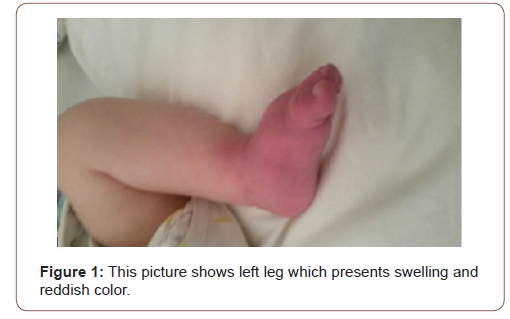
Our patient is the first child of an Albanian couple without consanguinity. Birth weight was 2400g Pregnancy and delivery were normal. This child was breastfed, but his mother noticed poor weight gain and several episodes of diarrhea. He was sent at our center at the age of 4 months. He presented failure to thrive and sweat test resulted 130 mmol/L confirming cystic fibrosis. No history of important disease in family. The rest of examination was quite normal except mild hypoalbuminemia and hyponatremia. After two albumin infusions and regulation of electrolyte disbalance this child was discharged at home with Creon, vitamin ADK supplement every day and vitamin E 100 mg/day in alternate days . A month later this child is returned to our service because of dehydration, poor feeding and developed swelling and tenderness of left leg . Skin over the affected area was turned reddish (Figure 1). He presented pain during leg movements associated with warm skin over affected zone . According to his mother treatment was not administrated properly because of frequent vomiting and child refusal.

Hemorrhage and coagulation test showed: hemorrhage time 2 min; coagulation time 3 min 30 sek; PT 89.5%; APTT C 27 sek.; D dimer was positive. Testing for secondary infections showed: CRP negative. Tests for viral and bacterial agents: negative. Other tests: Homocysteine 6.8 micro mol/l; Protein C 61% (N 70-110%); Protein S 67% (N 65-58%). Level of E vitamin was 0.8 mcg/mL (3- 18.4 μg/dL). We excluded antiphospholipid antibodies (aPLs), FV Leiden and antithrombin deficiency. Doppler ultrasound of the leg showed a blood clot at distal part of posterior tibial vein, but overall flow of the blood is not being constrained or restricted. We started immediately rehydration 3 l/m2 correcting hyponatremia. At the same time, we restarted creon, vitamin A,D,K and vitamin E 50 mg per day making sure a proper administration. We used antibiotics to prevent infection. Blood clot dissolved and leg took normal color 4 days later. During follow up this boy didn’t show any thrombosis phenomenon.

irispublishers-openaccess-pediatrics-neonatal-care

Discussion

The incidence of venous thromboembolism is increasing in pediatric population especially in hospitalized children [3]. Children with cystic fibrosis have an increased risk of thrombosis due to central venous catheters, as well as acquired thrombophilia secondary to inflammation or deficiencies of anticoagulant proteins due to vitamin K deficiency and liver dysfunction [4]. In our case we excluded these etiologies because our patient had not a central vein, he had a normal PCR excluding a severe inflammation. According to normal INR we logically can say that is excluded deficit of vitamin K Liver function was also normal. Most of our tests regarding thrombophilia resulted negative. It is well known that dehydration remains a serious cause of deep vein thrombosis such as in our case [5]. The second point which is not frequently described in literature is the deficiency of vitamin E as a cause of thrombosis. Vitamin E is all the following eight compounds alpha, beta ,gamma, and deltatocopherol and alpha, beta , gamma , and delta-tocotrienol. Alpha - tocopherol is the only compound of eight that are known to meet human dietary needs. All of the vitamin E forms are absorbed in small intestine, and then liver metabolizes only alpha-tocopherol. The liver then removes and excretes the remaining vitamin E forms [6-9]. Vitamin E is a lipid soluble nutrient. Patient with cystic fibrosis fail to secrete pancreatic enzymes to absorb vitamins A,D,E and K. Vitamin E inhibits platelet adhesion by preventing oxidative changes to LDLs and inhibition of platelet aggregation by reducing prostaglandin E2 [10]. In our patient we found a level of vitamin E 0.8 μg/dL [11-13]. In infants a level of vitamin E lower than 3 μg/dL is considered as deficiency. In this context we started immediately a prompt administration of vitamin E 100 mg per day for 7 days and later 50 mg per day continuously. We didn’t start thrombolytic agents because of small age of our patient and lack of experience. Preferring a strategy wait and see the outcome of conservative treatment including aggressive rehydration, creon and vitamin supplements resulted very efficient.

Conclusion

Indications of Alfa Tocopherol Acetate (Vitamin E) in cystic fibrosis are well designed. Vitamin E is given by mouth 50mg/ once day adjusted as necessary in children 1 month to 1 years old with cystic fibrosis. Deficiency of vitamin E associated with other risk factor such as dehydration in our case may lead to deep vein thrombosis. A prompt treatment can avoid serious complications. Other studies should be done to confirm our findings.

To read more about this article....Open access Journal of Pediatrics & Neonatal Care

Please follow the URL to access more information about this article

https://irispublishers.com/gjpnc/fulltext/deficiency-of-vitamin-e-and-dehydration-as-acause-of-deep-venous-thrombosis-in-a-child.ID.000556.php

To know more about our Journals...Iris Publishers

To know about Open Access Publishers

Iris Publishers-Open access Journal of Biology & Life Sciences | Climate Change and its Impact on the Agricultural Sector

  Authored by  Zakaria Fouad Fawzy*, Abstract The agricultural sector is one of the sectors that will be negatively affected by this phenome...