Friday, September 29, 2023

Iris Publishers-Open access Journal of Ophthalmology & Vision Research | Pattern and Prevalence of Ocular Diseases in Secondary School Students in an Oil Producing Community in Rivers State, Nigeria


Authored by Elizabeth A Awoyesuku*,

Abstract

Aim: This study was carried out to determine the proportion of secondary school students with ocular disorders in two rural communities in Rivers State.

Study design: A cross sectional study Place and Duration of Study: Gokana Local Government area, Rivers State. Methodology: A cross sectional study on vision screening among secondary school students was carried out in Kegbara-Dere (K-Dere) and Begbara-Dere (B-Dere) Communities in Gokana Local Government Area of Rivers State between. A multistage sampling technique was used to recruit students aged 11-22 years (mean age 15.7 ± 2-08) in the 2 community secondary schools. A total of 263 students consisting of 146 (55.5%) males and 117(44.5%) females were screened. Statistical package for social science (SPSS 20) was used to analyze the data.

Results: Seventy one of the 263 students had ocular disorders giving a prevalence of 27.0%. Thirty-four (47.9%) of the students with ocular disorders were in the 14 - 16 years age range. Of the total students with ocular disorders, 45 (63.4%) were males and 26 (36.6%) were females. Males did not have statistically significant higher prevalence of ocular disorders (df=1, P=0.53). There were eight different ocular disorders observed in these students, refractive error (23; 8.7%) being the commonest followed by suspicious cupped disc (18; 6.4%). The prevalence of defective color vision and amblyopia were 4.6% and 1.4% respectively with male preponderance in defective color vision which was statistically significant (df=1, p=0.047). No student had a visual acuity < 6/18 or < 3/60 in the better eye. Only 32.4% (n=23) of those with ocular disorder actually complained of ocular symptoms.

Conclusion: The prevalence of ocular disorders in this study was 27% with uncorrected refractive errors ranking highest. School Eye Screening is necessary to reverse this trend.

Keywords: Ocular disorders; Rivers state; Rural communities; Secondary school students

Introduction

Vision screening in early childhood is a necessity as it enables a child attains his or her full potential; since most persons are visual learners, acquiring approximately 85 percent of all knowledge through vision [1]. it is therefore imperative that a possible visual problem be identified early and treated. However, in typical rural communities such as Kegbara-Dere (K-Dere) and Begbara-Dere (B-Dere) (in South-South Nigeria) where the poverty level is high and majority live on less than one dollar a day [2], access to an eye care specialist is limited and many children would have to continue with any poor eye condition they have into adulthood, sometimes dropping out of school altogether. If severe enough, many vision problems entail a variety of symptoms that greatly affect skills and systems related to learning [3]. It is known that youths from low income families suffer from a disproportionately high prevalence of educationally relevant vision problems and are at high risk of inadequate treatment of vision problem, in which low access to eye care services is a major contributing factor [4].

School eye screening has been acclaimed as one of the best ways to battle childhood blindness and one of the major means of attaining the aim of “Vision 2020 - The Right to Sight” which is a global initiative for prevention of avoidable blindness. The school presents a captive population that can be easily accessed, and intervention carried out where possible, as most children are commonly docile by nature [5,6].

About 80% of blindness in the world are due to avoidable causes [7]. Many of these ocular problems such as trachoma, glaucoma, cataract, refractive error that are amenable to interventional measures are detected through vision screening of selected population such as school children [8-10]. Eye surveys have the potential to promote normal vision, preserve healthy eyes and prevent blindness [11].

Screening is defined as the presumptive identification of individuals at risk in a population likely to be affected by asymptomatic or subclinical condition who can benefit by being further investigated [12]. It is quite disturbing that an estimated 500 000 children become blind each year and in developing countries up to 60% of them are thought to die within a year of becoming blind [7]. Almost half of all blindness in children-particularly those in the poorest communities is due to avoidable causes that are amenable to cost effective interventions [13]. Considering the concept of Disability--adjusted -life -years (DALY) which is described as a measure of the time lived with a disability and the economic loss incurred during the years,[14] It is therefore very important that children have very good vision considering they have a long time to live as the magnitude of associated economic loss is huge indeed [15].

One effective way to reduce this economic burden is prioritizing school eye survey by relevant authorities because vision screening can also serve as a public health intervention measure [16]. Available report shows that schools with long established tradition of engaging in vision screening have proven to be effective in detecting previously undiagnosed conditions including refractive errors [17].

Materials and Methods

A population - based cross-sectional Study. The sample size was determined by the Leslie-Kish formula: Araoye MO [18] to be 263 students. A multistage sampling technique was used to select students for this study. Health talk was given on the screening days. Pretested semi - structured questionnaires were administered to the students.

Visual acuity was determined separately for each eye at 6 meters outside the classroom using the Snellen’s chart. Where the visual acuity was 6/9 or less, a pinhole was presented for that eye and the test repeated. Ocular examinations were in one of the staff rooms of each school. Both eyes were checked for ocular alignment by Hirschberg Test.

Using ophthalmic loupe and pen torch, adnexa and conjunctiva were checked for abnormalities, the cornea for opacities. Pupil reaction to light was assessed and lens examined for any visible opacities. The anterior chamber depth was also assessed. Direct ophthalmoscopy in a chosen dark spot was done. The state of the retina and optic nerve were assessed.

Students with ocular disorders needing further investigation and treatment were referred to the University of Port Harcourt Teaching Hospital, Port Harcourt with a predesigned referral form.

• Inclusion Criteria

• Students enrolled in both secondary schools

• Enrolled students that parents/guardian gave consent

• 4.2. Exclusion Criteria

• Students above 22 years of age.

• Student whose parents/guardian did not give consent.

Results and Discussion

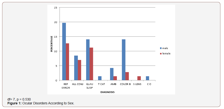
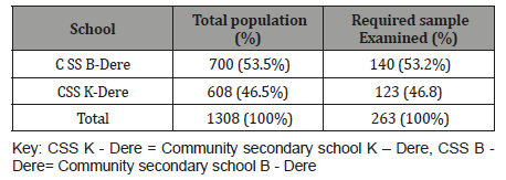
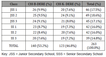
A total of 263 students were screened during a two-week period, giving 100% coverage of the estimated sample size for this study. (Table 1-6) (Figure 1).
irispublishers-openaccess-ophthalmology-vision-research

Table 1: School distribution of sampled students.
irispublishers-openaccess-ophthalmology-vision-research

Table 2: Distribution of Sampled Students According to Class.
irispublishers-openaccess-ophthalmology-vision-research

Table 3: Age and Sex Distribution of Sampled Students.
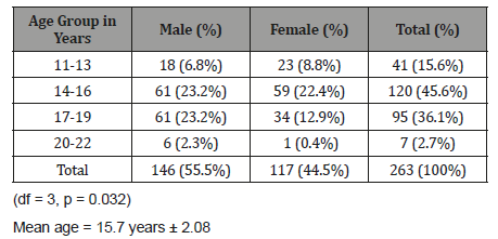
irispublishers-openaccess-ophthalmology-vision-research

Table 4: Ocular Symptoms among the Students.
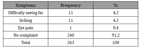
irispublishers-openaccess-ophthalmology-vision-research

Table 5: Types and Prevalence of Ocular Disorders.
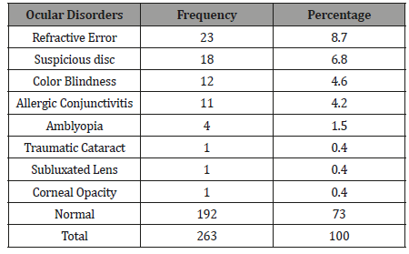
irispublishers-openaccess-ophthalmology-vision-research

Table 6: Ocular Diagnosis According to Age Group.
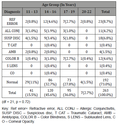
irispublishers-openaccess-ophthalmology-vision-research

Discussion

This study found the prevalence of ocular disorders in secondary school students in B-Dere and Kegbara-Dere communities to be 27.0%. This is similar to a study by Alakija W [19] involving secondary school students in Benin City where a prevalence of 25.6% for females and 19.9% for males was found. Other studies in Nigeria involving secondary school students by Ajaiyeoba AI, et al. [15], Adegbehingbe BO, et al. [16], and Abah ER, et al. [28] found a prevalence of 19.5%, 22.5%, and 22.6%, respectively. While Adio AO, et al. [21] found a prevalence of 9.4% among primary school pupil aged 5-15 years in Yenagoa, Bayelsa state. Wedner SH, et al. [22] in Tanzania reported a prevalence of 6.9% in a study among secondary school students. A strict comparison of these studies is difficult as there are some differences in the age groups studied, the geographic location, the case definition of eye disorders and sample size. In this study, the cut off level of defective vision was visual acuity <6/6 which was the same in the study by Alakija AI, et al. [19] while Adegbehingbe BO, et al. [16] used <6/9 as the cut off. In this study the age range was from 11 years to 22 years (mean age 15.7 years ± 2.08) which contrasts with Abah ER, et al. [20] (7-15 years).

The difference in sample size could also account for the differences in prevalence; in Ajaiyeoba AI, et al. [15], Adio AO, et al. [20], and Adegbehingbe BO, et al. [16] studies large sample sizes (1144, 1,242 and 1707 respectively) were used , compared to the small sample size of this study ( n = 263).The poor accessibility to eye care facilities in these rural communities could also account for the higher prevalence as most of the people in these communities are yet to fully utilize the existing community outreach program of the Department of Ophthalmology, University of Port Harcourt Teaching Hospital, which is often times disrupted because of incessant communal clashes.

In this study, (Figure 1) males had a higher prevalence (17.1%) of ocular disorders compared to females (9.9%). This difference was however not statistically significant (df =8, p=0.42). This contrasts with the studies by Ajaiyeoba AI, et al. [15] and Osunde EA, et al. [23] who reported a higher prevalence of ocular disorders among females, though more females were represented in their sample sizes, comparatively the finding of more ocular disorders among males in this study may also be due to their higher representation in the sample size. Most ocular disorders (Table 6) occurred in the 14-16-year age group (12.9%) which was not statistically significant (df = 24, p = 0.67), followed by the 17-19-year age group. This differs from the finding by Ajaiyeoba AI, et al. [15] who reported the highest prevalence among the 10-14-year age group, followed by the 15-19-year age group; again this variance may be due to difference in the age range and the age groups with largest sample size.

Only 32.4%(n=23) of those with ocular disorders actually complained of ocular symptoms this agrees with other studies by Alakija W [19] and Ajaiyeoba AI, et al. [15], where very few had ocular symptoms compared to disorders. The common ocular symptoms were ‘difficulty seeing far’ and itching. The most prevalent ocular disorder in this study was refractive error, followed by glaucoma suspect and color blindness. Similarly, Abah ER, et al. [20] also reported refractive error as the commonest ocular disorder followed by allergic conjunctivitis. However, the study by Ajaiyeoba AI, et al. [15] and Adegbehingbe BO, et al. [16] reported vernal conjunctivitis as the most common ocular disorder followed by refractive error.

The prevalence of refractive error in this study was 8.7%, which falls within the WHO range of 2-10% worldwide, [24]. most of the students (73.9% n =17) with refractive error had small refractive error (presenting VA 6/9 bilaterally). The observed prevalence of 8.7% in this study is lower than the prevalence of 13.5% and 13.9% reported in studies by Adegbehingbe BO, et al. [16] and Osunde ER, et al. [23] respectively. It was however closer to figures reported by Naido KS, et al. [25] in South Africa and that by Murthy (6.4%) Murthy GV, et al. [26] in India. The prevalence of suspicious disc of glaucoma (6.8%) in this study was higher than that reported by other studies [16,20]. This is about three times that reported by Pedro Egbe CN, et al. [27] in a study on the prevalence of glaucoma suspects in Ahoada East LGA Rivers state. This may be a true reflection of the disease in the communities studied.

The prevalence of congenital color blindness in this study was 4.6%, with a male preponderance which was statistically significant (df=1, p=0.047). This is expected considering that congenital defective color vision has X-linked pattern of inheritance, and may also be due to the higher representation of males in the sample. A lower prevalence of defective color vision has been reported by other workers [28,21,20]. The lower prevalence(4.2%) of allergic conjunctivitis in this study as compared to other studies may be due to the geographic location of these communities in the mangrove belt, where it rains all year round with few period of dryness and the age range falls into the group with lower prevalence in the natural history of allergic conjunctivitis. This finding agrees with the prevalence of allergic conjunctivitis reported by Adio AO, et al. [21] in study involving primary school pupils in nearby state of the same geographic zone.

The prevalence of Amblyopia was 1.5%(n=4) and was found within the range of 13-16 years in this study. This finding at late childhood can be explained by paucity of eye care service in the communities before the advent of the Outreach by University of Port Harcourt Teaching Hospital Ophthalmology Department. Subluxated lens, cataract and corneal opacity all shared a prevalence of 0.4%; they were not visually significant as visual acuity in the subluxated lens and the cataract (immature) was 6/9 which improved with pinhole to 6/6. Though subsistence farming is the main economic activity in both communities, the subluxated lens and cataract were due to domestic violence while for the corneal opacity (which was away from pupillary axis), a clear history could not be obtained, and the student was not aware of the pathology.

In this study no case of squint was observed which contrasts with the finding by Adio AO, et al. [21] (prevalence of 0.4%) in her study involving similar geographic zone, this may be explained by the difference in sample size.

The finding of students in their early twenties in this study, is not uncommon, this can be observed in other studies in Nigeria; [15,16,20] and Tanzania [22,29-39]. This late placement of children in schools may be dependent on the economic and educational status of their parents or guardians.

Limitations

This study was carried out limited resources; availability of portable slit lamp and Perkins applanation tonometer would have been of immense value in the clinical assessment of the sample population.

Conclusion

The prevalence of ocular disorders in this study was 27% with uncorrected refractive errors ranking highest. School Eye Screening is necessary to reverse this trend.

To read more about this article...Open access Journal of Ophthalmology & Vision Research

Please follow the URL to access more information about this article

https://irispublishers.com/wjovr/fulltext/pattern-and-prevalence-of-ocular-diseases-in-secondary-school-students.ID.000545.php

To know more about our Journals...Iris Publishers

To know about Open Access Publishers

Iris Publishers-Open access Journal of Pharmacy & Pharmacology Research | Is there any Relation Between Glucose in Urine with Music Listening During Travel?


Authored by Muhammad Imran Qadir and Maria Rizvi*,

Abstract

Music can be found almost everywhere being used for entertainment. People also listen to music while traveling. Glucose appears in urine due to hyperglycemia. Basic purpose of this study is finding any relationship between urine glucose and music listening during travel. Urine test was performed for this purpose. 5% students have glucose in urine and like music in their journey. 77% students also like music but have no glucose in urine. 18% students don’t like music and have no glucose in urine. Glucose in urine has no relation with music listening during travel.

Keywords: Hyperglycemia; Music listening

Introduction

Music is used everywhere for entertainment purpose. It can be played at work, home, gym, restaurant, cafes and during travel. People listen to music even for hours. Recent technology of mobiles has made access to music easier than before. As now people can listen to music wherever they are going and these days individuals are spending more time and money on it [1]. Music listening while traveling is a popular activity which is increasing day by day. Approximately 90% of the transport has musical manifestation. It influences a person traveling in a stress condition. Soft music helps to get rid off from anxiety and feel relaxed. But hard rock or heavy metal music puts negative effect on one’s mood [2]. Renal damage of proximal tubules or hyperglycemia causes glucosuria. Normally, all the filtered glucose is reabsorbed by these proximal tubules. Diabetes mellitus and stress causes hyperglycemia. Glucose starts appearing in urine when ultrafiltrate concentration and blood concentration increase from the absorption level of proximal tubules. Detection of glucose is done by enzymatic reaction of glucose oxidase that is specified for glucose. Shelf life of reagent pads is limited. So sunlight protection should be given. If concentration of glucose is low and ascorbic acid is in high concentration in urine than ascorbic acid interferes with this test and false negative results are produced. Formaldehyde also produces false negative results. For false positive results, sample must be contaminated with hypochlorite, chlorine or hypochlorite [3]. Basic purpose of this study is finding any relationship between urine glucose and music listening during travel.

Materials and Methodology

Required equipment for this test is gloves, urine sample, test strips, watch, clean surface, tissue paper and waste bag. Test was performed on hundred students selected randomly. Gloves were put on before starting the test. Test strip was completely soaked in urine for 2 seconds and then it was extracted from the container having urine sample. To remove the excess urine from the strip it was supported on the container’s mouth. Strip was placed on tissue paper to avoid mixing color pads. Time was observed through watch. About after one minute, reactions on reagent strip were appeared. Then this strip was matched with colors of strip container and all the results were recorded. After that all the used materials were thrown in the waste bag and hands were washed completely. Positive results mean there is some abnormality in the person. While negative results mean the person is normal.

Result

Relation of music listening while traveling with urine glucose is provided in Table 1. About 2% male students and 3% females like music and have glucose in urine while14% males and 63% females also like music but they don’t carry glucose in their urine. On the other side, 5% males and 13% females don’t like music during travel and have no glucose in urine (Table 1).

Table 1: Relation of urine glucose with listening to music while traveling.

irispublishers-openaccess-pharmacy-pharmacology

Conclusion

Urine glucose has no affinity with music listening while traveling.

To know about Open Access Publishers

Wednesday, September 27, 2023

Iris Publishers-Open access Journal of Rheumatology & Arthritis Research | Atypical Griscelli Syndrome Presenting with Immune Dysregulation, Systemic Granulomatosis and Normal Pigment Secondary to A Structural Variant in RAB27A


Authored by Gunderman L*,

Introduction

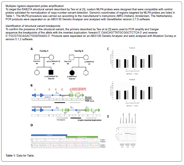
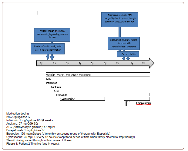
Griscelli Syndrome Type 2 (GS2) is a rare autosomal recessive disease characterized by organ granulomas, central nervous system inflammation, hemophagocytic lymphohistiocytosis (HLH) and partial albinism. Patients have immune dysregulation secondary to dysfunctions in natural killer (NK) cell and T cell cytotoxicity as a result of poor vesicular transport [1-2]. Typically, patients also have abnormal skin and hair pigmentation from accumulation of melanosome clumps in hair shafts and melanocytes. Homozygous or compound heterozygous pathogenic variants in RAB27A are associated with GS2. However, patients with atypical GS2 harbor a structural rearrangement in RAB27A not detected by typical genetic sequencing [3]. We present two cases of atypical GS2 (below and in supplement) characterized by systemic granulomatosis, neuroinflammation and normal skin and hair pigment, secondary to a structural variant (SV) in RAB27A and a second pathogenic sequence variant on the other allele of the gene.

Patient Case

Our patient (patient 1) is a previously healthy Caucasian female of distant Lithuanian descent who presented at age 13 with pneumonia requiring IV antibiotics and supplemental oxygen. Despite antibiotics, her respiratory status worsened. She was unable to wean from oxygen until the addition of high-dose corticosteroids. Extensive infectious workup was negative. Immunology workup revealed mild hypogammaglobulinemia, persistent lymphopenia and mild neutropenia. Biopsy of a skin nodule showed non-specific mild perivascular and interstitial dermatitis. Bone Marrow biopsy showed a hypo-cellular marrow with progressive multi-lineage hematopoiesis, but no evidence of leukemia. Nodular opacities were visualized on Chest CT. Of interest, the lung biopsy portrayed intra-alveolar macrophages and extensive non-necrotizing intraalveolar granulomas, concerning for interstitial lung disease (ILD).

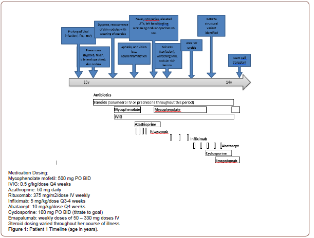
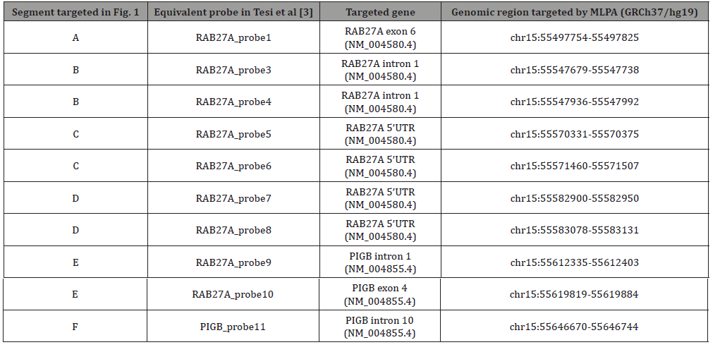
After discharge, she re-presented with dyspnea despite continued steroids. Intravenous Immunoglobulin (IVIG) and mycophenolate (a steroid-sparing agent) were started for suspected GLILD (granulomatous lymphocytic interstitial lung disease) and steroids were decreased. However, attempts to further wean steroids led to reoccurrence of hypoxia and skin nodules. The disease course was further complicated by an episode of aphasia, unilateral loss of peripheral vision and severe headache. Brain MRI revealed numerous subcortical, cerebral white matter and basal ganglia lesions concerning for an inflammatory or demyelinating process. Due to concerns for relapse, Mycophenolate was switched to Azathioprine and Rituximab, standard treatment for GLILD. Around this time, singleton WES resulted in multiple variants of uncertain significance (VUS), including a heterozygous likely pathogenic variant in RAB27A (c.259G>C, p. Ala87Pro). She was not diagnosed with Griscelli Syndrome because of the normal skin pigmentation and lack of a second pathogenic variant in the RAB27A gene. and Mycophenolate was restarted for possible better disease control. Yet after 4 weeks on Mycophenolate, episodes of confusion developed and nodular skin lesions worsened. The confusion was attributed to seizures based on Brain MRI results showing acute diffusion restriction in the left temporal lobe and insula, and she began antiepileptic therapy. In the following 6 months, the patient developed asymptomatic anterior uveitis of the eye, retinal granulomas and vasculitis. Prolonged steroid use caused significant side effects including insulin resistance, short stature, central obesity and mild compression fractures. She switched from Rituximab to Infliximab to Abatacept for immune suppression; all failed to gain disease control. Seizures worsened with attempts to wean steroids. Labs became significant for low CD107a (NK cell degranulation) and elevated serum neopterin (62 nmol/L, Reference range: <10.0 nmol/L), despite the normal soluble IL-2 receptor. Therefore, evaluation for a RAB27A SV was conducted via a multiplex ligation-dependent probe amplification (MLPA) assay using 10 probes in coding and non-coding regions of RAB27A and the nearby PIGB gene (Table 1). A complex duplicationinversion SV was found in the 5’ untranslated region (5’UTR) of RAB27A and confirmed by Sanger sequencing. The SV was classified as pathogenic. This, in conjunction with the likely pathogenic RAB27A variant found on WES, resulted in the diagnosis of atypical GS2. Parental genetic testing revealed the variants were inherited in trans. After diagnosis, she was switched from Mycophenolate to Cyclosporine for immune modulation, and Abatacept was discontinued after a 3-month trial. Despite changes in therapy, the neuroinflammation worsened and neopterin levels (76.3 nmol/L) were found elevated in the CSF. Given this progression, she started anti-interferon gamma therapy (Emapalumab; a cytokine therapy) as a bridge to stem cell transplant to reduce inflammation and control disease progression. While on this medication, the CSF neopterin levels declined and repeat MRI was stable. Stem cell transplant was completed. Unfortunately, the patient (Patient 2) did not survive complications from the post-transplant period (Figure 1).

irispublishers-openaccess-rheumatology-arthritis-research

Discussion

The RAB27A gene encodes the Rab27a protein and is important for vesicle transport and docking in a variety of cell types (platelets, leukocytes and melanocytes). Variants in RAB27A lead to GS2, resulting in an immune dysregulatory phenotype with increased susceptibility to infection, cytopenias, HLH, neurologic involvement and complete to partial albinism [2]. Lack of pigment and immunodeficiency in GS2 are linked as both result from a dysfunction in secretory vesicles, a component essential for the proper functioning of cytotoxic T lymphocytes, natural killer cells and melanocytes.

Unlike the variants in RAB27A associated with typical GS2, RAB27A structural variants associated with atypical GS2 are not easily detected. The SV found in the 5’ untranslated region (5’UTR) of RAB27A in our patients was previously reported by Tesi et al., in 5 children from 5 unrelated families of the Baltic Sea population who had characteristic findings of GS2 (immune dysregulation, including neuroinflammation, skin granulomas, and late-onset HLH) but the atypical finding of normal skin and hair pigment [3]. In these families, heterozygous carriers of the SV were not affected by disease, though those homozygous or compound heterozygous for the SV and a separate pathogenic RAB27A variant were affected by atypical GS2 [4]. Expression studies in a homozygous carrier of the SV, using RNA from peripheral blood mononuclear cells, provided evidence that the inverted duplication interrupts the transcription start site of the longest RAB27A transcript (NM_183235.2) [5]. This transcript is the predominant RAB27A isoform in lymphocytes but is expressed to a lesser extent in melanocytes, according to FANTOM CAGE data and expression studies in melanocytes from control individuals [6].

The custom designed targeted MLPA assay used to detect the SV in RAB27A was key to the diagnosis in these patients (Table 1). Typical next-generation sequencing (NGS) based assays are unable to detect the SV as non-coding regions of the genes are not routinely sequenced in panel testing or WES studies. Without the MLPA analysis, it would have been challenging to recognize that these patients had atypical GS2, due to lack of pigment abnormalities and also differences in age of presentation and variability in HLH as a predominant feature. Patients who present with skin granulomas, neurologic involvement, concern for HLH and ancestry in the Baltic Sea region should be evaluated for structural RAB27A variants, even with the absence of pigment abnormalities. If a RAB27A variant is found on NGS and phenotypic features of GS2 are present, a MLPA assay is necessary to evaluate for this SV. This case outlines the importance of early diagnosis in atypical GS2, reiterates the usefulness of targeted genetic testing for early diagnosis and offers a playbook of therapeutic options, including both the success and failures of targeted therapies.

irispublishers-openaccess-rheumatology-arthritis-research

Table 1:MLPA probes used in this study.

irispublishers-openaccess-rheumatology-arthritis-research

Iris Publishers-Open access Journal of Anaesthesia & Surgery | A Rare Cause of Naso Endotracheal Tube Obstruction: A Case Report


Authored by Walid Atmani*,

Abstract

Nasotracheal intubation is often indicated in oral and maxillofacial surgical procedures. It allows better use of the oral and intraoral sphere as well as surgical comfort, however, various complications are associated with nasotracheal intubation. We present a case of a complete obstruction of the nasal endotracheal tube by a nasal tissue during blind nasal endotracheal intubation in tumoral oral surgery

Keywords:Nasotracheal intubation; Probe obstruction; Sniff test

Introduction

Nasotracheal intubation is often indicated in oral and maxillofacial surgical procedures. It allows the manipulation of the mandible into occlusion with the maxilla without endotracheal tube interference and provides for easier visualization of the intraoral structures. various complications are associated with nasotracheal intubation including obstruction of the endotracheal tube with mucus, blood, pus, debris, granulation tissue, teeth, tonsillar tissue, or other soft tissue or bony tissue [1]. We present a case of a complete obstruction of the naso-endotracheal tube by a nasal tissue during blind naso endo tracheal intubation (BNTI) in tumoral oral surgery.

Case Report

A 34-year-old female patient was admitted to oral and maxillofacial clinic and scheduled for mandible tumor resection, The medical history was unremarkable; and physical exam was within normal limits.; Pre-anesthetic evaluation found a patient ASA 1 in good health, This patient presents a predictable intubation difficulty else where the mouth opening was normal. The cervical spine has normal mobility and thyro-chin distance is more than 6 cm, Mallam Pati II, a sniff test is carried out to detect the permeable nostril, which is right, the choice of nasotracheal intubation is above all for surgical comfort given the intraoral manipulation: cardiac and respiratory evaluation without abnormality

On the day of surgery, the patient was taken to the operating room and placed in the supine position on the operating room table. The patient underwent standard monitoring, electrocardiogram blood pressure and pulsatile saturation.

The patient had a heart rate at70 bpm, blood pressure at 110 /81 mmHg and 100% saturation in ambient air Premedication with 2mg of midazolam was given. Perfusion of 250 ml of serum saline 0,9 % through and intravenous line 18G, after check list verification of difficult intubation equipment (laryngeal mask, Eschmann guide, armed probe of different size 4-6 mm, plus tracheotomy table). A phenylephrine spray was applied to the nares bilaterally, After induction of anesthesia with propofol 3 mg/kg, fentanyl 3 μ/kg, the patient was easy ventilated by mask, rocuroniumwasgiven0.6 mg/kg.

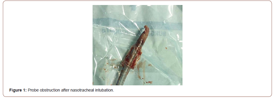
The dilation of the right nares after lubrification with xylogel proved difficult, orientation of the bevel of the probe towards the nasal septum, without forcing on the tube; after an audible cracking the passage of the probe proved much easier. Intubation with armed probe n° 6,5was confirmed with laryngoscopy, the end of the probe was not visible due to blood and secretions, the checkout of tooth positions without abnormality, then patient was manually ventilated with a feeling of resistance, then the patient flinched in mechanical ventilation, however, the breath sounds, chest excursions were absent no sibilant rale on auscultation, there were no capnography course. After one minute of verification, addition of 50 mg of propofol and 50 ug of fentanyl, a paroxysmal increase in airway pressure to the 50 cm H20 range was noted. Oxygen saturation remained at 94%; there was no cyanosis, and breathily ways absent. Endotracheal tube obstruction was thought to be the most probable cause, verification of the freedom of the probe was not carried out for fear of migration of a plug, the extraction of the intubation probe objectified the obstruction by soft tissue (Figure 1) a ventilation with the mask was carried out allowing an oxygenation increasing the oxygen saturation to 100% then reintubation by the same nares with armed probe n° 6 mm without Difficulty. To prevent laryngeal oedema a bolus of 120 mg solumedrol, Intubation was confirmed with bilateral equal breath sounds, good chest excursions, and capnography. Maintenance of anesthesia was performed by sevoflurane 1,5 % and oxygen 50%.

irispublishers-openaccess-anaesthesia-surgery

The Hemodynamic status was stable during the operation without episodes of hypotension or neo synephrine injection. Surgical procedure allowed complete excision in an operation that lasted 2h30 min Patient then admitted posting interventional surveillance room de curarized by neostigmine and extubated without incident. The SPO2 was at 99% in ambient air without dyspnea nor dysphonia.

Discussion

Nasotracheal intubation is the common method used to induce anesthesia in oral surgery patients. It has a distinct advantage of providing good accessibility and better isolation for oral surgical procedures. It’s indicated in Intraoral and oropharyngeal surgery [2, 3], Oral route of intubation not possible due to trismus, In ICU as an alternative to tracheostomy for longer ventilation periods, Surgery of maxillofacial cases needing better surgical access, tonsillectomies, rigid laryngoscopy and micro laryngeal surgery. Its contraindications include previous history of old or new skull base fractures, Bleeding disorders predisposing NTI to epistaxis.

The occurrence of an unexpected desaturation in the operating room is a critical situation requiring a mucinous evaluation of the airway as well as a checklist of the anesthesia equipment and apparatus; it can occur during induction, after intubation or at a distance, Several causes can be related to an intraoperative desaturation. They can be related either to the anesthesia station: failure, lack of oxygen supply, or related to circuits: leaks in the circuit, esophageal intubation, extubation, selective intubation or an obstruction of the probe or related to the patient: bronchospasm, mismatch. The analysis of pressures, spirometry, capnography, and clinical examination of the patient can identify the responsible cause. The speed of analysis and treatment largely conditions the prognosis.

Blind nasotracheal intubation is a procedure frequently performed in otorhinolaryngologic surgery and maxillofacial, Complications associated with the introduction of the naso endo tracheal tube include: epistaxis; submucosal laceration or dissection; obstruction of the endotracheal tube with mucus, blood, pus, debris, granulation tissue, teeth, tonsillar tissue, or other soft tissue or bony tissue ;tracheal laceration with subcutaneous emphysema, mediastinitis, or even pneumothorax; cuff perforation by turbinate projections or with McGill forceps; septal hematoma; and esophageal intubation[5-7].

The obstruction of the intubation probe by a nasal turbinate inserted and blocked at the distal end of the probe is reported by several authors [8]. Knuth reported a case of obstruction by a turbinate which then migrated to the left lung bronchus and cause atelectasis of the entire left lung. Other causes of obstruction have been reported such as: polyp, blood clot and tumor fragment [9].

Harvey and Amorosa [10] have shown that in the case of nasotracheal intubation, the probe is contaminated by the pharyngeal tissues and secretions in 33% of the cases. This incidence increases to 57% if resistance is offered to the passage of the tube, and 69% of cases when there is bleeding and to 86% of cases when the last two factors are combined.

To prevent complications of nasotracheal intubation, several measures have been described, namely at the beginning the test of the permeable nostril which allows the use of the freest nostril, the nebulization of vasoactive drug to reduce bleeding and the use of gel to facilitate the introduction of the probe which should be as small as possible.

There is another factor that should be considered before nasal intubation. In a large proportion of such patients, one nasal passage is smaller than the other. Thus, there is a 50% chance of inserting a nasal trachea tube in the narrower of the two. Having the patient sniff test [11] and inspection of the nares will facilitate the choice of the permeable nostril and avoid complications like bleeding and mucus laceration.

In a study realized by smith to identify the patent nostril for nasotracheal intubation , 61% of the 75 patients presenting for nasotracheal intubation had one nostril that was more suitable for intubation than the other on fibre optic examination, The tests comprised estimation of the rate of air flow through each nostril during expiration by palpating the passage of air when the contralateral nostril was occluded, and asking for the patient’s assessment of air flow through the nostrils, following the administration of a vasoconstrictor. After each test, noses were classified as left or right nostril clearer or nostrils equally clear. After the induction of general anesthesia, bilateral naso endoscopies were performed and videotape recordings of these were later analysed by an otolaryngologist who had no knowledge of the test results. Intranasal abnormalities were identified, and noses were again classified as left or right nostril clearer or nostrils equally clear. There was no significant difference between the overall diagnostic success rates of the two tests (44% and 47%, respectively) [12].

For that Rector suggested a series of measures to prevent obstruction of the intubation tube:[13] sniff test use of an appropriate diameter. Orientation of the bevel of the probe towards the nasal septum, and its return to normal position once the horns are crossed. Avoid forcing on the tube ensure a laryngoscopy of the freedom of the distal end of the probe before it crosses the vocal cords. Kawamoto suggested the protection of the light of the probe by a Foley probe during its nasotracheal passage. Intubation under a fiberscope, by allowing visual control of all stages of intubation, is avoided when this type of complication is indicated [14].

Conclusion

All these precautions should make it possible to avoid a rare complication, which should be considered in the event of ventilatory disorders occurring during the course of nasotracheal intubation.

To read more about this article...Open access Journal of Anaesthesia & Surgery

Please follow the URL to access more information about this article

https://irispublishers.com/asoaj/fulltext/a-rare-cause-of-naso-endotracheal-tube-obstruction-a-case-report.ID.000551.php

To know more about our Journals...Iris Publishers

To know about Open Access Publishers

Iris Publishers-Open access Journal of Biology & Life Sciences | Climate Change and its Impact on the Agricultural Sector

  Authored by  Zakaria Fouad Fawzy*, Abstract The agricultural sector is one of the sectors that will be negatively affected by this phenome...