Authored by Walid Atmani*,
Abstract
Nasotracheal intubation is often indicated in oral and maxillofacial surgical procedures. It allows better use of the oral and intraoral sphere as well as surgical comfort, however, various complications are associated with nasotracheal intubation. We present a case of a complete obstruction of the nasal endotracheal tube by a nasal tissue during blind nasal endotracheal intubation in tumoral oral surgery
Keywords:Nasotracheal intubation; Probe obstruction; Sniff test
Introduction
Nasotracheal intubation is often indicated in oral and maxillofacial surgical procedures. It allows the manipulation of the mandible into occlusion with the maxilla without endotracheal tube interference and provides for easier visualization of the intraoral structures. various complications are associated with nasotracheal intubation including obstruction of the endotracheal tube with mucus, blood, pus, debris, granulation tissue, teeth, tonsillar tissue, or other soft tissue or bony tissue [1]. We present a case of a complete obstruction of the naso-endotracheal tube by a nasal tissue during blind naso endo tracheal intubation (BNTI) in tumoral oral surgery.
Case Report
A 34-year-old female patient was admitted to oral and maxillofacial clinic and scheduled for mandible tumor resection, The medical history was unremarkable; and physical exam was within normal limits.; Pre-anesthetic evaluation found a patient ASA 1 in good health, This patient presents a predictable intubation difficulty else where the mouth opening was normal. The cervical spine has normal mobility and thyro-chin distance is more than 6 cm, Mallam Pati II, a sniff test is carried out to detect the permeable nostril, which is right, the choice of nasotracheal intubation is above all for surgical comfort given the intraoral manipulation: cardiac and respiratory evaluation without abnormality
On the day of surgery, the patient was taken to the operating room and placed in the supine position on the operating room table. The patient underwent standard monitoring, electrocardiogram blood pressure and pulsatile saturation.
The patient had a heart rate at70 bpm, blood pressure at 110 /81 mmHg and 100% saturation in ambient air Premedication with 2mg of midazolam was given. Perfusion of 250 ml of serum saline 0,9 % through and intravenous line 18G, after check list verification of difficult intubation equipment (laryngeal mask, Eschmann guide, armed probe of different size 4-6 mm, plus tracheotomy table). A phenylephrine spray was applied to the nares bilaterally, After induction of anesthesia with propofol 3 mg/kg, fentanyl 3 μ/kg, the patient was easy ventilated by mask, rocuroniumwasgiven0.6 mg/kg.
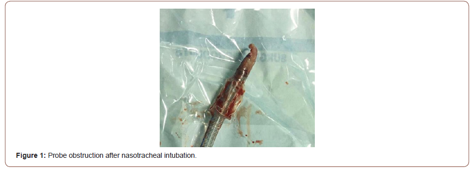
The dilation of the right nares after lubrification with xylogel proved difficult, orientation of the bevel of the probe towards the nasal septum, without forcing on the tube; after an audible cracking the passage of the probe proved much easier. Intubation with armed probe n° 6,5was confirmed with laryngoscopy, the end of the probe was not visible due to blood and secretions, the checkout of tooth positions without abnormality, then patient was manually ventilated with a feeling of resistance, then the patient flinched in mechanical ventilation, however, the breath sounds, chest excursions were absent no sibilant rale on auscultation, there were no capnography course. After one minute of verification, addition of 50 mg of propofol and 50 ug of fentanyl, a paroxysmal increase in airway pressure to the 50 cm H20 range was noted. Oxygen saturation remained at 94%; there was no cyanosis, and breathily ways absent. Endotracheal tube obstruction was thought to be the most probable cause, verification of the freedom of the probe was not carried out for fear of migration of a plug, the extraction of the intubation probe objectified the obstruction by soft tissue (Figure 1) a ventilation with the mask was carried out allowing an oxygenation increasing the oxygen saturation to 100% then reintubation by the same nares with armed probe n° 6 mm without Difficulty. To prevent laryngeal oedema a bolus of 120 mg solumedrol, Intubation was confirmed with bilateral equal breath sounds, good chest excursions, and capnography. Maintenance of anesthesia was performed by sevoflurane 1,5 % and oxygen 50%.

The Hemodynamic status was stable during the operation without episodes of hypotension or neo synephrine injection. Surgical procedure allowed complete excision in an operation that lasted 2h30 min Patient then admitted posting interventional surveillance room de curarized by neostigmine and extubated without incident. The SPO2 was at 99% in ambient air without dyspnea nor dysphonia.
Discussion
Nasotracheal intubation is the common method used to induce anesthesia in oral surgery patients. It has a distinct advantage of providing good accessibility and better isolation for oral surgical procedures. It’s indicated in Intraoral and oropharyngeal surgery [2, 3], Oral route of intubation not possible due to trismus, In ICU as an alternative to tracheostomy for longer ventilation periods, Surgery of maxillofacial cases needing better surgical access, tonsillectomies, rigid laryngoscopy and micro laryngeal surgery. Its contraindications include previous history of old or new skull base fractures, Bleeding disorders predisposing NTI to epistaxis.
The occurrence of an unexpected desaturation in the operating room is a critical situation requiring a mucinous evaluation of the airway as well as a checklist of the anesthesia equipment and apparatus; it can occur during induction, after intubation or at a distance, Several causes can be related to an intraoperative desaturation. They can be related either to the anesthesia station: failure, lack of oxygen supply, or related to circuits: leaks in the circuit, esophageal intubation, extubation, selective intubation or an obstruction of the probe or related to the patient: bronchospasm, mismatch. The analysis of pressures, spirometry, capnography, and clinical examination of the patient can identify the responsible cause. The speed of analysis and treatment largely conditions the prognosis.
Blind nasotracheal intubation is a procedure frequently performed in otorhinolaryngologic surgery and maxillofacial, Complications associated with the introduction of the naso endo tracheal tube include: epistaxis; submucosal laceration or dissection; obstruction of the endotracheal tube with mucus, blood, pus, debris, granulation tissue, teeth, tonsillar tissue, or other soft tissue or bony tissue ;tracheal laceration with subcutaneous emphysema, mediastinitis, or even pneumothorax; cuff perforation by turbinate projections or with McGill forceps; septal hematoma; and esophageal intubation[5-7].
The obstruction of the intubation probe by a nasal turbinate inserted and blocked at the distal end of the probe is reported by several authors [8]. Knuth reported a case of obstruction by a turbinate which then migrated to the left lung bronchus and cause atelectasis of the entire left lung. Other causes of obstruction have been reported such as: polyp, blood clot and tumor fragment [9].
Harvey and Amorosa [10] have shown that in the case of nasotracheal intubation, the probe is contaminated by the pharyngeal tissues and secretions in 33% of the cases. This incidence increases to 57% if resistance is offered to the passage of the tube, and 69% of cases when there is bleeding and to 86% of cases when the last two factors are combined.
To prevent complications of nasotracheal intubation, several measures have been described, namely at the beginning the test of the permeable nostril which allows the use of the freest nostril, the nebulization of vasoactive drug to reduce bleeding and the use of gel to facilitate the introduction of the probe which should be as small as possible.
There is another factor that should be considered before nasal intubation. In a large proportion of such patients, one nasal passage is smaller than the other. Thus, there is a 50% chance of inserting a nasal trachea tube in the narrower of the two. Having the patient sniff test [11] and inspection of the nares will facilitate the choice of the permeable nostril and avoid complications like bleeding and mucus laceration.
In a study realized by smith to identify the patent nostril for nasotracheal intubation , 61% of the 75 patients presenting for nasotracheal intubation had one nostril that was more suitable for intubation than the other on fibre optic examination, The tests comprised estimation of the rate of air flow through each nostril during expiration by palpating the passage of air when the contralateral nostril was occluded, and asking for the patient’s assessment of air flow through the nostrils, following the administration of a vasoconstrictor. After each test, noses were classified as left or right nostril clearer or nostrils equally clear. After the induction of general anesthesia, bilateral naso endoscopies were performed and videotape recordings of these were later analysed by an otolaryngologist who had no knowledge of the test results. Intranasal abnormalities were identified, and noses were again classified as left or right nostril clearer or nostrils equally clear. There was no significant difference between the overall diagnostic success rates of the two tests (44% and 47%, respectively) [12].
For that Rector suggested a series of measures to prevent obstruction of the intubation tube:[13] sniff test use of an appropriate diameter. Orientation of the bevel of the probe towards the nasal septum, and its return to normal position once the horns are crossed. Avoid forcing on the tube ensure a laryngoscopy of the freedom of the distal end of the probe before it crosses the vocal cords. Kawamoto suggested the protection of the light of the probe by a Foley probe during its nasotracheal passage. Intubation under a fiberscope, by allowing visual control of all stages of intubation, is avoided when this type of complication is indicated [14].
Conclusion
All these precautions should make it possible to avoid a rare complication, which should be considered in the event of ventilatory disorders occurring during the course of nasotracheal intubation.
To read more about this article...Open access Journal of Anaesthesia & Surgery
Please follow the URL to access more information about this article
To know more about our Journals...Iris Publishers
To know about Open Access Publishers


No comments:
Post a Comment