Friday, July 30, 2021

Iris Publishers- Open access Journal of Cardiology Research & Reports | Lp (A) and Aortic Stenosis


Authored by Scott W Shurmur*

Lipoprotein (a), commonly called Lp “little a”, is composed of an LDL-like particle, with an apolipoprotein B component covalently bound to apo (a). The structure of the apo (a) portion is somewhat similar to plasminogen, though number and repetition of the “Kringle” portions differs [1]. The atherogenicity of Lp (a) is increasingly appreciated. Recent genetic study confirms its strong association with clinical atherosclerosis. Additionally, some iso form of Lp (a) are strongly associated with calcific aortic stenosis (Figure 1).

Specifically, SNP rs 10455872 is strongly associated with markedly elevated Lp (a) levels (greater than 50 mg/dl) and the only monogenetic risk factor linked to calcific aortic valve stenosis in multiple racial groups. Mechanistically, autotaxin, which is involved in the lysophosphatidylcholine pathway, appears to be a promoter of inflammation, fibrosis and cell motility [2]. Several clinical trials have shown an association of elevated Lp (a) levels, and increased rate of progression of calcific aortic stenosis. Therapies targeting Lp (a) are in development, including highly specific antisense oligonucleotides [1].

To read more about this article.....Open access Journal of  Cardiology Research & Reports

Please follow the URL to access more information about this article

https://irispublishers.com/ojcr/fulltext/lp-a-and-aortic-stenosis.ID.000576.php

To know more about our Journals...Iris Publishers

To know about Open Access Publishers

Thursday, July 29, 2021

Iris Publishers- Open access Journal of Complementary & Alternative Medicine | Cleansing


Authored by Viviana Siddhi*

Short Communication

The happiness that we seek through accomplishments is more readily available to you when you let go of meeting outside expectations and begin to pay attention to what your mind, body and spirit actually need. You are likely to discover that the most important achievements of your life come relatively easily when you are truly taking care of yourself.

“Among the discoveries Dr. Bates made was how visual clarity changes – in the same person – from good to bad and back again, depending on that person’s physical and emotional state. He concluded that vision is not a static condition, but one that changes constantly.

Dr. Bates was the first ophthalmologist to make a scientific study of this phenomenon. His research shows how vision defects can be created and/or worsened by the stress of every-day situations. He also proved that these problems can be corrected by conscious and correct visual behavior [1].”

In the past I attended Meir Schneider’s workshop “Natural Cleansing” and later on I conducted several workshops “How to Improve Your Vision?”. In three months, my vision improved for one diopter. We must be conscious of doing eye exercises all the time. We should work on them in a way that makes them more alive. In every situation, there is a better way to use your eyes. There are many opportunities each day to practice new visual habits. I believe that you will find that even spending a few minutes daily with some of eye exercises will yield measurable results, in the form of diminished eye strain, clearer vision, and a greater ability to shift your focus without loss of clarity. It has been my experience that, when people do these Cleansing exercises correctly and regularly – mild vision problems improve rapidly, and poor vision improves noticeably over a longer period. We can see improvements in few months; however, it takes longer to stabilize these gains.

Poor vision, like most other sight problems, is caused by muscular imbalance and some nerve malfunction. It shows up at its worst in crossed eyes or double vision. In less severe cases, it may produce a slight blurring, headaches, or simply a feeling of strain. Even perfect vision is not perfect all the time.

“Before you undertake any techniques for acquiring perfect fusion, your eyes must be relaxed; so, first sun, palm, and do the long-standing swing. Do not attempt the fusion techniques at all when you are tired, ill, or under mental strain. When you undertake them, practice them in the order in which they appear bellow, progressing from easy to advance.

A. Sit facing a wall about six feet away, and hold a ruler or yardstick vertically, narrow edge forward, a foot from your nose.

B. Look up and down the stick three or four times, then up and down the wall the same number of times. As you scan the wall, the stick will seem to become two sticks separated by a space.

C. Alternate between stick and wall for three minutes at the start.

D. After a few days, lengthen the time and vary the distance from the wall.

Like all procedures for improving fusion – and sight in general – this technique should be practiced slowly, gently, in a state of relaxation [2].”

To prepare yourself for enjoyable, helpful experience, be sure you have all tight clothes loosened, and then get yourself in just as comfortable a position as you can. Close your eyes and inhale deeply and hold it for three seconds and then exhale slowly. Again,

breathe in deeply and exhale with a long, slow breath. Keep doing it several times. As you inhale, you bring more oxygen into your body, and as you exhale it causes your body to keep relaxing more and more. Now you can continue breathing easily and freely and can feel yourself becoming calmer and more peaceful. You are revealing signs that indicate you are moving into a very deep, peaceful state of relaxation, you can keep relaxing more peacefully, just happy to continue becoming calmer, more peaceful, and more at ease, continuing to breathe easily and freely. Rest the optic nerve, relax your nervous system and bring more circulation to your eyes. Your eyes need the conscious, active relaxations that comes with palming. It is impossible to palm too many times or for too long.

Simple instruction for palming:

A. Darken the room and sit at a table with a cushion on it. The point is to be able to hold your hands to your eyes without straining any part of your body and without putting pressure on your eyes and face.

B. Warm your hands by rubbing them together. Drop your shoulders and relax them.

C. Close your eyes. Lightly rest the heels of your hands on your cheekbones and cover your eyes with the palms of your hands. Your hands should not actually be touching your eyes.

D. Start to imagine an ever-deepening blackness.

Some people can improve their vision only with palming. The purpose of palming is to nurture your eyes in conscious way. It gives your eyes a rest in a way that even sleep does not. When you sleep, you are passive and are not in control of the visual images, mental activity, and rapid eye movement that occurs. When you palm, you are focusing on relaxing your body, and resting your eyes as they experience a complete absence of light. Palming is done with slow and deep breathing, relaxed shoulders, and completely relaxed hands. In some ways, palming is similar to energy massage or Reiki in that the heat and radiating energy of your hands can bring relief to tired and depleted eyes.

Palm at least 15-30 min several times per day. However much or little tension you have been able to release, continuing to practice palming will lead to a lifetime of healthier eyesight.

“Hypnosis is 90% belief, because the subject has to cooperate so much. Therefore, if the subject doesn’t believe that something can be fixed or accomplished using hypnosis, chances are, it won’t happen.

Be sure that your body language, vocal tone and attitude all reflect the possibility, probability and certainty of the suggestion you’re giving the subject, otherwise the subject will pick up on your incongruence [3].”

Trusting the process is essential. Healing work may cause temporary distressing symptoms that are all part of healing. The life-force and healing process work with complexity and wisdom that are beyond our conception or comprehension. Keep running the energy. Keep your breathing going. Connect your breathing to the sensations in your hands. The person in need of healing the eyes is a healer. The energy follows the natural intelligence of the body to do the necessary healing. Pay attention to body intelligence. With each breath you can track how the sensations in your hands change. As long as you are doing the breathing and connecting it to the energy, everything you try will work.

“We now stand on the brink of extraordinary breakthroughs in the art of hands-on healing. Human abilities that heretofore may well have been considered “science fiction” are in fact quite real and can withstand the rigors of scientific scrutiny [4].”

Each of our organs is specialized: it has its own particular function to perform and it can convey only its own particular type of sensation. In order to see clearly reality and truth, we need life experiences.

“The brain is capable of great things, but on condition that the solar plexus keeps it supplied with energy. The source, therefore, screen which manifests, expresses and publishes projected whatever the plexus feeds to it. The pictures projected onto the screen of the brain come from the plexus. Whether good or bad, they are all produced on the screen, just as at the cinema. The only difference is that, at the cinema, the masculine principle is the cameraman or his projector which projects the images onto the screen, and the screen represents the feminine principle, the matter onto which the spirit projects forces and energies [5].”

A number of factors can cause eyestrain, including, overuse from driving, reading, watching television, or working at a computer monitor. Air pollution and fatigue can also cause or aggravate eyestrain. When your eyes feel achy or strained, it is often a signal that you’re under stress and your whole body is tired. Other fatigue symptoms that usually accompany eyestrain include headaches, irritability, and tension in the back of the neck and shoulders.

Self-help techniques and acupressure points for relieving eyestrain can help you feel better when you are weary, overworked, or tense. Wash your hands with soap and water to prevent eye infections before you begin any eyes’ routine. Concentrate on breathing slowly and deeply throughout all of self-acupressure massage points. Deep breathing increases circulation to every part of your body, washes away tension, relieves depression, and infuses your body with vitality.

In just 10 minutes you can oxygenate your body and relieve depression by practicing deep breathing and relaxation (Figure 1).

“According to Dr. Serizawa, a Japanese physician, who regularly uses acupressure in his medical research and practice:

The ailments from which (acupressure) can offer relief are numerous and include the following:

Symptoms of chilling; flushing; pain, and numbness; headaches; heaviness in the head; dizziness; ringing in the ears; stiff shoulders arising from disorders of the automatic nervous system; constipation; sluggishness; chills of the hands and feet; insomnia; malformations of the backbone frequent in the middle age and producing pain in the shoulders, arms, and hands; pains in the back; pains in the knees experienced during standing or going up or down stairs [6].”

“This supremely important exercise creates the relaxation that amplifies the effects of all the other exercises. It will:

a. Rest the optic nerve

b. Relax your nervous system

c. Bring more circulation to your eyes [7].”

We can improve eyesight with regular eye exercises that should become again a healthy habit. Children has good healthy movements and we have to re-learn them as follows:

a. Blinking several times per second;

b. Using neck muscles instead of over strength eyes’ muscles;

c. Breathing deeply.

The human eye goes out of shape and bad vision occurs when the muscles around the eye become too tight. Alkalizing the body helps tremendously. A high percentage of living foods is called for as well as detoxification.

It isn’t the actual experience that shapes your self-image but the act of imagining yourself in a certain way that affects your selfimage. Your mind and body react to your internal self-image. So, if your self-image is that of a person with poor vision, your body will do everything that it can to make that true.

“Become a Doctor of Soul

One doctor says: “This is a case of appendicitis.”

Another doctor says: “This is a clear case of pleurisy.”

Third doctor says: “this is Hepatitis”.

When doctors differ, patients die.

If the patient dies, it is cholera or pneumonia.

If the patient survives,

It is simple gastritis or simple bronchitis.

Doctors still grope in darkness.

They make experiments and kill the patients.

A doctor of soul alone is infallible.

He is full of illumination and wisdom.

Therefore, become a doctor of soul [8].

To read more about this article.....Open access Journal of Complementary & Alternative Medicine

Please follow the URL to access more information about this article

https://irispublishers.com/ojcam/fulltext/cleansing.ID.000573.php

To know more about our Journals...Iris Publishers

To know about Open Access Publishers

Monday, July 26, 2021

Iris Publishers- Open access Journal of Archives in Biomedical Engineering & Biotechnology | Importance of Cumulus Cells in Oocyte Maturation and Conservation Strategies to Improve Vitrification Results


Authored by Fahiel Casillas*

Abstract

Vitrification is not a recent technique, it was firstly described in 1934, but was not successful until years later because the effects of the cryoprotectants (CPAs) used were unknown. Later, in 1985, Rall and Fahy [1] resumed the application of ultrafast cryopreservation by reducing the toxicity of the media and decreasing exposure times, modifying temperatures in order to improve their results [2]. In this way, the cryopreservation basis has been broadened with the aim of optimising conditions for the development of this process. Some of the main concerns are, the formation of intracellular and extracellular ice crystals, the CPAs toxicity and the osmotic changes during vitrification [3].

The large amount of lipids in porcine oocyte cytoplasm and the presence of the surrounding cumulus cells (CC), which are necessary for the meiotic resumption through gap communicating junctions, have been linked to less CPAs cell permeability and protection leading to cell damage [4]. However, previous studies indicate that the presence of the CC is essential to protect oocytes against damage induced by the vitrification procedure compared to the vitrification of denuded oocytes [5]. Also, it was reported that cumulus-oocyte complexes (COCs) vitrification reduces the viability of the CC while preserving oocyte viability [6]. This is important to be considered since these cells establish communication with the oocyte for the exchange of molecules such as: cAMP, ions, pyruvate, among others, which are required for the acquisition of its mitochondrial function, maturation and fertilizaton capacity. Therefore, maintaining the viability of the CC is a key factor for the vitrification of immature oocytes. Consequently, vitrification of immature porcine oocytes has been more difficult to achieve than other species such as sheep, cattle, hamsters, mice, human, among others [7-10] and this is why it still remains a challenge for cryobiologists.

Introduction

Vitrification can be used for the cryopreservation of cells, tissues, organs and even organisms. However, its current use is mostly associated to gametes due to its importance in human assisted reproduction techniques and animal production. This technique requires the use of CPAs added to media in high concentrations and devices with high vitrification-warming rates to reduce ice crystals formation. Due to their chemical characteristics, CPAs are highly toxic. If these substances are misused, they can cause cell damage and consequently cell viability loss.

Recently, several studies have been carried out to improve vitrification procedures with the aim of increasing viability and embryo production rates for obtaining live offspring. However, few studies have evaluated the effects on oocytes viability in the germinal vesicle (GV) stage after vitrification. This is because one of the main factors that impact on viability is the nuclear state of the cell. It is known that GV oocytes are more sensitive to cooling compared to those in metaphase II (MII) or embryonic stages [11]. This impact on viability is due to structural changes in the membrane of porcine oocytes as the transition temperature of the lipid phase is affected. It is currently known that the cell lipid content varies between species. In porcine the triglyceride content is higher compared to other domestic species. Therefore, due to the high amount of lipids and the presence of the CC, immature porcine oocytes have low CPAs permeability into their intracellular space [4]. Several studies have implemented techniques to reduce the lipid content in porcine, these techniques include mechanical delipidation, polarization of cytoplasmic lipid droplets by centrifugation and partial removal of lipids by micromanipulation [10,12,13]. This suggests that the low viability rates are due to insufficient permeability of CPAs.

Oogenesis

In mammals the oogenesis is produced by mitotic and meiotic divisions. In the first division process the chromosome number is maintained diploid (2n) but later by meiotic division processes it is reduced to half to produce haploid (n) gametes. Ovogonies are diploid cells (2n) that are produced from the primordial germ cells during embryonic development. Later, these cells undergo mitosis to become primary oocytes. The onset of first meiotic division, in most mammals, is arrested at the stage of diplotene at prophase I during fetal life and resumes until puberty is reached. The nucleus of the oocyte is known as the GV. During puberty, the action of gonadotrophins allows the process of meiotic resumption and extrusion from the first polar body at the stage of MII. Again, the meiosis is stopped until the processes of ovulation and fertilization are generated. If the oocyte is fertilized, the second meiotic division is completed, which includes the expulsion of the second polar body and the formation of the pronuclei. Finally, a zygote is obtained, which again presents a diploid chromosome number (2n) [14].

Oocyte Maturation

During the oogenesis, the oocytes gradually acquire nuclear and cytoplasmic maturation during their growth. Meiotic competence, which is the ability of the oocytes to resume meiosis and become nuclear mature, is acquired during folliculogenesis and is related to the formation of the antrum, where the oocytes reach approximately 80% of their final size. The development of the oocyte competence is related to the cytoplasmic maturation and refers to its capacity to be fertilized and develop an embryo capable of continuing its development to term and produce a living individual. Cytoplasmic maturation is acquired after the oocyte is meiotically competent. An oocyte that has acquired meiotic competence does not necessarily acquire cytoplasmic maturation. Meiotic maturation involves a cascade of processes, initiated with a surge of LH, promoting the progress of the oocyte to MII and to the extrusion of the first polar body. After the LH surge, an event that is essential for the resumption of meiosis, is the expansion of the CC, which is caused by the production of hyaluronic acid from these cells in response to gonadotropins [15].

Meiotic Arrest

Mammalian oocytes, unlike amphibians or echinoderms, have spontaneous maturation when released from the follicles and cultured in vitro. Therefore, it has been proposed that the follicular cells that surround the oocyte, the CC, are responsible for this arrest through the production and release of molecules such as cAMP or purines such as adenine and hypoxanthine. The inhibition of the follicular cells can be transmitted to the oocyte through the follicular fluid, by the communicating junction as it is the case of the mouse and the rat [14]. High levels of cAMP, supplied mainly by the CC, allow the oocyte to be held in prophase I and only released from the inhibitory effect when the cell communication is interrupted, and the cAMP levels drop. This decrease will inactivate cAMPdependent protein kinase (PKA) and through a signaling cascade, a phosphatase is activated that dephosphorylates the maturation promoting factor (MFP) [14].

Mechanism of Meiotic Arrest

The cAMP produced through the stimulation of the G-protein called Gs by the G-protein coupled receptor 3 (GPR3), is transported to the oocyte by the CC and/or captured by phosphodiesterase 3A inhibitors (PDE3A) in the follicular medium. AMPc-dependent protein kinase regulates MPF activity by CDC25 phosphatase, and Weel/Mytl kinase: cDC25 dephosphorylates cyclin-dependent kinase (CDK1), although Weel/Mytl phosphorylates it. High levels of cAMP result in the phosphorylation of CDK1 and the FPM complex, CDK1/cyclin B (CyB) becomes inactive, so that the oocyte remains arrested in the GV stage [16]. Another model proposed for the detention in prophase I is due to the action of purines, where adenosine stimulates adenylate cyclase through a membrane receptor, while hypoxanthine transferred by follicular cells through the communicating gap junctions prevents the hydrolysis of cAMP, by inhibiting cAMP-phosphodiesterase. The effect is that no resumption of meiosis is generated by the maintenance of high levels of cAMP [14].

Mechanism of Meiotic Resumption

Gonadotropins bind to their receptors on the CC producing an increase in the production of cAMP. Protein kinase A type II (PKAII) is activated by this increased cAMP in the granulose cells leading to the regulation of CREB in the transcription of the cre-gene, including the transmembrane precursors of epidermal growth factors (EGF), which are converted into mature peptides possibly by the protein kinase C (PKC) pathway. EGF factors bind to their EGFR receptor by activating mitogen-activated protein kinase (MAPK) using phosphatidyl inositol 3-kinase (PI3K)/protein kinase B (PKB) and down-regulation of steroidogenesis. Activation of MAPK and PI3K/ PKB decreases the level of cAMP in the oocyte by phosphorylation of connective 43 (Cx-43) and activation of PDE3A, respectively, followed by the FPM complex which is activated for meiotic resumption. The accumulation of cGMP by nitric oxide (NO) and/or natriuretic peptides by FSH stimulation serve to prevent premature oocyte maturation, while the decrease of this second message after LH treatment is involved in oocyte maturation and ovulation [16].

Follicular Cells and Oogenesis

The primordial follicles of mammalian ovaries are made up of an oocyte, detained at the prophase of the first meiotic division, surrounded by a single layer of follicular cells. The follicular cells emit cytoplasmic projections towards the oocyte establishing communicating unions. At the end of the follicular growth period, the oocyte stops growing and remains surrounded by several layers of follicular cells, known from that moment as CC or COCs [14]. Communication between germ cells and somatic cells is necessary for the survival of the oocyte and its development. Low-molecular-weight nutrient substances such as pyruvate and metabolic precursors such as amino acids and nucleotides are transferred through the communicating bonds. These substances make it possible to conclude the first meiotic division and acquire its fertilizing capacity. After an increase in LH levels, which induces ovulation, intercellular signals are interrupted by the production of hyaluronic acid by the CC, which causes its dispersion.

MPF was originally described in amphibian oocytes. It is a widely conserved protein complex from an evolutionary point of view, since it has been found in similar forms from yeasts to humans, both in somatic and germ cells. In mammals it has been found in species such as cattle and pigs. The fundamental role of MPF is to trigger the transition of cells from G2 to M phase in the cell cycle, but because cyclic changes in MPF activation are crucial in the regulation of the mitotic cycle in somatic cells, in oocyte maturation, it may involve similar cyclic changes in MPF activity in oocyte maturation [14].

Hormonal Role of Follicular Cells

Under the influence of gonadotropins, the follicles synthesize steroid hormones such as androgens and estrogens, which contribute to follicular development by inducing the proliferation and differentiation of granulosa cells through androgen receptors (AR) and estrogen receptors (ER), respectively. Theca cells produce androgens such as androstenedione under the influence of an LH stimulus, while granulose cells produce estrogens such as oestradiol, using androgens as a substrate, under the influence of FSH. Androgens participate in the proliferation and survival of granulosa cells through AR. Female rats with an AR deficiency are subfertile with a reduced number of antral follicles and ovulated oocytes, with a high rate of apoptosis in granulosa cells. Thus, specific AR for granulosa cells are essential for follicular development and survival [17]. Oestradiol is the predominant estrogen in terms of estrogenic activity. One of the main functions of pre-ovulatory granulosa cells is the synthesis of oestradiol. Within the follicle, oestradiol is produced by the enzyme aromatase, increasing the response of granulosa cells to gonadotrophins. Oestradiol plays an essential role in gonadotrophin-induced follicular differentiation. The activins and inhibins produced by granulose cells play essential roles in paracrine functions by regulating the LH-induced synthesis of androgens produced by theca cells, and thus ensure the supply of oestradiol. The activin-inhibin system not only regulates the proliferation of granulose cells, but also their differentiation and oocyte maturation (accelerated by the action of activin A) [17].

To read more about this article...Open access Journal of Archives in Biomedical Engineering & Biotechnology

Please follow the URL to access more information about this article

https://www.blogger.com/blog/post/edit/102220482794846253/4782918574391201596

To know more about our Journals...Iris Publishers

To know about Open Access Publishers

Friday, July 23, 2021

Iris Publishers- Open access Journal of Archives in Neurology & Neuroscience | Is there still some reason for classic presentation of Amyotrophic Lateral Sclerosis described in the literature?


Authored by Marco Orsini*

Opinion

Obviously, a lot has changed regarding Amyotrophic Lateral Sclerosis (ALS). That old and exemplary definition made by Chacort, of a neurological disease, of a progressive, degenerative and inexorable character, with “isolated” depletion of the upper and lower motor neurons no longer seems so limited to words. ALS is already considered a systemic disease; not only because of new findings that circumvent motor manifestations, but because of genetic codes that signal a form of evolution of hereditary cases. Speaking of that 5-8 year survival after the onset of the first signs and symptoms, the increase in new technologies, associated with a different way of medicating and treating, changed the natural history of book chapters; even with the cruelty with which it strips and compromises patients in various topics related to their functional abilities. We are looking for a picture of Amyotrophic Lateral Sclerosis described in detail in the literature [1-2].

Charcot3, one of the best neurologists in the world, should “return” with his knowledge and unique clinical dissection of his cases, and to her the right to redescribe this new spectrum of presentation of ALS. We focus on what is known about ALS and where the research is going - from the small steps of extending longevity, improving therapies, conducting clinical trials and compiling population registries, to the global goals of establishing measures that protect against onset and finding the triggers for this neurodegenerative disorder [4]. In cases of hereditary ALS, much has become history with the advancement of molecular biology. In patients with ALS/FTD (Fronto-Temporal Dementia), for example, the neuroinflammation mechanism characterized by innate immune responses of tissue-resident glial cells is uniformly present in end-stage pathology. Human imaging studies and rodent models support that neuroinflammation begins at early stages of disease pathogenesis. In addition, changes in circulating immune cell populations and cytokines are found in ALS/FTD patients, and there is evidence of an auto-inflammatory state (humoral system) [5]. Increased levels of pro-inflammatory IL-6, IL-8, and nitrite and significantly decreased endogenous antioxidant GSH levels could identify these humoral constituents as systemic biomarkers for ALS. However, systemic changes in IL-2, IL-5, and IL-6 levels determined between visits in ALS patients might indicate adaptive immune system responses dependent on current disease stage [6]. ALS has broad genetic and hypothesized environmental causes and phenotypic variability. And what about sporadic ALS cases? These are even more unpredictable. In clinical practice, diagnostic difficulties mostly arise with patients who present either with only upper motor neuron, or with only lower motor neuron signs. In addition, patients with ALS may also have other nonmotor findings, such as changes in sensitivity, cognition problems, dysautonomia. It may be difficult to distinguish ALS with clinically predominant lower motor neuron involvement from alternative diagnoses including spinal atrophies of adult onset, Kennedy’s disease, inclusion body myositis and motor neuropathies with conduction blocks 4-[7]. Although the degeneration predominantly affects the motor system, cognitive and behavioural symptoms have been described for over a century. In ALS, it is not possible to define a standard, nor to dare to scrutinize the survival of these patients. We have a certain medical rationality coupled with experience and dedication to the study of this merciless disease; but we still know little about its mysterious pathophysiological framework. We take advantage of and dedicate this opinion article to Professor Marcos RG de Freitas, neurologist, student and eternal apprentice of Professor Antonio Rodrigues de Mello.


To read more about this article...Open access Journal of Archives in Neurology & Neuroscience

Please follow the URL to access more information about this article

To know more about our Journals...Iris Publishers

To know about Open Access Publishers

Thursday, July 22, 2021

Iris Publishers- Open access Journal of Yoga, Physical Therapy and Rehabilitation | Use of Yoga Practices for Young Children


Authored by  Beth Elenko*

Abstract

The purpose of this opinion paper is to present the use of yoga practices in young children, primarily those younger than age 3 years old. The benefits that are seen in older children are just as beneficial to the young child who is typically developing as well as for those who are at risk or developmentally delayed who participate in early intervention. Young children can use yoga strategies to promote motor, and sensory skills in the early years. Even if modified from typical yoga poses to enhance their skills in a fun and creative way. Applying these yoga practices with young children has limitedly been studied, and further research on the benefits and uses is still necessary. Programs that are developing need to increase the evidence to support their practices.

Keywords: Young child; Yoga; Early intervention; Motor; Sensory development

Abbreviations: EI= early intervention Early interventionists: occupational, physical therapists, speech language pathologists or special educators toddlers -typically referred to as children ages 1-3 years.

Introduction

Yoga practices in children have been steadily increasing over the years. A 2017 National Health Interview Survey (NHIS) done every 5 years found that the use of yoga has increased in percentage almost tenfold in children ages 4-17 years [1]. Yoga is used with notable benefits throughout the lifespan. In children, yoga can help improve many skills such as decreasing anxiety, emotional regulation, improving body awareness, concentration, strength, flexibility, and behaviors that affect them. Through yoga we can improve our physical bodies, learn to breathe, and begin to take care of our bodies [2-5]. Most of the benefits of yoga have been demonstrated for school age children, even those with special needs [6-8]. Yet, yoga for young children under the age of 3 years has been studied the least in the literature [8-11], The purpose of this opinion paper is to increase awareness of using yoga practices with younger children.

Younger children especially toddlers, ages 1–3-year-olds can practice modified yoga poses. At a young age, children imitate the movements and behavioral practices of their caregivers whether it be grandparents, parents, teachers or siblings. It is what they naturally do and how they learn their basic developmental skills. Yoga itself benefits not only mindfulness, and reduces stress but improves skills in motor, attention, and behavior [5]. The benefits are growing and outweigh the negatives if there are any, although in one study it has been reported that yoga may be harmful to young children due to their lack of development [8]. Yoga not only benefits typically developing children, but children with developmental disabilities who receive early intervention (EI) (services for children who are at risk for or have developmental delays from birth to the age of 3 years old). I would argue that simple yoga postures mimic motor skills and promote weight bearing and elongation of muscles in young children as they develop. This can not only be beneficial, but essential to their sensory motor development. There are some children whether developmentally delayed or who engage in sedentary play that can engage in this an alternative activity. One that promotes their motor learning through yoga and that could be transferred to their development of motor skills.

While there has been little research on benefits of yoga for young children [8-11], there is much to support the use and benefits of yoga practices on improving mindfulness of children especially in schools [1-5]. These same benefits can apply to younger children, and they go beyond mindfulness. In young children, primary development is occurring in the first years of life. Yoga can promote and enhance what is already naturally occurring, while simultaneously teaching the child an activity to promote mindfulness as they grow. Not only can this be done as a family activity, but it can also be done to enhance skills that may be delayed in young children under proper direction from early interventionists [7]. Early interventionists promote motor and sensory skills to optimize development in young children which yoga can simultaneously nurture.

Motor

Think about babies first movements against gravity during the first year of life. Babies push on their arms and lift their heads as early developmental skills. As they gain these skills and begin to be more active in their movements, they strengthen their core muscles as a stable base for their arms and legs to move more freely. By the time they are physically ready to walk and stable in these muscles, they can begin to imitate simple yoga poses. Many yoga poses encourage these basic movements in creative and fun ways for toddlers [11].

Even those toddlers that are developmentally delayed need to work on these skills and practice them in a variety of ways. This can be done by imitating animals or silly postures. While doing this, it allows them to imitate postures that enhance their movement capabilities. Thus, strengthening their core muscles in their trunk, head and neck as well as their extremities. Toddlers can work on postural control, muscles, and joint stability as they maintain these postures. Toddlers who need to work on increased upper body stability through weight bearing benefit from a fun and creative way as they imitate these simple and modified yoga poses. The practice and repetition in learning these motor skills and yoga integrate beautifully while building their endurance for movements which they can carry over into their daily routines. Many poses focus on balance and stability of posture. The young child’s participation helps them with their balance and learning of these movement skills. This full body movement brings together their use of hands and feet enhancing bilateral integration and midline orientation. Overall, a toddler’s motor development can be enhanced by creative and fun mechanisms using yoga poses and principles at an early age [11].

Sensory

As the young infant develops control motorically in different postures, other important sensory skills are developing simultaneously. They develop skills in their visual system as they gain control of their head and neck muscles and begin to move in space. As the child grows and experiences control in more movement, they develop proprioception, a sense of body awareness to know where they are in space, in relation to the world around them and feel the surfaces their body touches in their environment. Yoga can provide this sense of body awareness as they hold various yoga positions which are essential for their sensory motor development. Their vestibular or movement system is enhanced by various postural changes, and head position as they explore different postures. The visual, proprioceptive, and vestibular systems are fundamental systems to development of a young child.

Discussion

While yoga has been known to improve mindfulness and decrease stress in persons across the lifespan, its benefits for learning have yet to be explored in young children [11]. Specifically, with young children with special needs in EI. Children today are affected by trauma and other stressors of their social-emotional development that would benefit from coping strategies [12]. These can include behavioral, breathing, focus, and meditation. Learning these at a young age can inspire children to implement practices across their lifetime. Modifications of yoga postures can be done to meet the needs of differing abilities as well. Many poses can be modified for young children who may not have the complete control or achieved the skill needed to maintain the poses.

Conclusion

Applying yoga principles to young children needs further research and continued practice to identify the benefits and strategies that work best for their young developing bodies. There is no reason to think that the benefits children ages 4 and up receive would not benefit typically developing children under 4. Further application for children with developmental delays under three with modifications is necessary under the guidance of their occupational and physical therapists. As programs develop and utilize these yoga poses to emulate benefits for younger children, it is critical that we increase the evidence to support them.

To read more about this article...Open access Journal of Yoga, Physical Therapy and Rehabilitation

Please follow the URL to access more information about this article

https://irispublishers.com/wjypr/fulltext/use-of-yoga-practices-for-young-children.ID.000551.php

To know more about our Journals....Iris Publishers

To know about Open Access Publishers

Wednesday, July 21, 2021

Iris Publishers- Open access Journal of Advances in Cancer Research & Clinical Imaging | A Simple Screening Test for Cancer


Authored by  AbulKalam M Shamsuddin*

Abstract

Comparative and correlative studies of the pathology and pathogenesis of colon cancer in animal models and human disease have resulted in conceptualization of ‘field effect” theory, and identification of a simple carbohydrate marker that is expressed early during carcinogenesis. This assimilated body of knowledge has resulted in development of a simple screening test for cancers of the lung, breast, colorectum, uterus, pancreas, and prostate. The marker galactose-N acetyl-galactosamine (Gal-GalNAc) is expressed in the cell surface and secreted glycoproteins of otherwise normal appearing tissues remote from cancer or precancerous lesions and is detected by enzymatic oxidation (10 minutes) followed by color reaction (1 minute). The high sensitivity, specificity, and cost-effectiveness of this point-of-care test makes it a great tool in our strategies for early detection, hence control of cancer. It would also reduce the number of unnecessary and expensive procedures, thereby decreasing the total national health-care cost to the societies and governments, globally.

Keywords: Screening test; Lung cancer; Breast cancer; Prostate cancer; Colon cancer; FOBT

Abbreviations: AOM: azoxymethane; FOBT: fecal occult blood test; Gal-GalNAc: galactose-N acetyl- galactosamine; GOS: galactose oxidase Schiff; MNNG: N-methyl-N’-nitro-N-nitroso-guanidine; NCI: National Cancer Institute; T-Ag or TF-Ag: Thomsen-Friedenreich antigen

Introduction

Regrettably, in 2020 approximately 10 million people were killed by cancer; now-a-days there is hardly a family that is not affected by cancer. Additionally, the healthcare cost, and loss of productivity owing to morbidity and poor quality of life, has been immensely draining to families, societies, and governments alike, everywhere. This scourge has been around for a very long time with no end in sight. As in SARS-Cov-2 now, in 1971 President Richard Nixon had declared ‘War on Cancer’ to combat this menace. Alas, 50 years later, conquering cancer remains an elusive goal despite progress in treatment, albeit modest. Again, akin to SARS-Cov-2, prevention is our best alternative. Cancer prevention includes (a) detection at the very early stage of the disease (secondary prevention) to reduce cancer mortality and increase the survival rate of patients; and (b) etiology prevention or primary prevention to reverse precancerous lesions or in situ carcinomas (a cancer that is still confined) to normal or stop them from progressing to invasive malignancies in populations at high risk – ‘nipping in the bud’. Thus, early detection is fundamental to prevention, and the key is to find the marker which is differentially expressed in highrisk tissues (cancer and precancer) but not in normal. Biomarker(s) meeting this criterion is/are logical choice for establishing accurate methods to detect cancers at infancy; it may also help in monitoring the efficacy of chemoprevention program by serving as intermediate endpoint marker [1].

Fundamental to the success of prevention programs of any disease is the detection of the problems. It includes identification of people with existing disease and those who are at risk of developing the disease. Identification of people with cancer is relatively simple for most cancers because of signs and symptoms from the disease; the individual, now a patient seeks medical attention. Extensive work-up including a battery of diagnostic tests are performed and appropriate therapy is administered; regrettably, it is usually too late for many, if not for most as it does not prevent the disease. Hence the adage: an ounce of prevention is better than a pound of cure. For an effective prevention program, we must actively seek individuals with cancer or high risk thereof from an apparently healthy noncompliant population. This is done by separating i.e., screening individuals into groups with high and low probability of cancer with the help of rapid, simple, accurate, and inexpensive tests (screening tests). Implicit in the definition of screening is a promise that there is a benefit for those who participate; they will be followed with further diagnostic tests and future management of the problem. But a screening test is never intended to give the full diagnosis, hence the distinction from diagnostic tests [2]. An individual who is screening test positive will need to undergo diagnostic procedures to confirm the presence of the disease. Ideal screening tests should have a high sensitivity (proportion of diseased subjects who are test- positive) and specificity (proportion of non-diseased subjects who are testnegative), be simple and non- invasive or minimally invasive, easy to administer – therefore enjoying a high acceptability amongst populations and of course cost-effective. It would not be successful if it is shunned for discomfort, cultural, religious, or other reasons or just too expensive [2]. Currently, except for colon cancer, there are no true good screening tests for other cancers at early stages. Imaging technologies such as mammogram, chest X-ray, low dose CT etc. cannot detect early cancers, for a tumor must attain a minimum size before it can be ‘visible’ by these. And that is too late!

The Galactose Oxidase-Schiff Test

The Galactose Oxidase-Schiff Test is one such screening test that fits all the criteria of an ideal screening test as it is rapid, simple, non-invasive, easy to administer, and inexpensive. Its high accuracy has been consistently and reproducibly demonstrated independently by numerous investigators in three continents. That is not surprising because of extensive scientific research on carcinogenesis and operation of i) field-effect of carcinogens and ii) a marker differentially and specifically expressed during carcinogenesis, shared by both cancer and precancer, but not by normal or regenerating tissue.

Field-effect

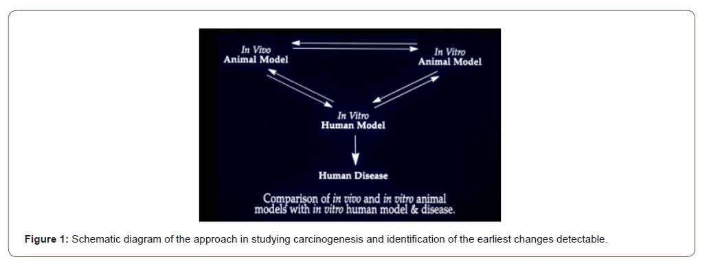
I studied early detection of cancer using colon cancer as a model since 1975 at the University of Maryland School of Medicine as my PhD dissertation. The advantages of colon as a model include a) relatively easy access to samples, b) well known precancerous changes (polyps) and conditions e.g., ulcerative colitis, Crohn’s disease etc.; c) well developed animal models with d) well characterized steps of carcinogenesis viz. initiation → promotion → progression; etc. Both in vivo and in vitro carcinogenesis experiments with rats and mice were performed to see how colon cancer forms in them, and to identify the earliest recognizable changes by microscopy and histochemistry [3,4]. Colon tissue from rats and human were cultured in the Petri dishes and exposed to the carcinogens N-methyl-N’-nitro-N-nitroso- guanidine (MNNG) for rat colon [3], and fecapentaene-12 a suspected human carcinogen, for human colon explants [5]. The findings were correlated with in vivo animal models, and ultimately to the human disease [6]: what does the human colon near and far from the cancer look like (Figure 1)?

irispublishers-openaccess-cancer-research-clinical-imaging

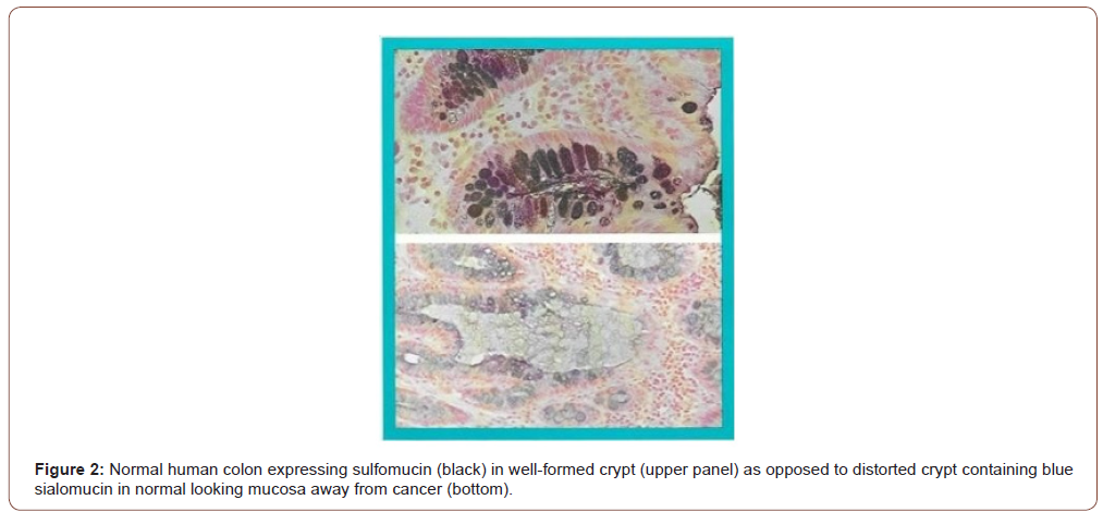
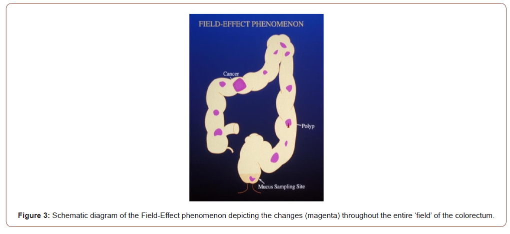
It has been observed that the earliest recognizable change in the carcinogen-exposed tissue is an alteration in the composition of secreted mucus that persists through precancerous and cancerous tissues. The microscopic and histochemical changes in the human colon away from cancer are identical to those seen in the colons of rats (mouse colon is distinctly different from human or rat colon [7]) treated with the carcinogen azoxymethane in vivo, or MNNG for rat explants and fecapentaene-12 for human explants in vitro in Petri dishes. These changes are therefore the earliest evidence (or markers) of cancer formation – even before a cancer has formed. Using high iron diamine alcian blue technique, the mucus in the colon away from the cancer that looked normal by naked eye appears as blue (Figure 2, lower frame) as opposed to black in truly normal human colon without cancer, in the upper frame (Figure 2). Also note that the colonic glands or crypts in a cancer bearing colon appear distorted – changes identical to the rat carcinogenesis models. It was reasoned that because of the generalized effect of the carcinogen throughout the entire field of the large intestine, it is most likely that the tissue away from an obvious cancer would be abnormal – Field-Effect. The carcinogens in our environment such as the air we breathe, the food we eat etc. expose the entire lung or large bowel. Therefore, it is logical that their effect would be observed throughout the exposed field. While the vast majority of the cells will undergo DNA repair, and other host defense mechanisms such as NK cell will prevent them to progress to cancer, a few cells will be promoted and even fewer progressed to precancer and cancer who may share the same early changes as the rest of the exposed tissue in the field. (Figures 2,3).

irispublishers-openaccess-cancer-research-clinical-imaging

irispublishers-openaccess-cancer-research-clinical-imaging

Field-Effect phenomenon indicates that the entire field bears the brunt of the carcinogenic assault and expresses variable changes throughout. In the schematic drawing of the large intestine (Figure 3), it is depicted that irrespective of where a cancer or a precancerous polyp may be, areas of the otherwise normal appearing mucosa will show expression of the marker perhaps in a patchy manner (it is not practical to sample the entire large intestine to see if every millimeter has the change; hence this assumption of “patchy”). Rectum being a part of the large intestinal “field” and a convenient sampling site is therefore likely to show the same changes. Since digital rectal examination is a part of routine physical examination (though not practiced diligently by many primary care physicians now-a-days), it is simple and noninvasive. It also allows the physician to examine the prostate in males, and uterus, cervix, and adnexa in females. Samples from lungs such as coughed up sputum, nipple aspirate from breast, endocervical mucus, or prostatic secretion are likewise simple and noninvasive.

The marker: gal-galnac

The observed mucin histochemical change has further been identified as due to a biochemical alteration in the cell surface and secreted glycoprotein – presence of the carbohydrate moiety D- galactose-ß-(1-3)-N-acetyl-D-galactosamine (Gal-GalNAc, also called Thomsen-Friedenreich antigen or T- Ag, or TF antigen, though it may be different). In normal cells, a terminal sialic acid blocks the T-Ag from being recognized by the lectin peanut agglutinin (PNA), or the enzyme galactose oxidase; in cancer and precancer owing to loss of the sialic acid it is now recognizable. D-galactose oxidase specifically oxidizes C-6 hydroxyl groups of D-galactopyranose and N-acetyl galactosamine residues of Gal-GalNAc, generating two vicinal aldehyde groups that react with basic fuchsin to give magenta/purple coloration. Thus, Gal- GalNAc can be visualized by a simple enzymatic reaction with galactose oxidase followed by Schiff’s reagent, resulting in the development of a simple test for early detection, initially of colorectal cancer [8] by using rectal mucus sample.

What is an Ideal Cancer Marker According to the National Cancer Institute (NCI)?

Kelloff et al of the Division of Cancer Control and Prevention (DCCP) at the National Cancer Institute [1] outlined six criteria for intermediate endpoint biomarkers of use in chemoprevention, and here is how Gal-GalNAc and STEDi live up to those expectations:

Is the intermediate biomarker differentially expressed in normal and high-risk tissue? YES!

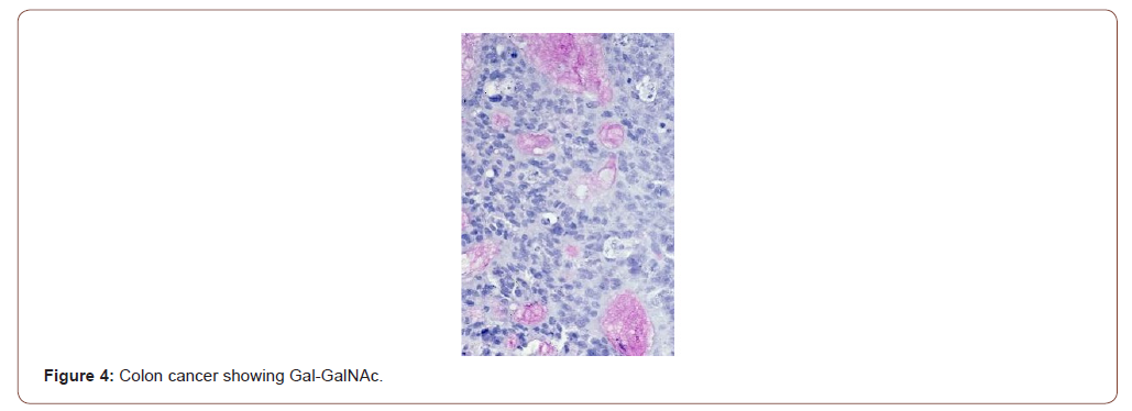
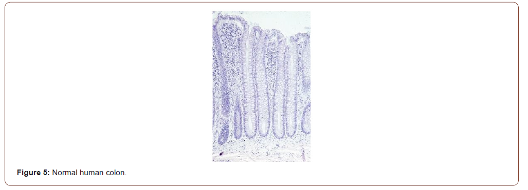
Figure 4 shows the expression of Gal-GalNAc (magenta) in a colon cancer but, not by the normal human colon (Figure 5). The normal tissues were obtained from healthy normal people without any cancer. Note the absence of magenta color in the mucus of normal colonic goblet cells. Also note that the colonic glands or crypts in truly normal humans appear uniform test tube shaped as compared to the those in cancer-bearing colon or in rats treated with the carcinogen azoxymethane (see Figure 4-6).

irispublishers-openaccess-cancer-research-clinical-imaging

irispublishers-openaccess-cancer-research-clinical-imaging

irispublishers-openaccess-cancer-research-clinical-imaging

At what stage of carcinogenesis does the marker appear?

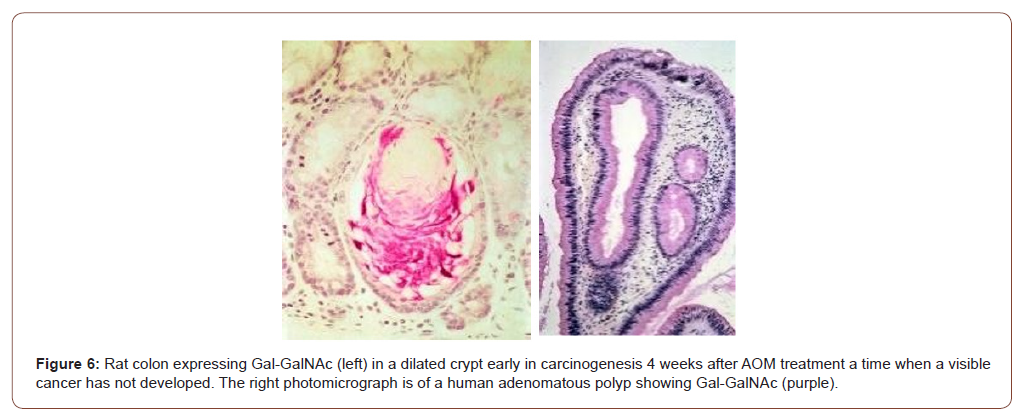
The earlier a reliable marker appears in the carcinogenic process, the greater is the chance for successful intervention. Answer: Gal-GalNAc is expressed very early during carcinogenesis. Not only the marker is expressed early during the carcinogenesis in rats in vivo (Figure 6 left panel) but also by human precancerous polyp (right panel appearing as purplish). The colors are slightly different owing to different batch of staining and tissue preparation. The mucus alteration is also observed in rat and human explants exposed to carcinogens in vitro [4,5]. Note the distorted appearance of the colonic glands as opposed to uniform test tube shaped ones in normal (Figures 5,6).

Does the marker and its assay provide acceptable sensitivity, specificity, and accuracy?

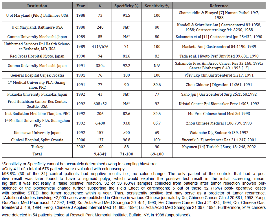
Answer: Both the marker [9,10] and the assay [11-14, Table I] enjoy 70-100% sensitivity and specificity. That it is not expressed by regenerating cells following wounding is an added evidence that Gal-GalNAc is carcinogenesis specific [10]. Please see the results on colon cancer in the following Table: results on other organ sites are provided after this: (Table 1)

Table 1: Performance Summary of GOS Test for Colorectal Cancer.

irispublishers-openaccess-cancer-research-clinical-imaging

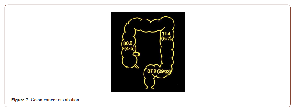
The proof of field-Effect: Schematic diagram of the human large intestine depicting the location of the cancers detected by the rectal mucus test. That the “field-effect” phenomenon is operational is proved by the detection of cancers in the various segments of the colon remote from the rectum where the mucus was sampled from. Note that 4 of 5 cancers (80%) of the ascending (or the right) colon, 5 of 7 (71.4%) of the descending (or the left) colon and 29 of 33 (87.9%) of the sigmoid colon were detected in the two studies by Sakamoto et al [11,12] (Figure 7). The widely used current fecal occult blood test (FOBT) for colon cancer screening has been notoriously inaccurate; “Occult blood testing is, at best an imperfect approach to the screening of colorectal cancer” concluded Dr. Ahlquist [15]. And that is not surprising since blood is not a marker of cancer or precancer. A newer test combining FOBT and DNA in stool appears to have better sensitivity and specificity albeit at a very high price.

irispublishers-openaccess-cancer-research-clinical-imaging

Gal-GalNAc is a common tumor marker: The usefulness of the tumor marker Gal-GalNAc in differentiating the benign from the malignant and pre-malignant lesions of the prostate was tested [16] yielding similarly high sensitivity and specificity (vide infra). Having determined that the principle is practicable in colon and prostate, studies were conducted on other cancers. The expression of Gal-GalNAc determined in a total of 133 tissue samples from 81 cases of the carcinomas of the breast, ovary, pancreas, stomach, and endometrium and 52 cases of respective normal controls [17]. None of the 52 cases of normal tissues (except 15 cases of stomach) showed expression of Gal-GalNAc. In contrast, 100% of adenocarcinomas from the breast (19 of 19), ovary (15 of 15), and pancreas (6 of 6), and 94.1% of stomach (16 of 17) cancers, and 91.7% (11 of 12) of uterine adenocarcinomas expressed Gal- GalNAc. The normal epithelia and their secretions in the vicinity of the carcinoma (within the “field”) in the breast, bronchus, endometrium, and pancreatic duct also expressed Gal-GalNAc in contrast to normal tissues obtained from non-cancerous individuals, which were totally non-reactive. Thus, the tumor marker Gal- GalNAc recognized by galactose oxidase-Schiff sequence was highly expressed not only by a variety of adenocarcinomas but also by the apparently normal-appearing epithelia and their secretions in the vicinity of carcinomas confirming the operation of a field effect phenomenon by carcinogenic agent(s) in these organs as well, setting the stage for identification of the marker in these secretions for mass screening for these cancers too [17].

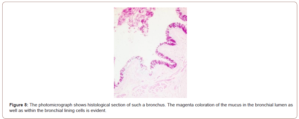
Studies on lung cancer: Twelve of 12 pulmonary adenocarcinomas expressed Gal-GalNAc. The bronchial tissue away from the cancer were available in 4 cases, all of whom also expressed the marker both in the epithelial lining cells as well as in the secreted mucus [17]. Coughed-up sputum therefore can be used to screen people for cancers of the lungs. Indeed, three clinical studies on lung cancer were performed on coughed up sputum [18-20]. Lai et al [18] reported the results of their study on sputum specimens from 116 healthy persons; and 216 cases of benign and malignant lung diseases were tested for the marker Gal-GalNAc. The result showed that 165 of the 182 patients (90.7% sensitivity) with lung cancer, confirmed by cytology and histology, had positive results, whereas 22 of 116 (19.0%) healthy controls were positive (81.0% specificity, Table 2). In 28 cases of patients whose sputum cytology showed various degrees of dysplasia-a precancerous condition that progresses to cancer, 21 were found Gal-GalNAc positive, of which 15 patients were identified to have lung cancer on further work-up! Thus, the concept of “false positive” as generally used does not apply to Gal-GalNAc and the GOS Test since they take the precancerous lesions and early cancers into consideration. In addition, three cases of early lung cancer in this study were also positive, supporting the fact that Gal-GalNAc is expressed at an earlier stage in the malignant process of the lung as well. The studies by Cox & Miller [19] and Miller et al [20] had smaller sample size and showed sensitivities of 64.7 – 88% and specificities of 77.8 – 93.6%. The test revealed 20 of 23 lung cancers among 76 patients. The other 53 patients were either healthy or had benign lung disease such as bronchitis. Even more germane to the issue of prevention is the fact that 13 of 15 cancers detected were early stage (Stage I and II). Of note is that a tumor must attain a minimum size before it can be detected by imaging techniques. And that is late! (Figures 8,9) (Table 2).

irispublishers-openaccess-cancer-research-clinical-imaging

irispublishers-openaccess-cancer-research-clinical-imaging

Table 2: Lung Cancer study.

irispublishers-openaccess-cancer-research-clinical-imaging

Breast cancer study: Gal-GalNAc is also expressed by the normal-looking breast tissue away from an obvious cancer by way of the field-effect phenomenon [17]. The marker was positive in 19 of 19 histological specimens of breast cancer giving it a 100% sensitivity. More importantly, and germane to our screening, the ducts away from the cancers and close to the nipple also express the marker both in the cells as well as in the secretions in the ducts. Thus, the nipple aspirate from a breast harboring a cancer should express the marker. Kumar et al [21] demonstrated that Gal-GalNAc (or TF antigen) is differentially expressed in nipple aspirate, albeit using a different technique – monoclonal anti- TF antibody. Nineteen of 25 cancer patients and none of the 25 healthy controls were positive yielding a 100% specificity and 76% sensitivity. The relatively low sensitivity of the test in nipple aspirate as opposed to tissue expression may be due to the use of different techniques for identifying the marker – galactose oxidase Schiff’s v monoclonal anti-TF antibody. Chagpar et al [22] also reported the utility of using nipple aspirate and Gal-GalNAc in screening for breast cancer. They investigated 23 women with biopsy confirmed, unilateral stage I or II breast cancer. They took samples (nipple aspirate by way of a suction cup attached to a syringe) from both breasts prior to surgery. Most, but not all the women were able to provide large enough fluid samples that could then be evaluated. Based on the resulting color of the test strips one could differentiate between a healthy and cancerous breast (Figures 10,11).

irispublishers-openaccess-cancer-research-clinical-imaging

irispublishers-openaccess-cancer-research-clinical-imaging

irispublishers-openaccess-cancer-research-clinical-imaging

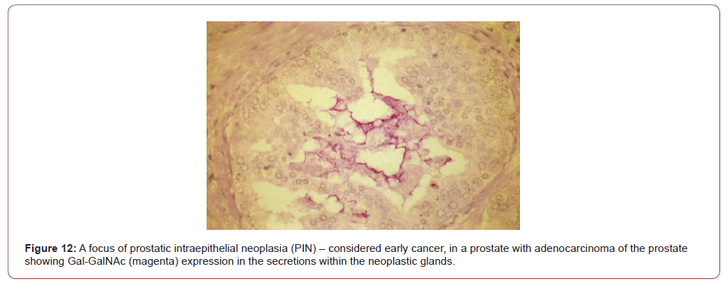
Prostate cancer study: The current screening test-PSA is inaccurate primarily because it is non-specific; its level goes up in benign conditions as well. The marker Gal-GalNAc is differentially and specifically expressed in prostate has also been observed in histological sections taken from 65 cases of adenocarcinoma [16]. While none of the 35 benign prostates and 11 foci of adenosis expressed Gal-GalNAc (100% specificity), 62 (95.4% sensitivity) of 65 adenocarcinomas expressed the marker. That the expression of the marker is not non-specific (meeting the NCI’s criteria #3) has been demonstrated by the absence in 25 samples of benign prostatic hyperplasia (BPH) and 11 of adenosis, neither of which are precancerous, nor were the normal prostates (10 samples) giving the marker a specificity of 100%. Notably, foci of prostatic intraepithelial neoplasia when present in some of the cancer specimens were also positive (Figure 12), demonstrating the useful of this marker in identifying early cancers [16]. As for colon, lungs, and breast, testing prostatic massage secretion or seminal fluid with GOS test should yield similar results (Figure 12).

Uterine cancer: Currently there are no screening tests for uterine cancers except for maybe the Pap smear wherein a uterine cancer could be detected by chance; but it is usually advanced when the cancer cells shed from endometrium are detected in Pap smear. Expression of Gal-GalNAc with high sensitivity (91.7%) was observed in 12 cases of uterine endometrial adenocarcinoma [17]. In all three cases where endocervical tissues was available, the endocervical mucus was positive for Gal- GalNAc. The photomicrograph (Figure 13) shows histological section of endocervix of a patient with endometrial adenocarcinoma; magenta coloration of the mucus in the endocervical glands and in endocervix is evident. Again, this supports the Field-Effect of carcinogenesis. More importantly, this makes it a simple, convenient, and non-invasive method of screening for endometrial adenocarcinoma while a gynecologist routinely collects samples for Pap smear (Figure13).

irispublishers-openaccess-cancer-research-clinical-imaging

irispublishers-openaccess-cancer-research-clinical-imaging

Pancreatic cancer: Pancreatic cancer is one of the deadliest of all cancers, primarily because of its silent inception; and owing to its location, signs and symptoms do not show till very late. All 6 of 6 cases of adenocarcinoma of pancreas were positive for Gal- GalNAc, and the normal ducts away from the cancers also showed positive reaction [17]. Thus, GOS test could be performed on fluid samples including pancreatic juice obtained during ERCP (endoscopic retrograde cholangiopancreatography) thereby providing additional value to the screening of the cancer. Figure 14 shows an adenocarcinoma of the pancreas with magenta color in the cancerous ducts and glands. (Figure14).

How easily can the marker be measured?

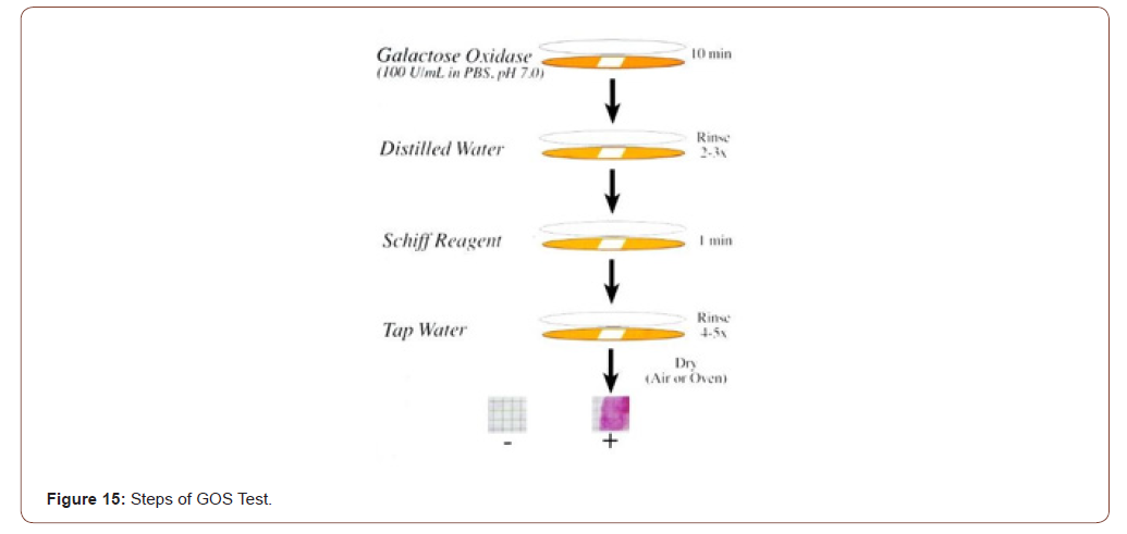
Answer: GOS test for detection of the marker is non-invasive done on mucus sample obtained during routine digital rectal examination, coughed-up sputum, nipple aspirate, prostatic massage secretion, endocervical mucus etc., and the entire assay period is ~15 min. This is the only point-of-care test for breast, lung, colon, prostate, and uterus; the results are available before the individual is ready to leave the doctor’s office. The test sample is placed on a special paper and reacted with galactose oxidase for 10 minutes, rinsed with distilled water, reacted with Schiff’s basic fuchsin for 1 minute, washed with tap water and dried. A pinkmagenta- purple color is positive, no color is negative (Figure 15). Positive indicates the presence of the marker Gal-GalNAc which is correlated with the presence of precancer, precancer or a high-risk thereof (Figure15).

irispublishers-openaccess-cancer-research-clinical-imaging

irispublishers-openaccess-cancer-research-clinical-imaging

Note: Though desirable, quantitative evaluation of the color reaction is neither practical nor meaningful as studies have shown no correlation between the intensity of color and the underlying disease.

Can the marker be modulated by chemo preventive agents?

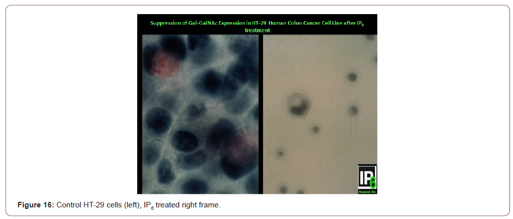
Answer: YES! Sakamoto et al. [23] and Yang & Shamsuddin [24] have demonstrated that; indeed Gal-GalNAc expression can be suppressed by the chemo preventive agent IP6 [25,26]. Figure 16 shows that HT-29 human colon cancer cells express the marker Gal- GalNAc (magenta color in mucus of cells in left panel). Following IP6 treatment, HT-29 cells terminally differentiate and produce mucin, yet not Gal-GalNAc, akin to normal goblet cells (right panel showing a differentiated HT-29 cell that has a mucus vacuole yet not expressing the marker [23,24]. Clinical studies are needed to validate this, but that would take a very long time. In this regard of particular importance is the study on colon cancer by Vucenik et al [13] where 32 of 53 (60%) samples collected from patients after tumor resection showed persistence of the biochemical change; 5 out of these 32 (16%) post-operative cases with positive GOS Test had tumor recurrence within a year. Thus, persistently positive test may serve as a predictor of tumor recurrence (Figure16).

Does modulation of the intermediate biomarker correlate with a decrease in cancer rate?

This would require a long time-years if not decades, and additional resources.

In conclusion, GOS test is a point-of-care screening test that is very simple, rapid, non-invasive, and inexpensive yet accurate for identifying asymptomatic people who may be at high risk of cancer or precancer of the lung, breast, colon, pancreas, prostate, and uterus. It is based on robust scientific background; researched and validated over three decades in >20 independent clinical studies in North America, Europe, and Asia. It exploits a disaccharide marker expressed early during carcinogenesis that persists in precancer and cancer and, satisfies all the practical rigid criteria set forth by NCI for an ideal marker. Given the fact that cancers of the lungs, breast, colon, prostate, and uterus comprise the most cancers, it is in the interest of public health that they are identified at a very early stage, or even before they are formed. The sooner it is used, the more lives could be saved from these cancers.

To read more about this article...Open access Journal of Advances in Cancer Research & Clinical Imaging

Please follow the URL to access more information about this article


To know more about our Journals...Iris Publishers

To know about Open Access Publishers

Iris Publishers-Open access Journal of Hydrology & Meteorology | Influence of Community Resilience to Flood Risk and Coping Strategies in Bayelsa State, Southern Nigeria

  Authored by  Nwankwoala HO *, Abstract This study is aimed at assessing the influence of community resilience to flood risk and coping str...