Tuesday, November 30, 2021

Iris Publishers-Open access Journal of Modern Concepts in Material Science | Heat Affected Zone in Welded Metallic Materials


Authored by Zakaria Boumerzoug*

Abstract

Welding is used extensively for pipe welding, aerospace, aviation, biomedical implants, fabrication of race cars, choppers, etc. Generally, the metallurgy of the welded joint performed by thermal fusion joining process can be categorized into two major regions, the fusion zone (FZ) and the heat-affected zone (HAZ). The heat-affected zone (HAZ) is a region that is thermally affected by the welding treatment. The main difficulty associated with welding is the prevention of unexpected deterioration of properties as a result of the microstructure evolutions which reduce the resistance to brittle fracture in the heat-affected zone (HAZ). Properties of the HAZ are different from those of the base material. According to the literature, the HAZ is the most problematic area in the high strength steels weld. For this reason, many research works investigated this critical zone in welded joint. The main research questions and results related to the HAZ will be presented.

Keywords: Welding; Heat affected zone; Microstructure; Mechanical properties; Thermal cycle simulation

Abbreviations: HAZ: Heat Affected Zone; Hv: Hardness Vickers; FZ: Fusion Zone; BM: Base Metal; T-HAZ: True Heat Affected Zone; PMZ: Partially Melted Zone

Introduction

Welding is a process of joining materials into unique piece. Welding is an enabling technology applied across almost all industries, from micro-joining of medical devices, electronics and photonics, to larger scale applications such as bridges, buildings, ships, rail, road transport, pressure equipment and pipelines [1]. Welding processes are divided into thermal fusion joining processes and solid-state joining processes. The most common processes of welding are thermal fusion joining processes such electric arc welding. This welding method is performed under high temperature conditions.

Heat generated during welding induces an important temperature gradient in and around the welded area. Generally, the metallurgy of the welded joint can be divided into two main zones, the fusion zone (FZ) and the heat-affected zone (HAZ). The HAZ is a zone which is outside the FZ of the welded joint that is thermally affected by the welding treatment. The HAZ is considered as a transition zone, because it is composed with the microstructure of the BM and the FZ. The properties of the HAZ are very important after performing a weld, because it is considered as a weaker zone, i.e.; the area of failure when the welded metal is submitted to hard conditions. For this reason, it is important to understand this critical zone in welded joint.

Microstructures of HAZ

The HAZ is the unavoidably heat treated area in the parent metal near the fusion zone during welding where structural transformations occur [2]. HAZ formed during welding is an area which some structural changes in the welded material take place as the result of experienced temperature [3]. Figure 1 shows how the HAZ in welded XC38 steel differs significantly from the base metal. There was a development of a recrystallization reaction in HAZ, with the partial dissolution of the colonies of pearlite (dark color).

Depending on the distance from the weld, the different parts of the HAZ can be affected differently during the welding process There are many descriptions of the HAZ, because it can be divided in different subzones and each subzone has its own microstructure. For example, It has been considered that the HAZ can be divided into four different zones [2], as shown in Figure 2, which are subjected to different heat treatments:

• Coarse grain zone

• The normalized zone

• The partially transformed zone,

• The annealed zone

However, according to Lippold [5], the HAZ was subdivided into two regions, the partially melted zone (PMZ) and the “true” heataffected zone (T-HAZ). The PMZ exists in all fusion welds made in alloys since a transition from 100% liquid to 100% solid must occur across the fusion boundary. According to Lippold [5], there are many possible metallurgical reactions in the HAZ: recrystallization, grain growth, phase transformations such as precipitation, and residual stress and stress relaxation.

Mechanical properties of HAZ

The mechanical properties and microstructures of the HAZ have its origin in the thermal heat treatment during welding and depend on the characteristics of the joint (position in the joint, thickness of the joint) and the heat input and the prior-heat treatment before welding (if it is applied) [6].

Controlling of HAZ

It has been concluded that by improving the microstructure of the HAZ, the properties of the welded joint can be improved [7]. The changes of microstructures in the HAZ depend on the level of thermal exposure and are varying with distance from the weld metal zone. High heat input increases the size of the HAZ which induces a low impact strength [8]. As reported by Gu et al. [9], the degradation in strength and toughness of welded joint, is generally happens in HAZ.

As many authors, Parmar and Dube [10] considered HAZ as the most complicated region. It is important to control its effects. HAZ is the most critical region in the welded joint as it affects the microstructure and grain size of weld bead. The main factors for improving welded joint quality are: welding process, material selection, and welding parameters. Concerning the welding parameters, they found that in order to study the heat-affected zone in welded carbon steel, following parameters were considered: welding current, welding speed, and arc voltage. These parameters can influence the Heat input and heat flow.

According to Lippold [5], heat input and heat flow conditions have an effect on the dimensions and nature of the HAZ. These dimensions are controlled by the temperature gradient from the fusion boundary into the surrounding base metal and the nature of the metallurgical reactions that occur over that temperature range. The size of HAZ has been studied by Śloderbach and Pająk [3]. They established an expression for determining the value of x of HAZ for a given time t and knowing diffusivity coefficient κ of given material:

[x/(2√κt )] ≈ 0.61

From this mathematic expression, the size of the HAZ x can be controlled by the time t during the welding. The x can be reduced by reducing the welding time.

Methods of Investigation of HAZ

It has been found that the study of the HAZ of real welded joints is not easy because of the narrowness of the HAZ [11]. Welding simulation is the appropriate technique to determine the different sub-zones in HAZ. This allows the prediction of the microstructure and the properties of these sub-zones [9]. Consequently, thermal cycle simulation in which the HAZ can be geometrically extended is the appropriate method in order to determine the different microstructures, which can be developed in real welded joints [12,13].

Hamza et al. [14] simulated HAZ of the welded stainless steel 304L by the thermal cycle simulation technique and compared it to the HAZ obtained from the real welded joint. They found that the simulated HAZ by the thermal cycle simulation technique has given more information. The HAZ is heterogeneous structure, because it is formed with different subzones. Raouache et al. [15], investigated the HAZ of welded 2014 aluminum alloy by the thermal cycle simulation of the base metal. They found that the HAZ is also a heterogeneous zone, because it is composed with different subzones and each subzone has a specific microstructure.

Conclusion

From the above literature, followings points can be summarized: • The HAZ is the critical zone in welded joint, for this reason it is necessary to control its effects.

• The HAZ is a heterogeneous zone and it can be divided into different subzones

• There are many possible metallurgical reactions in the HAZ

• The mechanical properties and microstructures of the HAZ depend on the heat input during welding

• Main factors for improving weld joint quality such as the welding process, the material selection, and the welding parameters.

• Welding simulation is a suitable technique for the investigation of the HAZ and to determine the various HAZ subzones.

To read more about this article....Open access Journal of Modern Concepts in Material Science

Please follow the URL to access more information about this article

https://irispublishers.com/mcms/fulltext/heat-affected-zone-in-welded-metallic-materials.ID.000566.php

To know more about our Journals...Iris Publishers

To know about Open Access Publishers

Monday, November 29, 2021

Iris Publishers-Open access Journal of Dentistry & Oral Health | Oral Cavity and HIV Infection


Authored by Marcelo Corti*

Introduction

The immunosuppression associated with the human immunodeficiency virus (HIV) and its consequence the Acquired immunodeficiency syndrome (AIDS), predisposes to a large series of opportunistic infections (OI) and neoplasms, such as Kaposi´s sarcoma (KS) and non-Hodgkin lymphomas [1,2]. These clinical complications are named as AIDS-defining diseases. AIDS defining illnesses include a group of pathologies whose incidence in the HIV individuals is much bigger compared with the general population. Oral cavity is a frequent engagement site in all stages of the natural history of HIV infection. The knowledge of these oral cavity clinical manifestations should suggest to the dentist the possibility of HIV infection and to investigate the serological status of the patient. For this reason, oral cavity should be carefully examined in all patients. Oral cavity manifestations of HIV infection should be classified in two groups; nonspecific clinical lesions and those directly related with the progressive immunosuppression.

Unspecific Lesions

Unspecific clinical lesions include the periodontal disease affecting the gums and dental support structures. These diseases have been classified as gingivitis, when limited to the gums, and periodontitis, when they spread to deep tissues. The linear gingival erythema (Figure 1), frequently observed in HIV/AIDS patients, is characterized by a red linear in the gingiva, near the dental arch. In patients with advanced stages of the retroviral disease ulcerative and necrotizing gingivitis can be observed. Clinical manifestations include fetid breath, gingival erosions, enamel alterations and tooth loosening [3]. HIV-infected patients can present different ulcerative lesions in the oral cavity. Recurrent aphthous stomatitis is characterized by single or multiple painful ulcerative lesions on the lateral edge of the tongue, oral mucosa and lips. These lesions can be treated with corticosteroids or thalidomide [4].

Sexually Transmitted Diseases with Oral Involvement

All the sexually transmitted diseases (STD) may have expression in the mouth; the mouth may be the site of frequent location of the syphilitic chancre (extragenital chancre) and the oral cavity can be the site of the secondary syphilis lesions. Mouth syphilitic chancre can be located anywhere into the mouth, including the tongue (Figure 2), the lips, the gums, the pharynx and the tonsils. Generally, syphilitic chancre is a single and painless lesion, but in conditions of immunodeficiency, as the HIV infection, the chancre lesions can be multiple. Symptoms of secondary syphilis include skin rash, swollen lymph nodes, fever and systemic symptoms. Secondary syphilis has multiple manifestations in the oral cavity, as the tongue papular lesions, the lichenoid lesions (opaline syphilis) (Figure 3), lingual depapilation, cracked papule at the lip commissure (Figure 4) and erosions in the oral mucosa [5].

Human Papillomavirus (HPV) is the etiologic agent of the most common sexually transmitted infection, named as genital warts or condylomata. Infection by the human papillomavirus (HPV) can also present intraoral warts (Figure 5) generally located in the oral mucosa [6]. Additionally, HPV is related with the pathogenic of 45% to 90% of oropharyngeal squamous cell carcinoma [7,8]. Generally, these malignancies appear after a long period of HIV infection; previous studies showed that HAART has not reduced the prevalence of HPV infection and has not declined the incidence of high grade anal, cervical or oral squamous epithelial lesions [9]. Gonococcal pharyngitis should be recognized and treated to avoid a reservoir of the Neisseria gonorrhoae.

Acute Primary HIV Infection

Acute primary HIV infection or acute HIV retroviral syndrome is symptomatic in more than 60% of patients. The most common clinical manifestation is a mononucleosis- like-syndrome with fever, headache, malaise, myalgia, arthralgia, a diffuse erythematous rash and a pseudomembranous or erithematous pharyngitis. Cervical symmetrical lymphadenopathy is also frequent. Mucocutaneous oral ulcerations and oral candidiasis have been reported in rare cases [10,11].

Opportunistic Infections of Oral Cavity

OI that can be seen in the oral cavity include bacterial, fungal, viral and parasitic diseases. The most frequent OI related with HIV is the oral thrush. Oral thrush is caused by an overgrowth of the fungus Candida albicans. Candidiasis is a prominent manifestation of HIV disease. This OI can be asymptomatic or produce dysphagia; is characterized by white or yellow patches of bumps on the inner cheeks, tongue, tonsils, gums, or lips (Figure 6); in some cases, oral thrush can affect the esophagus. Oral thrush is a manifestation of immunodeficiency in HIV individuals and should always make the suspicious of HIV status [12]. Same as the oral thrush, the hairy oral leukoplakia is other non-AIDS-defining disease with oral manifestation as a white lesion typically located at the edges and the tip of the tongue (Figure 7). Hairy oral leukoplakia is strongly related with the immunodeficiency of HIV infection and it can be seen in patients with less than 400 CD4 T-cell counts. Oral hairy leukoplakia is a condition related with the Epstein-Barr virus in his pathogenesis [13].

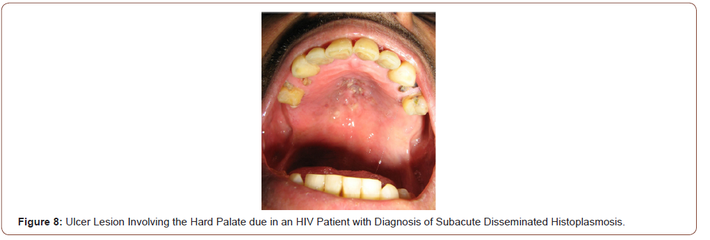
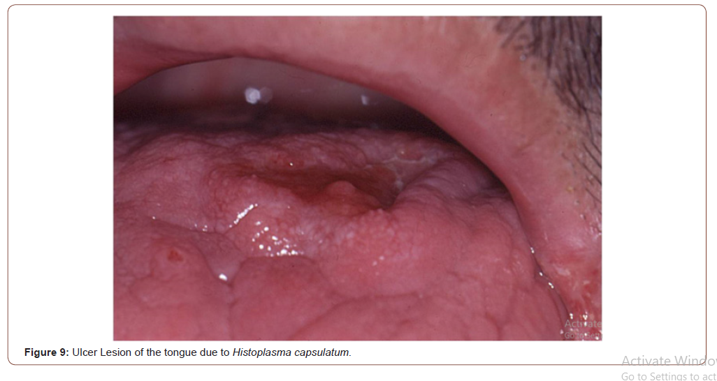
Histoplasmosis is an endemic mycosis caused by a dimorphic fungus, Histoplasma capsulatum. Histoplasmosis is a fungal disease most prevalent in patients with advance HIV/AIDS disease and CD4 +T cell counts less than 100 cells/μL. Acute and subacute disseminated forms of the disease result from reactivation of latent infection and are much more severe in patients with AIDS compared with other immunodeficiencies. The disseminated forms of the disease, affects the immunocompromised patients, especially HIV subjects with a CD4 T-cell count below 200 cell/μL, and may affect multiple organs: lungs, bone marrow, spleen, adrenal glands, liver, lymph nodes, gastrointestinal tract, central nervous system, skin and oral mucosa. In some cases, oral lesions appear to be the only primary manifestation of the disease and may compromise the tongue, palate or the oral mucosa (Figure 8, 9).

irispublishers-openaccess-dentistry-oral-health

Prolonged fever, bilateral lung involvement, papules and ulcerative cutaneous lesions and frequent oral mucosal lesions are generally observed. Oral cavity lesions can involve hard palate, uvula, tongue, gingival, tonsils and pillars with painful ulcers for several weeks. Diagnosis can be rapidly confirmed by the scarification or the biopsy of these lesions. Histologic findings of histoplasmosis with hematoxylin and eosin stain typically show diffuse lymphohistiocytic infiltrates with fungal elements about 2-4 μm in size detected within the cytoplasm of histiocytes and macrophages compatible with yeats of H. capsulatum. Well-formed granulomas are generally rare in HIV patients. Special stains, such as Grocott methenamine silver and periodic acid-Schiff (PAS) methods are also useful in visualizing the fungal elements and highlight the fungal organism cell wall. The clinical differential diagnosis of oral histoplasmosis is broad and ranging from non-specific ulcers to malignancy [14].

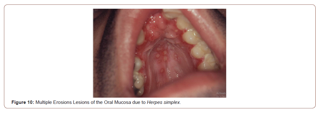
Viral infections such as Herpes simplex type I or II are to be considered in the differential diagnosis of erosive or ulcerative lesions that compromise the oral mucosa (Figure 10). Generally, the Tzanck cytodiagnosis demonstrate classic viral cytopathic effect, known as ballooning degeneration (viral syncics). Tzanck test is a quick and useful method in the diagnosis of different mucocutaneous conditions [15,16]. Bacillary angiomatosis is an unusual infectious disease, with angioproliferative lesions, typical of immunocompromised patients especially advanced HIV/AIDS disease. It is caused by Bartonella quintana and Bartonella henselae, two infectious agents of the genus Bartonella, which a variable clinical manifestations, including cutaneous vascular and nodular or tumor cutaneous violaceous lesions, regional lymphadenopathy, and even a systemic disease with visceral involvement. Purple angiomatous lesions can also locate in the oral cavity and the rhinopharynx (Figure 11). These lesions must be differentiated of the oral KS [17].

Parasitic infections, such as mucocutaneous leishmaniasis, may be rare in HIV patients, but should be investigated to exclude this unusual infection. The Tzanck cytodiagnosis can bring out amastigotes of Leishmania spp within the macrophages [14,16].

Neoplastic Diseases with Oral Involvement

KS is a malignant neoplasm, strongly associated with Human Herpes Virus 8 (HHV-8) in its pathogenesis. Lesions of KS can disseminate rapidly in severely immunocompromised HIV patients, with cutaneous, mucosal and visceral involvement. In advanced HIV/ AIDS disease, KS has a very aggressive clinical course with frequent involvement of lymph nodes, the lungs and the gastrointestinal tract in 50% of the cases. Lungs involvement occurs in 20% of the patients and is the most frequent cause of mortality. In the oral cavity, the most common sites involved by KS are the hard palate and the gums (Figure 12, 13). An intraoral examination generally revealed non-ulcerated, purple nodular lesions [18,19].

To read more about this article.....Open access Journal of Dentistry & Oral Health

Please follow the URL to access more information about this article

https://irispublishers.com/ojdoh/fulltext/oral-cavity-and-hiv-infection.ID.000556.php

To know more about our Journals...Iris Publishers

To know about Open Access Publishers


Thursday, November 25, 2021

Iris Publishers| Wish you Happy Thanksgiving Day


It’s time to wish on the occasion of Thanksgiving Day. We feel it as a merriment moment for each, so we Wish you Happy Thanksgiving Day to you and your family. Have a lovely joyful day!

Wednesday, November 24, 2021

Iris Publishers-Open access Journal of Otolaryngology and Rhinology | Evaluation of Saccular Function with cVEMP In Patients with Systemic Lupus Erythematosus


Authored by  Tuba Turkman*

Abstract

Introduction: Systemic lupus erythematosus (SLE) is a disease characterized by sensorineural hearing loss, tinnitus and vestibular disorders. Studies have shown that vestibular symptoms are quite common in patients with SLE. The aim of this study was to evaluate the relationship between cervical vestibular evoked myogenic potential (cVEMP) test and saccular dysfunction and the possible relationship between SLE and vertigo in patients with SLE.

Materials and methods: Forty-six SLE patients (aged 18-40 years) and forty healthy volunteers (aged 20-40 years) were included in the study. Patients with SLE were questioned about their duration of the disease, hearing status and balance. Pure-tone audiometry, tympanometry and cVEMP tests were performed to all patients in SLE and control groups.

Results: When P1 and N1 latencies were compared by the cVEMP in SLE and control groups, P1 and N1 latencies of the left and right ears of the SLE group were found to be significantly longer than the control group (p <0.05). In twenty-six SLE patients with vertigo, P1 and N1 latencies were significantly longer than patients with SLE without vertigo. There was also a significant relationship between vertigo and duration of the disease (p <0.05). It was found that the rates of saccular dysfunction were higher in patients with SLE.

Conclusion: This study showed a strong association between the balance disorders and SLE which is an autoimmune disease. Patients with SLE had a higher rate of saccular dysfunction, and there was a significant relationship between duration of the disease and vertigo.

Keywords: Systemic lupus erythematosus; Saccular function; cVEMP

Introduction

Systemic lupus erythematosus (SLE) is an idiopathic, chronic, autoimmune connective tissue disease that may affect all organs and systems. The disease usually begins between 16-55 years of age and the rate of prevalence among women and men is 1/9-10 [1]. In SLE patients, sensorineural hearing loss and ear symptoms were first identified by Kastanioudakis et al. [2] and Sperling et al. [3]. In patients with SLE, sensorineural hearing loss, tinnitus and vestibular disorders have been detected [4,5]. The clinical presentation of SLE may be moderate or severe. All organs are susceptible to the disease during the process [6]. In a questionnaire, the frequency of audio vestibular symptoms, including vertigo, has been found significantly higher in SLE patients than the control subjects [7,8]. In a histopathological study, the mean density of type I cells in peripheral vestibular sensory epithelium has been found significantly lower in patients with SLE [9]. The production of humoral antibody, immune complexes and autoantibodies circulating in SLE are the main causes of organ damage. The presence of vasculitis in the stria vascularis, spiral ligament and internal auditory artery may cause otologic symptoms [3].

The aim of this study was to evaluate the frequency of vertigo and saccular function with cervical vestibular evoked myogenic potential (cVEMP) test in patients with normal hearing who were diagnosed with SLE and had no other systemic disease.

Materials and Methods

This study was carried out with the consent of the cases in the Audiology Unit of the Department of Otorhinolaryngology. The ethics committee approval was obtained with the decision of the Ethics Committee of the Research on Humans to be held on people, dated 20/11/2018 and numbered 294626. Forty-four SLE patients between 18-40 years (mean 33.18±6.5 years) and forty healthy volunteers between 20-40 years (mean 31.3±6.2) were included in the study [10]. All patients were administered with steroid + hydroxychloroquine. Patients with SLE were divided into three groups according to the duration of the disease (Group 1: 1-5 years, Group 2: 6-10 years, Group 3: 11 years and above).

The inclusion criteria for the SLE groups were the definitive diagnosis of SLE, normal otological findings, immittance audiometry levels in normal range, bilateral acoustic reflexes and having a pure-tone average (PTA) within normal range [11]. The inclusion criteria for the control group were the absence of any known systemic and chronic diseases, normal otological findings, immittance audiometry levels in normal range, bilateral acoustic reflexes and having a pure tone average within normal range [11]. All of the patients in the SLE group and control group underwent ear examination, pure-tone audiometry, tympanometry and cVEMP tests. The duration of the disease, hearing status and vertigo in SLE patients were recorded.

Performing and recording of cVEMP test

In cVEMP tests; two separate waveforms were obtained and recorded by averaging 250 responses to the stimuli delivered at the repetition rate of 7.1/sec with an intensity level of 105dB nHL using the click stimulus at alternating polarity, 80 msec recording range, 10 Hz / 1kHz filter interval. In cVEMP measurements, Medelec Brand Synergy Model ABR device (Natus, A.B.D.) and standard intra-auricular TDH-49P headphones were used.

During recording, the gold-plated disc electrodes were placed as follows; the active electrode in the middle of the sternocleidomastoid (SCM) muscle, the reference electrode on the sternoclavicular joint where the SCM muscle attached to the sternum, and the ground electrode at the center of the forehead. During the application, the patient was asked to turn the head to the opposite side of the ear to be tested during the test while sitting. The impedance difference between the electrodes was below 3 kΩ. In our study, the change rate of mean peak latencies of P1-N1 biphasic waves were evaluated according to the ears, age, duration of disease and presence of vertigo with a stimulus delivered with a repetition rate of 7.1/sec and intensity level of 105 dB nHL.

Statistical analyses

The data were first transferred to Microsoft Office Excel and analyzed using SPSS 22.0 (IBM Corp. Released 2013. IBM SPSS Statistics for Windows, Version 22.0. Armonk, NY, USA) program. Categorical measurements were summarized as numbers and percentages, and numerical measurements as mean and standard deviation (median and minimum-maximum where necessary). The Kolmogorov Smirnov test was used to determine whether the variables showed normal distribution according to the groups. Independent Samples T test was used to compare numerical measurements between groups. Dependent T-test (paired t-test) was used to compare dependent numerical measurements. Kruskal Wallis test was used for general comparison between groups with more than two numerical measurements which are not normally distributed for the cases that were significant in this comparison, Bonferroni-corrected Mann-Whitney U-test was used for pairwise comparison of groups. The Pearson Correlation Coefficient and the corresponding p-value were obtained to examine the interaction between the numerical measurements. Statistical significance level was taken as 0.05 in all tests.

Results

Forty-four SLE patients and forty healthy volunteers were included in the study. No statistically significant difference was observed between the study and control groups in terms of patient ages. The disease duration of patients in the SLE group was between 1-25 years. The cVEMP results of the patients with 1-5 years, 6-10 years and 11 years and above of disease duration were evaluated. When P1/N1 latencies of SLE and control groups were compared, it was observed that the P1/N1 latencies of the right and left ears of the patients with SLE were significantly longer than the control group (p <0.05) (Table 1). No difference was found between right and left ear latencies of SLE patients and control group. In the SLE group, 26 (59%) of the 44 patients had vertigo. The P1 and N1 latencies were significantly longer in SLE patients with vertigo than those without vertigo. There was also a significant relationship between vertigo and disease duration (p <0.05) (Table 2).

When the groups were compared in terms of duration of the disease and frequency of vertigo, 6/18 (33.3%), 10/14 (71%) and 10/12 (83%) patients with a duration of disease between 1 and 5 years, 6-10 years, and 11 years and above had vertigo, respectively. It was found that the frequency of vertigo increased as the duration of the disease increased (Table 4).

Discussion

SLE is an extremely complex and multifactorial autoimmune disease with unknown etiology, caused by various genetic and environmental factors, characterized by the presence of autoantibodies, involving multiple organs [12]. Several studies have shown that vasculitis develops in the capillaries and arterioles due to autoimmune complex accumulation. Temporal bone studies have reported the presence of antiphospholipid syndrome and vascular thrombosis mechanisms that cause the accumulation of free radicals in the cochlea and stria vascularis [13]. Ear-related symptoms of SLE are chronic otitis media, progressive sensorineural hearing loss and vertigo. Viral infections, vascular lesions and immune mechanisms can all be effective in internal ear damage [14]. In addition, if the levels of circulating DNA-anti-DNA exceeds the level of cleansing capacity of the immune complexes, these immune complexes can accumulate in various tissues, including glomerulus, and may cause local damage [15].

Vestibular symptoms are quite common in patients with SLE [16]. In addition, the presence of audio vestibular disorder, endolymphatic and cochlear hydrops in patients with SLE have also been reported [17,18]. A strong relationship between SLE and balance disorders has been suggested, but the association of vestibular system in patients with SLE has not been fully investigated [19]. In our study, vertigo status and duration of the disease were investigated in patients with SLE and saccular function was evaluated by cVEMP. In a study, investigating the relationship between SLE and saccular hydrops, cVEMP has been performed in thirty patients and P1-N1 latencies were shown to be prolonged [20]. In another study, twenty patients with SLE underwent cVEMP testing and P1-N1 latencies were seen to be significantly prolonged [21]. However, no evidence of vestibular system has been found in both studies. Relationships between disease duration and latency of cVEMP have not been investigated in these studies.

In our study, when P1 and N1 latencies were compared between forty-four SLE patients and forty control subjects, P1 and N1 latencies of the left and right ears of the SLE patients were found to be significantly longer than the control group (p <0.05). In addition, 26 (59%) SLE patients with vertigo had significantly longer P1 and N1 latency values than SLE patients without vertigo. Also, there was a significant relationship between vertigo and disease duration (p <0.05).

In another study, electronystagmography (ENG) has been performed in SLE patients and abnormal findings have been found to be significantly higher than healthy subjects [18]. Another study confirmed the presence of abnormalities in the vestibular system via videonystagmography (VNG) and dynamic posturography in pediatric patients with SLE [16].

In a histopathological study, type I hair cells of the cristas in the three semicircular canals, saccular macula and utricular macula have been observed to be affected and the mean density of type I cells was lower in the SLE group when compared to the control group. However, type II hair cells were found to be unaffected. The intensity of type I and type II hair cells has not been associated with the duration of SLE [9]. In a study by Sone et al. [22], the temporal bone of the patients with SLE has been investigated using histopathologic methods and loss of spiral ganglion cells, loss of hair cells in varying degrees and atrophy in stria vascularis have been shown. Also, cochlear hydrops and stenosis in endolymphatic duct have been observed in seven cases. When the SLE group was compared with the control group, it has been observed that a significant amount of peripheral type vestibular pathology is present in the SLE group.

Regarding this information, we aimed to evaluate the saccular function causing balance disorder by performing cVEMP in patients with SLE and to investigate the effect of duration of the disease on vestibular system. Previous histopathological studies performed with VNG and dynamic posturography have shown that the vestibular system is affected. In our study, it was found that saccular dysfunction was more and latencies were prolonged in patients with SLE who underwent cVEMP test. We showed that as the duration of the disease increased, latencies were prolonged in cVEMP. The results revealed that the frequency of vertigo was higher as the disease duration increased. The most important limitation of this study is the low number of cases and the fact that the vestibular test battery has not been used in which the entire vestibular system is evaluated. However, there are only two studies in literature on cVEMP testing in patients with SLE and the relationship between SLE, duration of the disease and vestibular system and their effect on latencies have not been evaluated. This is a different aspect of our study.

Conclusion

As a result, it was found that saccular dysfunction was more and latencies were prolonged in patients with SLE who underwent cVEMP test. We showed that as the duration of the disease increased, latencies were prolonged in cVEMP. The results revealed that the frequency of vertigo was higher as the disease duration increased. it should be kept in mind that vestibular system findings may occur in patients with SLE and we think that evaluation of vestibular system should be performed at certain intervals.

To read more about this article....Open access Journal of Otolaryngology and Rhinology

Please follow the URL to access more information about this article

https://irispublishers.com/ojor/fulltext/evaluation-of-saccular-function-with-cvemp-in-patients-with-systemic-lupus-erythematosus.ID.000565.php

To know more about our Journals...Iris Publishers
To know about Open Access Publishers

Iris Publishers-Open access Journal of Otolaryngology and Rhinology | Carcinoma Lower Lip: Shaving and Reconstruction under Local Anesthesia


Authored by Md Ashraful Islam*

Abstract

The lips are one of the most important features of the face; and are functionally and aesthetically very important. It serves as border of the oral commissure, provides access to oral cavity and contributes to oral competence. It is important for verbal expression and fundamental for facial expression and overall appearance of face [1]. Excessive sun exposure and tobacco use, may develop cancer. The cancer can occur anywhere along the upper or lower lip but is most common on the lower lip. Most lip cancers are squamous cell carcinomas. Surgery is the main stay of treatment and lip shave is an established modality for superficial lesion followed by reconstruction. Extensive surgery may be necessary for larger lesion. Careful planning and reconstruction can restore eating and speaking normally, and also achieve a satisfactory appearance after surgery. Evidence supports that the concept of lip reconstruction started as early as 1000 BC in the sacred texts of great Susruta, India [2]. Lip mucosa is very special and it is recommended to reconstruct the surgical defect with lip tissue whenever possible, as any other tissue is never comparable or can replace the lip tissue [3]. This is the example of this presented case.

Keywords: Lip squamous cell carcinoma (SCC); Lip shaving; Reconstruction

Introduction

The lips are complex and specialized structures and most mobile elements of lower part of face. It has aesthetic and functional role that cannot be replaced by other tissue of body. It is always better to replace the lip tissue with ones’ own tissue whenever possible. Majority of lip defect occurs due to trauma or tumor excision. Basal cell carcinoma (BCC) is relatively common in upper lip and SCC is more common in sun exposed part of lower lip. Both are common in elderly people. Small defect should be primarily closed locally but the decision making relies on a defect occupying more than half. Superficial defect involving vermilion of lip can be closed by vermilion mucosa only, if necessary, some mucosa can be undermined or advanced to cover the defect without any obvious deformity [3]. In this case, we would like to describe repair of such a defect created after shaving of the lower lip for a lip lesion. Several flaps have been described throughout the literatures but here we have mentioned the simplest method by which one can close a defect in an effective acceptable way with oncological safety.

Case Report

A 65-year-old lady reported to otolaryngology outpatient department (OPD0 with the complaint of a swelling in left half of lower lip for 6 months duration. She noticed a small nodular swelling in her left half of lower lip 6 months ago which was gradually increasing. She had no local pain or burning sensation. According to the statement of the patient, she developed a small similar swelling in the lower lip 5 years back which had disappeared with local homeopathy treatment. Again, she initially tried with the same homeopathy treatment for the present lesion with no improvement at all. On the contrary, the lesion started increasing in size with whitish discoloration of entire oral cavity including lower lip. There was alteration of taste and slight burning sensation in the oral cavity during taking meal. She was hypertensive but nondiabetic. She used to take betel leaf with slacked lime and nuts regularly. The lady was housewife and did not give any history of prolonged sun exposure.

On local examination there was an ulcero-prolipharative lesion in left half of lower lip measuring 2.5×1.5 cm, non-tender, indurated with irregular cauliflower like surface. It did not bleed on touch. The lesion was 1cm lateral to the angle of mouth and 0.5cm distal to the vermillion border. On intraoral examination there was extensive leukoplakia involving inner aspect of lips, buccal mucosa, palatal mucosa and tongue. There was no palpable lymph nodes in the neck (Figures 1-16).

(Figures 17-19) Fine needle aspiration cytology (FNAC) was suggestive of chronic inflammatory lesion. Clinically it was a suspected case of SCC of lower lip. The incision line was infiltrated with 2% lidocaine with 1 in 200000 adrenaline solution. Planned excision with 6mm healthy margin all around was done under local anesthesia with a slice of orbicularis oris muscle was done. After excision, the area became a rectangular shape which was 3.5cmx2cmx1.5cm. the defect was reconstructed with a nearly 1.5cm×1.5cm advancement lip mucosa flap from the medial aspect and 0.5 cmx1.5 cm from the lateral aspect of the excision area. More than half of defect was covered with advancement flap. Outer (vermillion border) and inner incision line (gingival mucosa) was undermined to take some additional tissue. A portion of orbicularis oris muscle was raised and stitched in between outer vermillion border and inner gingival mucosa to fill the rest of the defect. The wound was closed in 2 layers with 3/0 vicryl suture without any significant tension. The patient was on oral cefuroxime for 10 days with antibiotic mouth wash for 5 days. She was allowed spoon feeding from the day of surgery. Stitches were removed on 5th POD.

On subsequent follow up there was no gap in between the lips during closer of mouth. It was cosmetically well acceptable. The lesion was histopathologically confirmed as Invasive Squamous Cell Carcinoma grade-I. The margin was free of tumor. There was no vascular invasion.

Discussion

Global estimates suggest increasing incidence of oral SCC. SCC constitutes 90% of oral malignant tumors. SCC is most common in lips after skin in the Head-Neck region. Among oral carcinoma, lip cancer is most common comprising nearly 35% of all. It is most likely due to sun exposure, although tobacco, alcohol, diet also plays role in the formation of Oral SCC. Majority of lip SCC arises in the vermillion border [4]. Lip cancer can arise both from superficial layer or full thickness (including muscle, skin) of lip. Treatment varies according to the site and extension of the lesion. For a superficial lesion involving lower lip- vermilionectomy [3,5] or “lip shave” [3,5] or “lip scalp” operation is an established treatment though most surgeons are accustomed to operate in full thickness for a superficial, medium to large sized lesion and to reconstruct the defect with different described flaps.

Lip shave is a simple procedure and can be done under local anesthesia. Aged patients with comorbidity can be the ideal group for local anesthesia in whom there is risk of General anesthetic drugs. Reconstruction with local lip mucosal advancement flap is easier. Gingivo-buccal mucosa can be undermined to take some additional tissue for reconstruction of local defect. There are several flaps described in the literature for reconstruction of lower lip defect like Abbe lip switch flap, Estlander flap, Karapandzic Flap and all these [6-10] is described to reconstruct full thickness lower lip defect with excellent outcome.

In the majority of cases, superficial defect of the lower lip best treated with full thickness wedge excision and primary closer in involve skin. up to one third of lip can be removed or excised by simple wedge excision and the defect can be closed primarily in layers [11,12]. Though in elderly patients it is much easier due to the tissue laxity and it can be safely done under local anesthesia without any anatomical defect [3].

Conclusion

Case selection is very important, as oncological outcome should have the ultimate priority rather reconstruction. One should have adequate case to case preoperative reconstruction planning. Whenever possible, reconstruction should be done with lip mucosa as it gives very good functional and aesthetic outcome.

To read more about this article....Open access Journal of Otolaryngology and Rhinology

Please follow the URL to access more information about this article

https://irispublishers.com/ojor/fulltext/carcinoma-lower-lip-shaving-and-reconstruction-under-local-anesthesia.ID.000564.php

To know more about our Journals...Iris Publishers
To know about Open Access Publishers

Iris Publishers-Open access Journal of Hydrology & Meteorology | Influence of Community Resilience to Flood Risk and Coping Strategies in Bayelsa State, Southern Nigeria

  Authored by  Nwankwoala HO *, Abstract This study is aimed at assessing the influence of community resilience to flood risk and coping str...