Authored by Marco Orsini*,
Introduction
Multiple exostosis is an inherited, autosomal dominant condition, characterized by multiple cartilaginous lesions, developed from the metaphysis of long bones, mainly in the proximal and distal regions of the femur [1].
Case Report


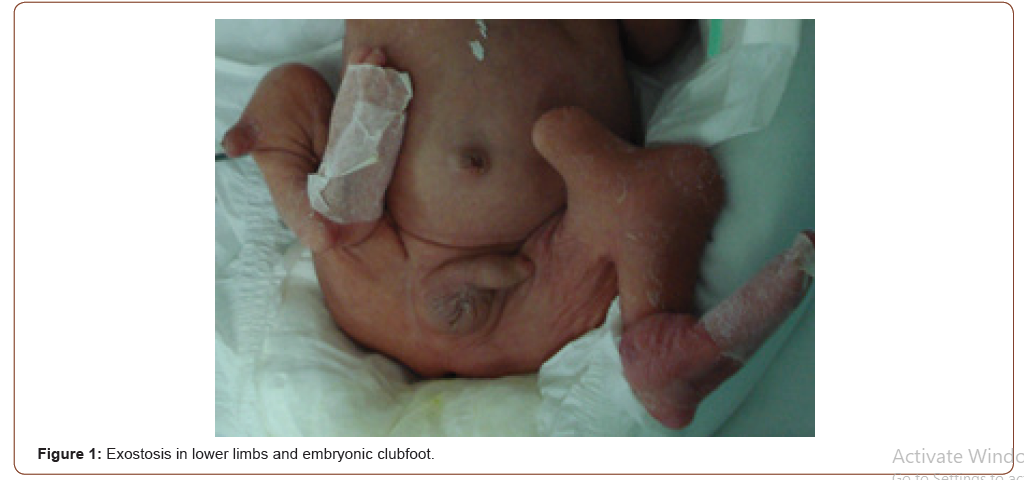
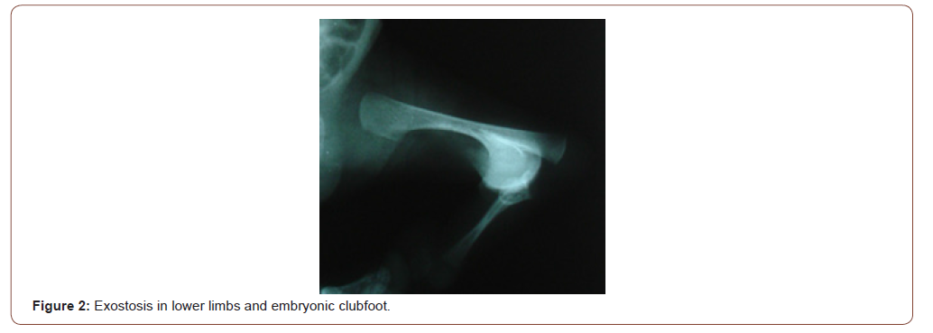
Premature, male newborn, 27 weeks of gestational age, birth weight 1215 kg, Apgar score in the first, fifth and tenth minutes was, respectively, 2/6/8, vaginal delivery, blood group A. He had structural abnormalities in lower limbs compatible with multiple exostosis and embryonic clubfoot (Figure 1), evidenced by simple radiography (Figure 2). Mother without prenatal care, with membranes’ rupture for over a month, Gestation I / Delivery 0, HIV / VDRL / Coombs direct negative, blood group A [2].
Discussion
The exostosis can be asymptomatic or associated with complications caused by compression of adjacent structures [3]. The diagnosis is made in the first life decade, based on clinical evaluation and radiological exams, changes are rarely present at birth [1,2].
Conclusion
Multiple exostosis is not classified as a true neoplasm, however, it is configured by the appearance, in childhood and adolescence, of bony protuberances surrounded by hyaline cartilage. The primary involvement occurs in the tubular bones, iliac wing and scapula, however, the entire skeleton can be compromised. It is known that less than 5% of cases evolve to the malignant form: chondrosarcoma. Congenital clubfoot, in turn, is characterized by generalized deformations of the musculoskeletal tissues distal to the knee. Until the current date, there are no reports of a causal relationship for both injuries, and further studies on this relationship are necessary.
To read more about this article....Open access Journal of Neurology & Neuroscience
Please follow the URL to access more information about this article
To know more about our Journals...Iris Publishers


No comments:
Post a Comment