Friday, March 31, 2023

Iris Publishers-Open access Journal of Anaesthesia & Surgery | Covid-19 Acute Respiratory Distress Syndrome And Acute Myocarditis Saved With Immunoglobulin


Authored by Jebbar Nourddine*,

Abstract

The novel corona virus disease (COVID-19) becomes a major public health. It was declared as pandemic by the World Health Organization on March 12, 2020.Myocarditis and myocardial injury are considered as one of the leading causes for death of COVID-19 Patients. The use of immunoglobulin seems to have an important role in the favorable evolution of these patients with cardiac involvement.

Background

Coronavirus disease 2019 (COVID-19) is a global pandemic. As of March 21, 2020, infected patients were present in 167 countries and region around the world and there were more than 285000 cases worldwide with nearly 12000 fatalities [1].

Besides focusing on the severe condition of pneumonia, the novel coronavirus may attack many important organs and cause multiple organ failure with cytokine storm. The heart is one of these most important organs and it is highly suspicious that viral myocarditis could be involved and even be considered as one of the leading causes of death of COVID-19 patients [2,3].

Case report

47-year old female patient, with chronic urticarial and corticosteroid in self-medication, having a stay in an endemic area (Egypt, 1 week before admission), admit to the emergency department of our hospital on 16th March 2020 for fever with asthenia and dry cough evolving for 8 days before admission. The patient benefited of a complete biological assessment objectifying a lymphopenia, moderate elevated ferritin and lactate dehydrogenase, C reactive protein at 180 mg/l and positive polymerase chain reaction (SARS COV 2).

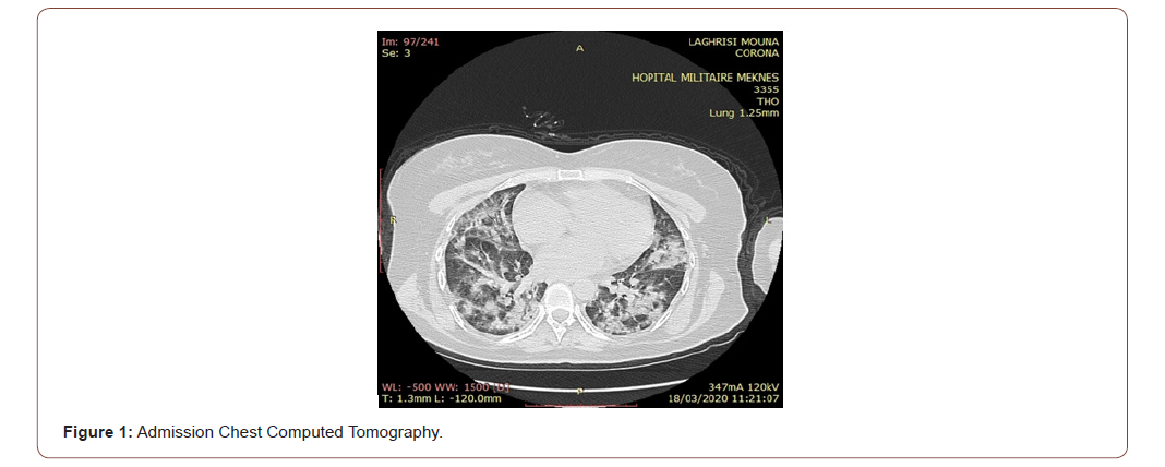
The patient was put on chloroquine, paracetamol, ceftriaxon and ciprofloxacine. 1 day before her admission in the intensive care (ICU) unit (18th March 2020), the patient presented worsening of her respiratory state made of dyspnea, 90% desaturation with a respiratory frequency at 30 cycles per minute and tachycardia at 120/min. Cardiac assessment was normal. Thoracic chest computed tomography (Figure 1) examination indicated a condensation syndrome multifocal affecting more than 60% of the pulmonary parenchyma necessitates an increased need for oxygen and her admission in the ICU. The patient was put on Lopinavir/Ritonavir, omeprazole to inhibit gastric acid and preventive anticoagulation.

The patient was intubated, 2 days after her admission in the ICU, on neurological and respiratory criteria and developed an acute respiratory distress syndrome (ARDS) and put on water restriction and diuretic (furosemide). A biological assessment objectifying a lymphopenia and C reactive protein at 188 mg/l. Blood gas analysis showed a report pao2/fio2 at 130.
irispublishers-openaccess-anaesthesia-surgery

8 hours after, the evolution was marked by a hemodynamic instability made of arterial tension at 70/30 mmhg and a heart rate at 130/min requiring the use of vasoactive agents (dobutamine, norepinephrine) with significant improvement under invasive monitoring.

The electrocardiogram showed sinus tachycardia. Echocardiography showed a diffuse myocardial dyskinesia, systolic dysfunction of the left ventricle, mitral annular plane systolic excursion at 7mm with left ventricular ejection fraction (LVEF) estimated to 35%, a small pericardial effusion at 5mm, inspiratory impedance threshold valve (ITV): 6-8cm, no pulmonary hypertension and dilated inferior vena cava. Troponin was at 0,06 μg/l (four times normal) and C reactive protein at 336 mg/l.

The diagnosis of this patient is coronavirus acute myocarditis with cardiogenic shock and ARDS. Treatments include methylprednisolone to suppress inflammation (200mg/day, 4 days), immunoglobulin to regulate immune status (20g/day, 4 days), dobutamine to raise blood pressure, diuretic (furosemide) to reduce cardiac load, prone position to improve the oxygenation and the zinc.

After treatment, the patient’s symptoms improved significantly after 48 hours with improvement in hemodynamic parameters and progressive withdrawal of vasoactive agents. Marker of myocardial injury (troponin) dropped after 4 days and the c reactive protein dropped to 96 mg/l after one day and to 35mg/l two days after.

Echocardiography showed that the function of the heart had returned to normal and the patient was extubated after hemodynamic stabilization and a report pao2/fio2>300. The patient benefited of non-invasive ventilation sessions for severe hypercapnia.

Otherwise, the patient presented confusion after her extubation. We performed a cerebral magnetic resonance imaging returning without abnormality. After one week, the marker of myocardial injury had fully recovered to the normal range.

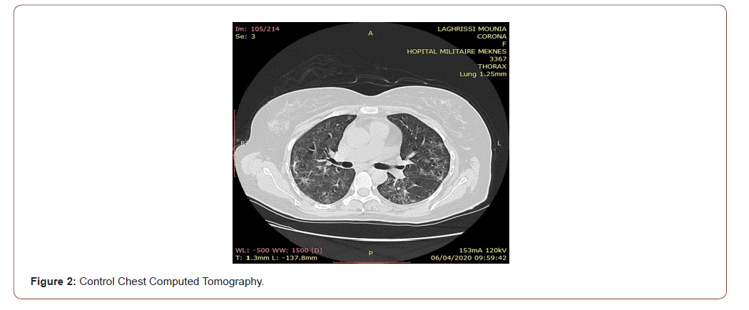
Thoracic chest computed tomography (Figure 2) objectifying an improvement in lung damage.

Then the patient was transferred to the observation ward. Negative PCR on day 27 and left the hospital with full recovery.

Discussion

American Heart Association defined myocarditis [4] by

1. Elevation in cardiac troponin I (over 0.12 ng/ml)

2. Abnormality on echocardiography: LVEF < 50% or segmental wall motion abnormality, or left ventricular wall thickening > 10mm and/or presence of pericardial effusion ≥ 5mm

3. Abnormalities on electrocardiogram (ECG): ST segment elevation/ ST-T changes

During the management of our patient, only one case of myocarditis linked to COVID-19 has been described in the literature before, indeed it is a Chinese article [5] on 37-year old male patient, admitted to hospital on 14 January 2020 with chest pain and dyspnea for three days. His blood pressure decreased to 80/50 mmhg. Chest computed tomography indicated pulmonary infection, enlarged heart and pleural effusion. The ECG suspected ST-segment elevation acute myocardial infraction. An emergency CT coronary angiography revealed no coronary stenosis. Markers of myocardial injury were elevated. Echocardiography revealed an enlarged heart and a decrease in ventricular systolic function. The diagnosis of this patient is coronavirus fulminant myocarditis with cardiogenic shock and pulmonary infection. Treatments include methylprednisolone (200 mg/day, 4days), immunoglobulin (20g/day, 4days), norepinephrine, diuretic, piperacillin sulbactam and pantoprazole. After treatment, the patient symptoms were improved. One week later, echocardiography showed that the size and function of the heart had returned to normal and markers of myocardial injury dropped significantly.

A second case [6] was published on 63-year old male, admitted to the hospital for cough with white sticky sputum, fever and shortness of breath. He had a recent history of travel to Hubei Province, China. Blood gaz analysis showed respiratory acidosis. Markers of myocardial injury were elevated. An electrocardiogram showed sinus tachycardia. Echocardiography showed an enlarged left ventricle, diffuse myocardial dyskinesia with a low LVEF (32%) and pulmonary hypertension. The diagnosis was considered severe pneumonia, ARDS fulminant myocarditis. The treatment regimen was ventilator support, high-flow oxygen, lopinavirritonavir, interferon α-1b, methylprednisolone, immunoglobulin, piperacillin-tazobactam and the ventilator was unable to maintain oxygen saturation, extracorporeal membrane oxygenation (ECMO) was used. After treatment, the LVEF gradually recovered to 68%, the left ventricle returned to its normal range.

A third published case [7] of a 21-year old female patient admitted for fever, cough, diarrhea, and shortness of breath. Markers of myocardial injury were elevated. Electrocardiogram showed nonspecific intraventricular conduction delay and multiple premature ventricular complexes. Echocardiography showed severe left ventricular systolic dysfunction. On the cardiac computed tomography, the coronary arteries were normal, and the myocardium was hypertrophied combined with a subendocardial perfusion defect on the lateral left ventricle. Cardiac magnetic resonance imaging revealed diffuse high signal intensity in the left ventricle myocardium on T2 short tau inversion recovery image and myocardial wall which suggests myocardial wall oedema. Myocarditis combined with COVID-19 was confirmed by multimodality imaging.

Conclusion

COVID-19 patients may develop cardiac complications such as myocarditis. This is the first report of COVID-19 complicated with acute myocarditis in our country. Benefit of troponin dosing and echocardiography for diagnosis. Immunoglobulin can play a very important role in the management of this situation.

To know about Open Access Publishers

Iris Publishers-Open access Journal of Cardiology Research & Reports | Brazilian Lipid Cardiovascular Risk Pre and During the Covid 19 Pandemic in Asymptomatic and Severely Affected Groups


Authored by Tania Leme da Rocha Martinez*,

Abstract

The COVID 19 pandemic still keeps investigators, public health representatives and patients in a state of extreme worry all around the world. Investigations of the hazardous influences played by the comorbidities have to be carried on with more details than ever opening new avenues for it’s’ understanding. In this paper there was a clear statistical response for worse in the lipids and lipoprotein profiles starting from the prepandemic to the pandemic, both asymptomatic groups, maintaining the worsening in the very severely affected that had to be admitted to the Intensive Care Units, analyzing the use of hypolipidemic medications as well. The modern use of encrypted emails sending with confidentiality all the results to one specific software center enables the investigators to carry on a research with a certain similarity to a national survey. The means of all lipid analytes were significantly different between the pre and post-pandemic cohorts (2018+2019 and 2020). It also shows that the frequency of participants with altered results for total cholesterol, triglycerides, and HDL-c and with dyslipidemia were also significantly different between the two cohorts. The year of 2020 had a higher mean of lipid analytes than the pre-pandemic years of 2018 and 2019 combined. The frequencies of participants with total cholesterol above 190 mg/dL and triglycerides above 175 mg/dL were both higher in 2020 than 2018 and 2019, and the frequency of those with dyslipidemia was also significantly greater for that cohort. Besides all the medical information for each of the patients this database in full can be used for public health policies.

Keywords:19; Total Cholesterol; LDL Cholesterol; HDL Cholesterol; VLDL Cholesterol; Non HDL Cholesterol; Pandemic changes; Pre pandemic

Abbreviations:HDL: High Density Lipoprotein; HDL-c: HDL Cholesterol; LDL: Low Density Lipoprotein; POCT: Point-of-Care testing; TG: Triglycerides; VLDL: Very Low Density Lipoprotein

Introduction

In a recently accepted communication to the European Atherosclerosis Society [1] we presented the information of cardiovascular risk factors themselves in patients admitted to the Intensive Care Unit and on Hemodialysis with a main focus on the presence of dyslipidemias, while contributing to the severity of COVID 19 patient’s evolution. In this paper we demonstrate the state of the matter highlighting the lipoprotein profiles pre and post the pandemic in groups from all over Brazil, not disregarding the presence of the main modifiable risk factors. Adjourning the public health condition, the comparison was completed with the profiles of the same COVID 19 patients. The consequence of having all the comorbidities listed gives us the reason why so importantly patients are not being diagnosed in their heart conditions; either being afraid of contagiousness in health facilities or unfortunate lack of condition to be assisted. In order to demonstrate this situation in a scientific scenario this article was planned and executed.

Method

In this study, 4767 blood samples were collected from patients over twenty years (age between 20-93 years) in the Brazilian health care service. The patients, who went spontaneously to the collection points, totaled 2501 between March 2018 and November 2019 (prior to the COVID19 pandemic) and 2266 in the year 2020 (during the COVID19 pandemic). The number of service providers involved was 370 (6 patients per unit) between 2018 and 2019, and 513 (4 patients per unit) in 2020. The lipid profile test was performed on the Point-of-Care testing (POCT) platform, using a colorimetric test strip and the result is obtained in a few minutes. The POCT equipment Hilab (Hitechnologies, Curitiba/PR) uses internet Of Things (iOT) technology, which recognizes the unique QR code and sends the reaction information via cloud to the company’s laboratory in Curitiba, where a trained professional validates the reaction. The analysis of the reaction that arrives, regardless of the collection location, will ensure the absence of interferences to issue the signed report to those who made the registration and sample collection, at the health care service location, as well as to the patient’s smartphone, in up to 10 min.

From each outpatient presenting at the health service location, forty microliters (40 μl) of whole blood fingerstick sample were collected in capillary pipette, according to the instructions to avoid pre-analytical interference. The Hilab equipment uses PTS Diagnostics lipid panel test strips that employs dry-chemical testing for measurement of total cholesterol, cholesterol from High Density Lipoprotein (HDL-C) and triglycerides (TG) in whole blood. Initially, a membrane removes the red blood cells, and via horizontal flow the test strip analyzes plasma lipid concentrations. Total cholesterol and HDL-C use the same enzymatic reaction for the evaluations. The HDL lipoproteins are separated from lipoproteins Low Density Lipoprotein (LDL) and Very Low Density Lipoprotein (VLDL) using phosphotungstic acid and a magnesium salt layer above the membrane fractionation layer. The resulting HDL fraction in plasma reacts with surfactants and enzymes for measuring cholesterol concentration. The linear range in this lipid panel test strip are in mg/dL: total cholesterol >120 and <400, HDL-C >20 and <100, TG >120 and <400. LDL Cholesterol (LDL-C) values were obtained by calculating Martin’s formula (Fried Ewald’s formula modification) and non-HDL-C values by subtracting the HDL-C value from cholesterol total value. The evaluation of TG is carried out by a colorimetric enzymatic method using lipoprotein lipase, glycerol kinase, glycerol phosphate oxidase and peroxidase [2-12]. The Hilab equipment employs reflectance photometry.

We worked with a retrospective database from March 2018 to December 2020. The results of the lipid profile are reported in mg/dL. The system of information exchange is encrypted in several layers. Personal information is dissociated from the results (different databases), making breach of confidentiality extremely difficult. Statistical tests were applied in various comparisons: chi square test, student t test and ANOVA test.

Result

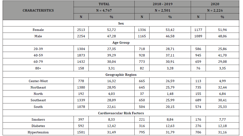
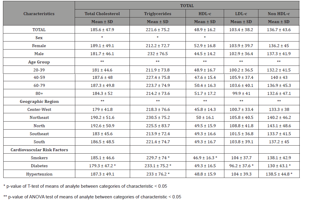
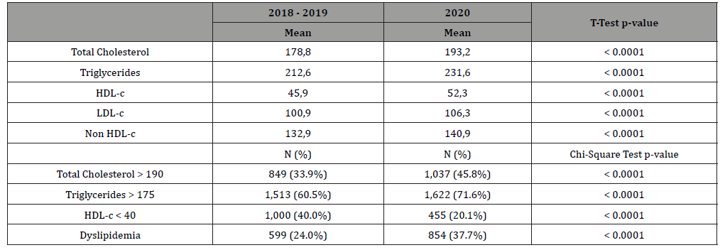
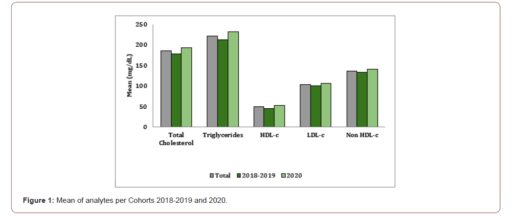
In the total sample, there was a significant difference of means of all lipid analytes between the different age groups and between the different Brazilian geographical regions (Table 1 & Table 4). Participants between the ages of 40 and 59 years old presented the highest levels of all analytes and the lowest levels of HDLcholesterol. Participants residing in the North region presented with the highest total cholesterol and LDL-c means, closely followed by the Northeast (Table 2). Those in the Northeast also presented with the highest mean of TG levels. There was a significant difference in the means of TG and HDL-c between those who identified as smokers and those who did not. Smokers presented higher TG mean and lower HDL-c mean (Table 3 & Table 4). The means of total cholesterol, TG and HDL-c were all significantly different between males and females (Table 4). Table 8 shows that the means of all lipid analytes were significantly different between the pre and postpandemic cohorts (2018+2019 and 2020). It also shows that the frequency of participants with altered results for total cholesterol, TG and HDL-c and with dyslipidemia were significantly different between the two cohorts. The year of 2020 had a higher mean of lipid analytes than the pre-pandemic years of 2018 and 2019 combined (Figure 1 & Figure 2).

Table 1: Number of participants (N) per year and per valid ranges.

irispublishers-openaccess-cardiovascular-research

Table 1: Number of participants (N) outside the range of the test.

irispublishers-openaccess-cardiovascular-research

Table 3: Distribution of participants by demographic characteristics and cohort - 2018 + 2019 and 2020.

irispublishers-openaccess-cardiovascular-research

Table 4: Mean and standard deviation of lipid analytes’ values by demographic characteristic of participants.

irispublishers-openaccess-cardiovascular-research

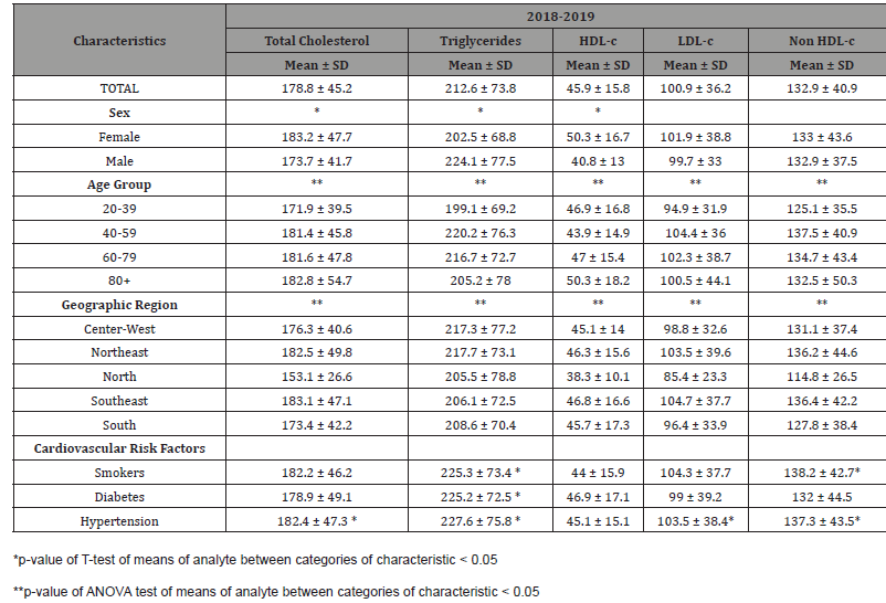
Table 5: Mean and standard deviation of lipid analytes’ values by demographic characteristic of participants by cohort - 2018+2019.

irispublishers-openaccess-cardiovascular-research

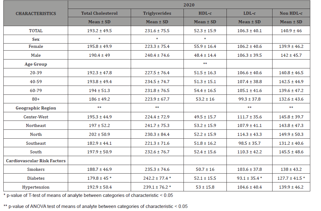
Table 6: Mean and standard deviation of lipid analytes’ values by demographic characteristic of participants by cohort - 2020.

irispublishers-openaccess-cardiovascular-research

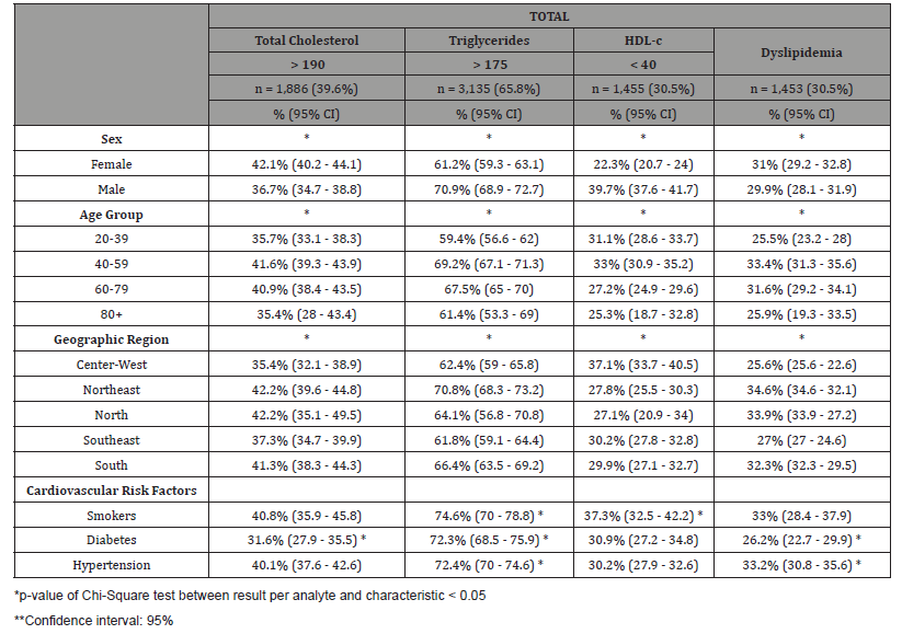
Table 7: Percentage and confidence interval of altered results of total cholesterol, triglycerides, HDL-c and dyslipidemia per demographic characteristic of population.

irispublishers-openaccess-cardiovascular-research

Table 8: Percentage and confidence interval of altered results of total cholesterol, triglycerides, HDL-c and dyslipidemia per demographic characteristic of population per cohort - 2018+2019.

irispublishers-openaccess-cardiovascular-research

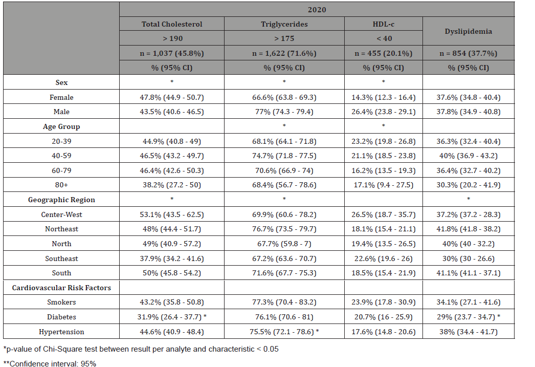
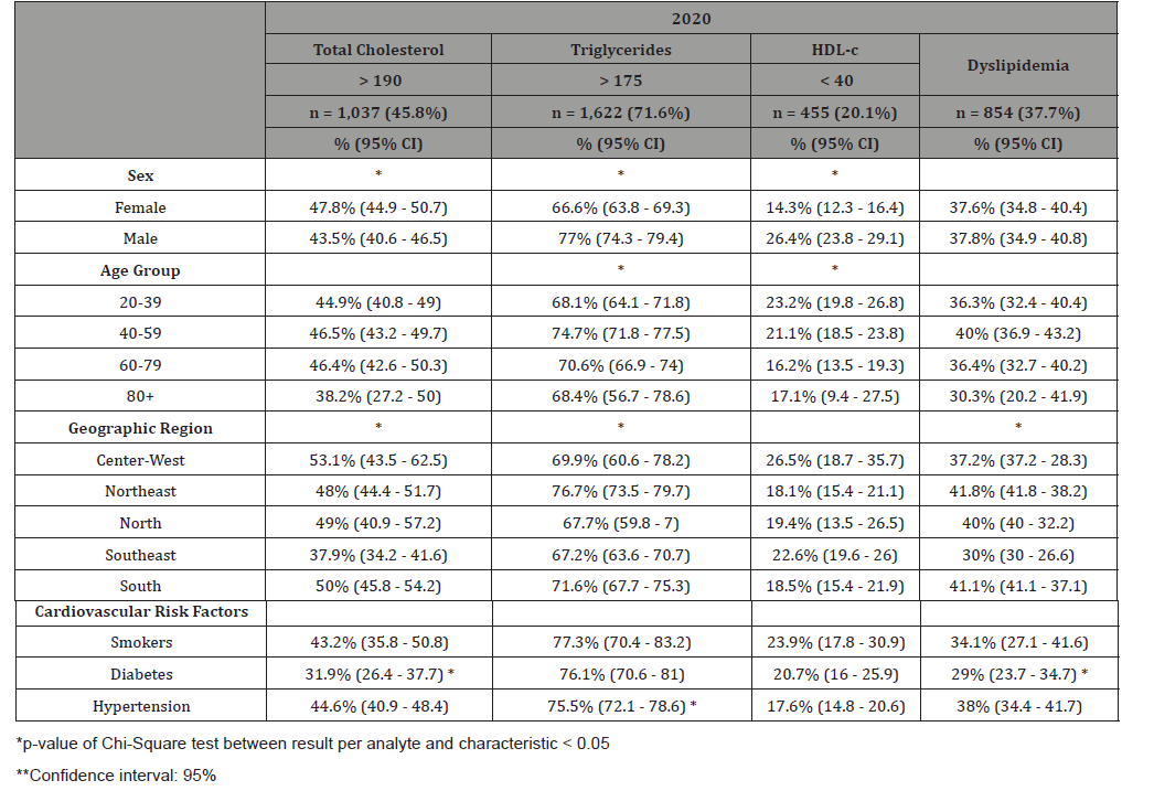
Table 10: Percentage and confidence interval of altered results of total cholesterol, triglycerides, HDL-c and dyslipidemia per demographic characteristic of population per cohort - 2020.

irispublishers-openaccess-cardiovascular-research

Table 11: Means of analytes and frequency of altered results between cohorts 2018+2019 and 2020.

irispublishers-openaccess-cardiovascular-research

irispublishers-openaccess-cardiovascular-research

irispublishers-openaccess-cardiovascular-research

The frequencies of participants with total cholesterol above 190 mg/dL and TG above 175 mg/dL were both higher in 2020 than 2018 and 2019, and the frequency of those with dyslipidemia was also significantly greater for that cohort. For both pre-pandemic and post-pandemic cohorts, there was a significant difference in the means of males and females for total cholesterol, TG and HDL-c. But the difference seen in the total sample, between different age groups, was only true for the 2018-2019 cohorts (Table 5A). The significant difference between Brazilian geographical areas stayed true when stratified by the different cohorts, except for HDL-cholesterol in 2020 (Table 5B). In Table 6, we can see that the majority of our total sample presented with TG above 175 mg/dL (65.8%). And 39.6% had total cholesterol levels above 190 mg/dL. In general, males, more frequently than females, had altered TG and total cholesterol levels. Males were also more likely to have HDL-c levels below 40 mg/dL (Table 6). Participants between 40 and 59 years old, presented higher frequencies of altered TG and total cholesterol levels. In this group, 69.2% of participants had TG levels above 175 mg/dL and 41.6% had total cholesterol levels above 190 mg/dL (Table 6). Between the different Brazilian geographical regions, the Northeast had significantly more participants with altered TG levels. It also, along with the North, presented with the highest frequency of participants with total cholesterol above 190 mg/dL.

The percentage of participants with altered total cholesterol levels increased from 33.9% in 2018-2019 to 45.8% in 2020. That difference for altered TG levels was from 60.5% to 71.6% during the pandemic (Table 8). Even after stratification by cohort, the Northeast region of Brazil maintained the highest frequency of participants with altered TG levels. However, the Southeast presented with the highest frequency of participants with altered total cholesterol in 2018-2019 (Table 7A) and, in 2020, the highest frequency was in the Center-West region of Brazil (Table 7B).

Conclusion

As expected, there was a worsening of several situations: fewer people went to check their lipids for fear of contamination and the effect of their results for their lipids and lipoproteins profiles were all signaling the same-change for worse interplaying with the other comorbidities and leaving the patients with less chances of partial or total recovery. The COVID 19 pandemic still keeps investigators, public health representatives and patients in a state of extreme worry all around the world. Investigations of the hazardous influences played by the comorbidities have to be carried on with more details than ever opening new avenues for its’ understanding. In this paper, there was a clear statistical response for worse in the lipids and lipoprotein profiles starting from the prepandemic to the pandemic, both asymptomatic groups, maintaining the worsening in the very severely affected that had to be admitted to the Intensive Care Units, analyzing the use of hypolipidemic medications as well. Besides all the medical information for each of the patients this database in full can be used for public health policies [13-22].

To read more about this article...Open access Journal of Cardiology Research & Reports

Please follow the URL to access more information about this article

https://irispublishers.com/ojcrr/fulltext/brazilian-lipid-cardiovascular-risk-pre-and-during-the-covid-pandemic-in-asymptomatic.ID.000606.php

To know more about our Journals...Iris Publishers

To know about Open Access Publishers

Thursday, March 30, 2023

Iris Publishers- Open access Journal of Gastroenterology & Hepatology | A Large Gastroesophageal Junction Mass in an Elderly Caucasian Male


Authored by Nathaniel A Parker*,

Case Description

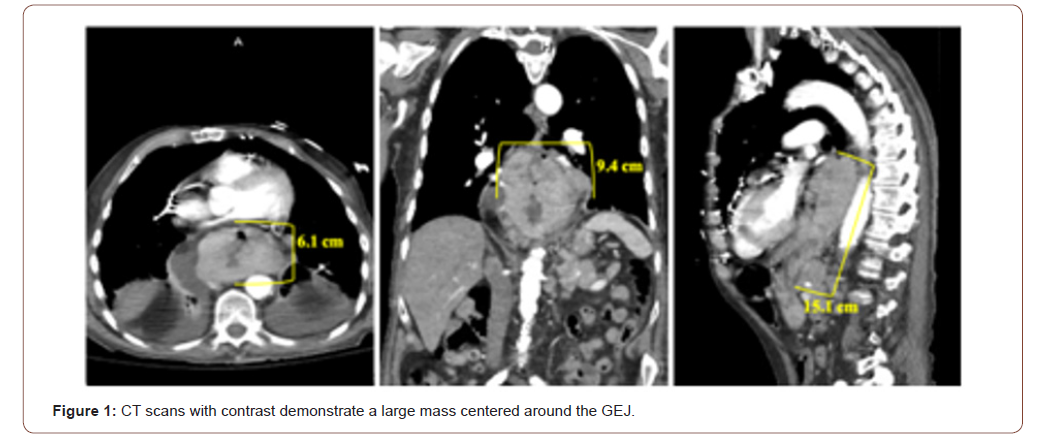
A 90-year-old Caucasian male presented to the emergency department with the chief complaint of solid-food dysphagia and unintentional weight loss. Symptom onset began three months prior to his initial presentation and had been progressively worsening. Vital signs and measurements, and physical examination were unremarkable. Serum laboratory evaluation was primarily nonrevealing, except for his hemoglobin which was approximately 8 gm/dL (reference range 12 – 16 gm/dL) and positive stool guaiac testing. Chest and abdominopelvic CT scans with contrast were obtained for further elucidation of the patient’s symptoms. Imaging showed a large, invasive esophageal mass. The mass originated at the level of the carina in the mid-esophagus and extended approximately 15 cm inferiorly into the gastric cardia, fundus, and body. At its maximum dimensions of 6 x 9.4 cm coronally, the mass was centered at the GEJ, and was associated with regional lymphadenopathy concerning for metastatic disease (Figure 1). Subsequently, he underwent an esophagogastroduodenoscopy which revealed a large, ulcerated mass at the GEJ resulting in near total esophageal occlusion. Core biopsies were collected during upper GI endoscopy to confirm the diagnosis. Histopathology revealed sheets of malignant cells. IHC staining was positive for MART-1. Pankeratin, CD56, synaptophysin, chromogranin, CDX2, p63, and PSA immunostaining was negative. Together with imaging evidence and pathology, primary melanoma of the GEJ was diagnosed. The patient was dismissed from the hospital and instructed to follow up with his local oncologist for a PET/CT scan and genetic testing. However, the patient expired before these subsequent tests could be performed.

irispublishers-openaccess-gastroenterology-hepatology

Discussion

Primary mucosal melanoma is an exceedingly rare phenomenon. Although dispersed normally in low numbers, melanocytes are present in non-sun-exposed regions of the body, such as the oral and nasal cavity, paranasal sinuses, larynx, esophagus, anorectal canal, vagina, and cervix [1]. However, the etiopathogenesis of benign melanocytes transforming into melanoma remains poorly understood. Less than 3% of all melanomas are non-cutaneous and diagnosed with a unknown primary site [2]. Recently, morphologically identical but characteristically diverse melanoma variants have emerged. Mucosal melanoma, melanoma of unknown primary, and melanoma of soft parts represent three unique melanoma variants with distinct differences in typical location of origin, pathogenesis, progression, aggressiveness, genetic profile, response to therapy, and survival rates. Melanoma of unknown primary occurs more commonly in middle-aged males, and typically is diagnosed in lymph nodes and subcutaneous tissue (70 – 90%) [2]. In contrast, the peak incidence of melanoma of soft parts occurs in the second through fourth decades of life. This variant preferentially involves the extremities, especially the legs, and is often found to be deeply infiltrative and adherent, such as being bound to surrounding tendons [3]. This case not only lacked the opportunity for PET scans to be used for staging and evaluation of distant metastasis, but also genomic profiling could not be performed. However, no universal staging system has been established for any of the non-cutaneous melanoma variants. Imaging supported extensive, local gastroesophageal tumor involvement. The patient denied previous skin excisions or current cutaneous lesions, and there was no radiologic evidence of lymphadenopathy in the sun-exposed lymph node groups (e.g. head and neck, axillary, and inguinal). Thus, although it is difficult to be entirely dogmatic on this point, the lesion most likely was a primary mucosal melanoma.

Primary mucosal melanomas are exceedingly rare, and account for approximately 1% of all melanomas [1]. Mucosal melanomas can arise from the gastrointestinal tract, but this is uncommon, whereas nearly 50% originate from anorectal tissue [1]. Gastroesophageal melanoma is an extraordinarily rare entity that represents less than 10% and 0.1% of primary mucosal and all melanomas, respectively [1]. They preferentially occur in females, which is thought to be due to a large majority of mucosal melanomas arising from the genital tract [4]. Although less pronounced compared to cutaneous melanoma, a Caucasian predominance exists [5]. Likely due to inordinately low incidence rates, no risk factors for development have been identified. Viral carcinogenesis studies have yet to find any associations [1]. However, chronic exposure to tobacco smoke and formaldehyde have been postulated to be a possible risk factor for oral and sinonasal mucosal melanoma, respectively [1].

Melanoma of unknown primary is predominantly hypothesized to develop from the spontaneous regression of melanoma from a known primary site [2]. Thus, with cutaneous melanoma accounting for more than 97% of all melanomas, melanoma of unknown primary and cutaneous melanoma typically share similar genetic profiles consisting of BRAF and NRAS mutations [2]. Either GNAQ or GNA11 gene mutations almost exclusively occur in uveal melanomas [6,7]. However, mucosal melanomas harbor a unique genomic profile characterized by alterations in the c-KIT gene [8- 9]. Since the discovery of c-KIT aberrations in primary mucosal melanomas, KIT-inhibitors like imatinib and sunitinib have provided the opportunity for targeted therapy. However, not all mucosal melanoma tumors harbor c-KIT mutations [1]. Furthermore, due to this entity’s exceedingly low incidence rates, large clinical trials studying the efficacy of targeted therapeutic agents are unlikely feasible. KIT-inhibitors have been shown to be effective in certain studies, but overall, the five-year survival for mucosal melanoma is significant less than cutaneous melanoma [10-12].

Conclusion

Written informed consent for publication of their clinical details and clinical images was obtained from the patient.

To know about Open Access Publishers

Iris Publishers-Open access Journal of Aging & Geriatric Research | Neuropsychological Assessment in Times of Social Distancing: What Can Telemedicine Offer to Older Adults?


Authored by María Josefina Gonzalez Aguilar*,

Abstract

Neuropsychological assessment is responsible for exploring cognitive performance through the administration of standardized tests. It collaborates with the clinical diagnosis and delivers objective information on cognitive deficits and abilities. Telemedicine is a tool that can be used to distribute medicine in a different and novel way to any part of the world and is especially useful for places where physical access to hospitals is difficult or impossible. The use of technology for neuropsychological evaluation is growing at an accelerated rate, both due to the advancement of technological facilities and, currently, due to the present health emergency context. Older adults where the first group to get confined, so they are protected from the daily activities that may be associated to the COVID-19 virus, but an interesting paradox has arisen: older adults are confined at home for protection, but with this measure, many of them are not able to receive medical attention. Given the growing demand to provide services in virtual mode due to the COVID-19 pandemic, it is important to review the usefulness teleneuropsychology and its benefits for attending older adults with cognitive complaints or a dementia diagnosis. Teleneuropsychology is a novel approach and the literature on this professional practice is still very limited, since to date there are few research studies on the use of this modality.

Keywords: Cognitive complaints; COVID-19; Neuropsychological assessment; Older adults; Telemedicine

Introduction

Origin and objectives of teleneuropsychology

Telemedicine is a tool that can be used as a way to distribute medicine in a different and novel way to any part of the world, and it is especially useful for inaccessible places where people are not able to visit a specialist or a nearby hospital [1]. Telemedicine was originally thought as a way to assist people living in rural areas. This sort of clinical attention offered the possibility of connecting these people with specialized medical care, avoiding the transport of patients, allowing the reduction of economic cost and loss of time [2].

As time went by, advances in technologies and the availability of broadband connections in most homes and workplaces expanded telemedicine services bringing a whole new paradigm on how clinical attention may be offered [3]. For the past decade, the use of internet in the daily work of neuropsychologists acquired more relevance, and the use of technology in the assessment-treatment process became more and more common in the professional practice [4]. Psychological treatment via telemedicine has been demonstrating its efficacy and satisfaction comparable to that of face-to-face attention in a wide variety of clinical settings and with specific populations, in the same way that its use has been shown to be effective for the diagnosis and treatment of various mental disorders [5]. The effectiveness and feasibility of the use of technology in psychological evaluation, diagnosis and treatment has promoted its use in other areas of psychology, such as neuropsychology. Teleneuropsychology is defined as the use of audiovisual technology to establish clinical contact with patients to carry out neuropsychological assessments and treatments, recognizing that this has reduced accessibility problems and has allowed the provision of health services in contexts of social distancing [6]. Neuropsychologists perform a variety of roles in different hospital and outpatient settings, either virtually or in person. When a neuropsychologist assesses a patient with a cognitive complaint or a suspected brain damage, the main objectives are to detect and characterize cognitive performance, guide a differential diagnosis and offer recommendations (to the patient and to the family) on daily life activities [7]. Computerized and virtual neuropsychological practices have been slowly integrated into research and professional activities bringing with it the development of computer-based versions of tests that until then could only be carried out using paper and pencil [8.9].

COVID-19, social distancing, and neurocognitive assessment

The COVID-19 pandemic is a global health crisis that has created sudden and unique challenges within the field of clinical neuropsychology. In this context, added to the novelty of this type of virtual approaches, studying the use of teleneuropsychology is relevant and necessary. Currently, the COVID-19 pandemic has profoundly impacted the world, causing significant changes in the daily functioning of society. The policies of social distancing have had enormous repercussions in the health sector, being that these had to be incorporated in a continuous process of modernization, where the field of neuropsychology had to evolve rapidly to incorporate evaluations to be carried out virtually. The various circumstances due to the health emergency confinement required that this care modality, which was before seen just as an alternative, had to become more effective in the face of the high demand [10]. In this way, the pandemic has pushed neuropsychology to become a discipline to evolve beyond traditional settings. In this context, teleneuropsychology could make it possible to measure and monitor cognition performance from home and may also help to identify the optimal time for a comprehensive face to face assessment [11]. Social distancing requirements associated from the COVID-19 pandemic persisted for a long time (and still persist in some countries), and future sanitary contexts may require patients and professionals to have a solid communication system to continue treatment virtually. In this sense, it is essential to have virtual alternatives available for neuropsychology which will improve the access to neuropsychological services [12].

The impact of lockdown and the social distancing policy in older adults’ health

The COVID-19 outbreak disrupted violently in the healthcare systems and caused a deep economic and social depression [13]. During early 2020, lockdown and social distancing were the first policies that most countries adopted to slow down the spread of the virus, while massive communication services advised the general population to avoid going out if it was not urgent [14]. Older adults were considered part of the most vulnerable population and these policies were targeted mainly to them. These measures might have prevented older adults from getting infected, but also impacted negatively in the follow-up of previous health conditions and the need of attention of new medical needs, such as novel cognitive complaints. In this way, professionals had to think of a way to offer continuity of care while protecting this vulnerable population from getting infected [15,16]. During lockdown, non-urgent procedures and most face-to-face visits were suspended, leaving many patients in front of a different dangerous situation: a drastic drop in spontaneous and non-urgent visits to the health care centers [14]. In this context, telemedicine has become critical when providing care and continuity to this vulnerable groups [17,18], and raised concerns about the way illnesses are categorized: is Alzheimer disease or Parkinson disease more or less urgent to attend than a respiratory syndrome? When should older adults with cognitive complaints or cognitive disabilities attend to a medical care health center?[15,17]. Mild cognitive impairment is an intermediate state between normal cognitive ageing and dementia and is a risk factor to progress to dementia in the years following the diagnosis [19]. In this way, early assessment and treatment of mild cognitive impairment in older adults is critical and must be addressed as a pending priority that must be revised in the actual context [16]. Reports around the world show that many patients with chronic diseases delayed their face-to-face visits to their doctors because of fear of getting infected [20-23]. The neglect of patients with neurologic and cognitive complaints has been alarming from early 2020 onwards, and leave health care professionals facing four main pending issues [15]: a) manage the morbimortality of the COVID-19 pandemic, b) consider how non-COVID-19 risk factors (such as hypertension, diabetes, stroke, etc.) must be approached, c) think how we may compensate the care of chronic diseases in older adults (such as cognitive impairment) that were mistreated during times of social distancing, and d) face the post COVID-19 stress effects in health professionals and patients.

Usefulness and limitations of teleneuropsychology

Currently, the applications of teleneuropsychology are growing, but the literature published over the years is still not conclusive regarding the representativeness of cognitive performance through virtual administration and the correlation between this setting and the face-to-face setting [24]. At present, teleneuropsychology is still in development and, despite its many benefits, it has not become a part of the routine of the health care professionals [25]. There is evidence that neuropsychological assessments can produce reliable and valid evaluations [26-33]. In the other hand, some studies have shown subtle differences in task performance when comparing face-to-face assessments with those of tele neuropsychology [34- 38]. The benefits of teleneuropsychology include convenience, user satisfaction, potential cost reductions, and improved access (for geographic reasons). In this way, research findings suggest greater patient acceptability with virtual methods, with 15% of older adults feeling less anxious, 7% finding it easier to concentrate, and a 29% reporting that the assessment was more interesting and fun by participating in video-based assessments [39]. Justice-related problems also arise in terms of equitable access to care through teleneuropsychology: while having access to technology (electronic devices, internet connection) can be a barrier to virtual services, difficult access to transportation may be a barrier to face-to-face services [16].

Conclusion

During the last two years and especially due to the COVID-19 pandemic, telemedicine and particularly teleneuropsychology have been showing several benefits in the possibility of offering health care services to older adults that cannot access face-to-face visits. Future studies should profoundly analyze local validity and representativeness of teleneuropsychology protocols to facilitate neurocognitive assessment utilizing virtual platforms [16]. Before the COVID-19 pandemic, the American Psychological Association and the Joint Task Force for the Development of Telepsychology Guidelines addressed this issue and gave professionals some guidelines for the adequate offer of virtual services, but the acute nature of the actual context left clear that those guidelines were somehow limited and insufficient [16]. In this context and due to the unprecedent need of novel ways to treat our patients, teleneuropsychology is showing a strong will of expansion, even after the COVID-19 pandemic. Future studies must address the analysis of validity and reliability of this type of attention compared to the face-to-face traditional evaluation, as teleneuropsychology has arrived to stay as a novel form of healthcare delivery.

To read more about this article...Open access Journal of Aging & Geriatric Research

Please follow the URL to access more information about this article

https://irispublishers.com/gjagr/fulltext/neuropsychological-assessment-in-times-of-social-distancing-what-can-telemedicine-offer.ID.000517.php

To know more about our Journals...Iris Publishers

To know about Open Access Publishers

Iris Publishers-Open access Journal of Hydrology & Meteorology | Influence of Community Resilience to Flood Risk and Coping Strategies in Bayelsa State, Southern Nigeria

  Authored by  Nwankwoala HO *, Abstract This study is aimed at assessing the influence of community resilience to flood risk and coping str...