Authored by Isaac Montoya Hernández*,
Abstract
The high jugular bulb is the common anatomical variation of the jugular bulb in the temporal bone. Encounters with jugular bulb abnormalities during ear surgery are a rare but recognized problem. The case of a 60-year-old male with a subtotal dry eardrum perforation who underwent and elective tympanoplasty and unexpected brisk hemorrhage in external acoustic canal incisions is presented.
Keywords: Case report; Jugular bulb; Jugular bulb dehiscence; Temporal bone abnormality; Middle ear surgery; Tympanoplasty
Abbreviations: HJB: High jugular bulb; JB: Jugular bulb; CT: Computed tomography; ENT: Ears-nose-throat
Introduction
The jugular bulb is a dynamic structure that forms after 2 years and stabilizes in adulthood. The determinants of its exact position and size are multifactorial and likely are dependent on postnatal events [1]. The high jugular bulb (HJB), which indicates its superior (either medial or lateral) position to the cochlea, is the most common anatomical variation of the jugular bulb in the temporal bone [2]. This abnormality can be present in 10%-15% of individuals and can be dehiscent in 0.5%-1.7% [3, 4]. These variations are rarely symptomatic, presenting pulsatile tinnitus, conductive hearing loss and vertigo, depending on position [5-7]. Encounters with jugular bulb abnormalities during ear surgery are a rare but recognized problem [8-12]. The aim of this article is to describe an unexpected bleeding during a middle ear surgery, its management, radiological findings and discuss relevant literature.
Case Presentation
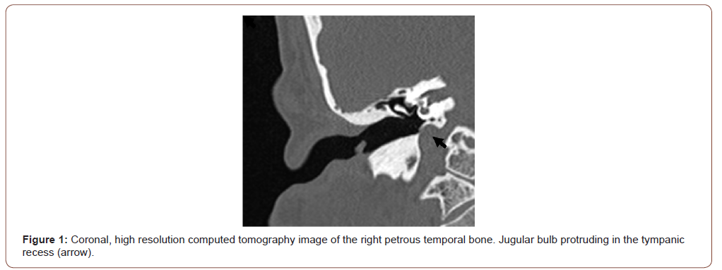
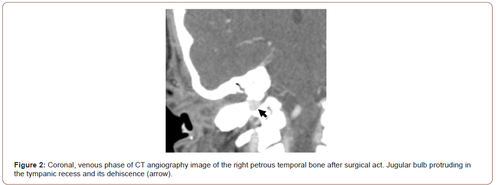
A 60-year-old male presented with four-year history of rightsided interment non pulsatile tinnitus, ipsilateral otalgia, and a mild conductive hearing loss, without any associated otorrhea, dizziness or facial weakness. Examination of the right ear revealed a subtotal dry perforation of the tympanic membrane. Computed tomography scan of the patient’s temporal bones demonstrated no signs of chronic process and structural anomalies were not reported. The patient was admitted for an elective tympanoplasty. During the surgery with postauricular approach, brisk venous bleeding was encountered at the point of the annulus upon removal of the skin of the anterior wall of the external acoustic canal. The hemorrhage arose from a dehiscent high riding jugular bulb lying immediately lateral to the annulus, in the pretympanic recess. Hemorrhage control was achieved through suction, extraluminal technique applying absorbent hemostatic gauze with hemostatic gelatin sponge, and replacing the anterior skin flap. Further review of the CT scan revealed a high, jugular bulb bulging into the external ear canal. Patient was discharged three days following procedure and still under tight follow up to monitor his condition (Figure 1, 2).


Discussion
Dehiscent jugular bulb is defined as a lack of bone over the jugular bulb, which occurs in 1-7% of bulb changes that affect the middle ear [13-15]. Although debatable, it seems that it also predominates on the right side, without differences regarding sex and being able to present in any age range [1, 13, 14]. In the clinical case presented, the location was right, though there was also an anormal presentation in contralateral temporal bone.
The anomalies of the jugular bulb are generally asymptomatic. However, the symptoms of suspected HJB may go unnoticed in the presence of a chronic otitis media process, as in our patient, and found directly in the surgery. If we have an imaging test, we must look for this type of alterations, because in general the jugular bulb area does not usually arouse the interest of the radiologist and the ENT, more pending the presence of cholesteatoma, position of the sigmoid sinus, tegmen tympani , ossicular chain, semicircular canals and facial nerve [4].
HJB might be injured during surgical act. Case reports have described damage and resulting profuse hemorrhage as a result of myringotomy [8, 9], elevation of a tympanomeatal flap [9, 10, 12], elevation of the inferior annulus [12], and removal of granulation tissue during middle-ear surgery [11]. Indeed, intrusion of a high jugular bulb into the middle ear has been well described. The only reported case of damage to a high jugular bulb during external ear canal surgery has been described by Moore [12]. In the clinical case presented, hemorrhage arose on removal of the skin of the anterior wall of the external acoustic canal, lateral to the annulus.
The management strategies in case of incidental surgical injury of a high and dehiscent JB, the postauricular approach is recommended to have a greater domain of the field3, as in our clinical case, in which the approach was originally performed for our planned procedure. When massive bleeding occurs during otologic surgery, it is recommended to place patient’s head lower below the level of the heart to avoid air embolism and compression with gauze or absorbable material [9-12]. With this action, sometimes it is possible to complete the expected surgery, although the most frequent is, as in our current case, that it must be delayed after achieving hemostasis.
To read more about this article....Open access Journal of Otolaryngology and Rhinology
Please follow the URL to access more information about this article
To know more about our Journals...Iris Publishers


No comments:
Post a Comment