Authored by Mosaab Akram Elshaer*,
Abstract
Background
For decades, many investigators have been trying to find the “factors” that increase the risk of coronary artery disease , A lot of cases reported for Asymptomatic patients or very mild symptoms and tendence to be coronary artery disease and in many cases, it could be critical situation.
Despite these massive efforts and findings, still some young individuals suffer from myocardial infarction without any “traditional” risk factors.
Coronary calcium score has emerged as a reliable tool to add to the already known mix of the risk factors to assess someone’s future risk of acute coronary syndromes by identifying atherosclerosis. Atherosclerosis, an inflammatory process in the arterial wall, starts at young ages and results in the formation of “plaques” in the arterial wall. As we age, the process of calcification begins in these plaques. We can identify the calcium in x-ray images, which confirms the underlying plaque in the arterial wall. These “plaques” are the main precursors of myocardial infarction. The CT can play important role in detection the coronary artery disease and evaluate the severity of the stenosis and its location.
Case Presentation
• Male patient, 50 years old, Mediterranean background, not diabetic, not hypertensive, nor hyperlipidemic, no significant medical history and takes no medications. He does not use tobacco, alcohol, or illicit drugs. No family history of medical disease.
• The patient has very mild occasional chest discomfort that’s not related to activities and has no precipitating factors. No dyspnea, no orthopnea.
• On physical examination: Temperature is 37c (99.5F) Blood pressure 120/80 mm, pulse is 90/m and respiration are 16/min. On Auscultation no heart sounds, no murmurs, The Lungs are clear. There is no peripheral edema.
• Electrocardiogram shows no significant changes. Echo shows Ejection fraction 56%, No resting wall motion abnormalities.
C.T Coronary angiography Findings
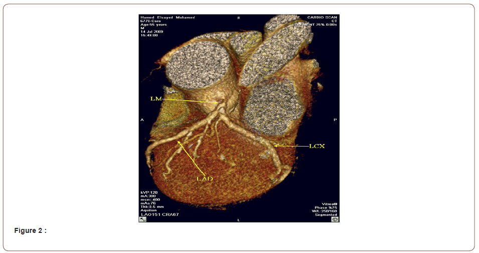
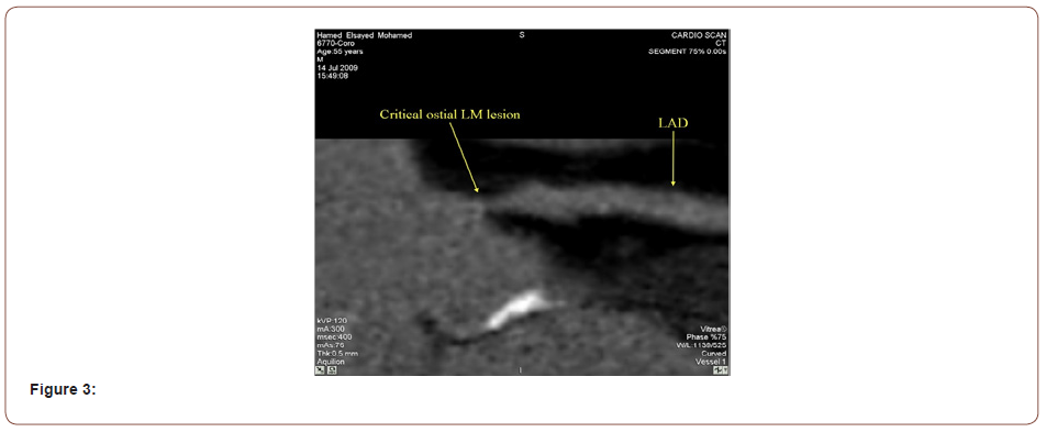
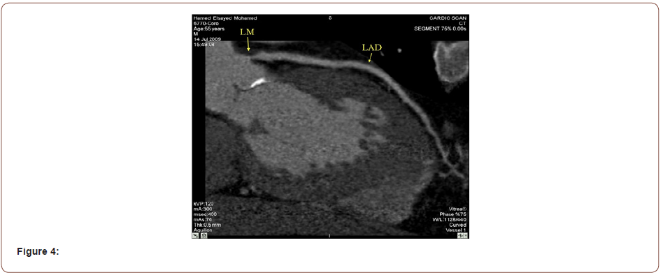
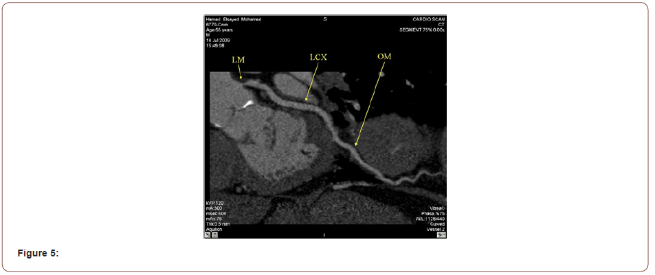
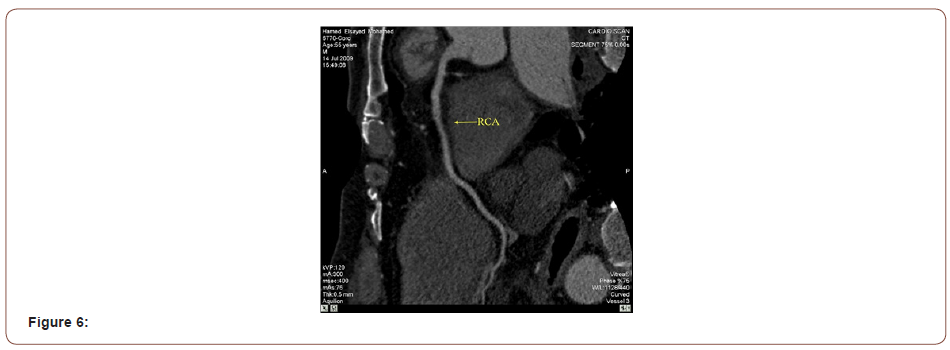
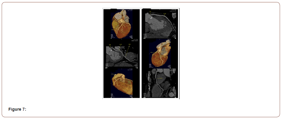
Left main coronary artery (L.M): is an average caliber atherosclerotic vessel showing an ostial non calcified plaque causing critical lumen narrowing; the LM is seen originating normally from the left coronary cusp, it ends by bifurcating into LAD and LCX. Left anterior descending coronary artery (LAD): is an average caliber atherosclerotic long vessel showing several noncalcified plaques throughout its course with no significant stenoses. It gives origin to three average caliber normal diagonals; the LAD ends by wrapping around the apex. Left circumflex coronary artery (LCX): is an average caliber non-dominant atherosclerotic vessel, giving origin to an average caliber long OM branch then the LCX continues as a small caliber vessel, the LCX and its OM branch are free of significant lesions. Right coronary artery (RCA): is a dominant average caliber atherosclerotic vessel, it gives origin to a small conal and RV branches and ends by bifurcating into average caliber PDA and PLB; the RCA and its branches show several nonocclusive plaques. Coronary Impression: Atherosclerotic CAD. Critical ostial LM stenosis with no other significant lesions.
C.T coronary calcium score report
(Table 1) This is the total amalgamation of a calcium score of 37 in the left main coronary artery, 8 in the right coronary artery, 0 in the left anterior descending coronary artery, 0 in the circumflex artery and 0 in the posterior descending artery.
Table 1: Calcium Plaque Burden.

Calcium Percentile Score
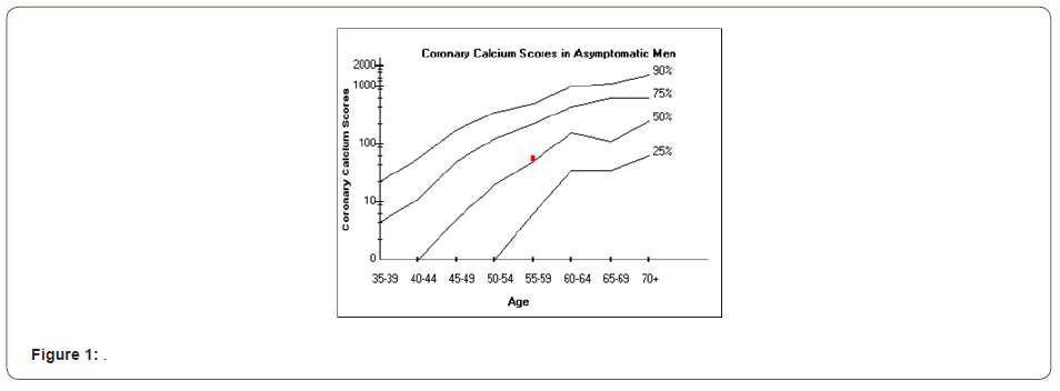
Total calcium score of 53 is between the 50th and 75th percentile for men between the ages of 55 and 59
This means that 50 percent of people this age and gender had less calcium than was detected in this study. The following graph shows the distribution of total calcium scores for each age group by percentiles. Your calcium score, relative to other age groups, is indicated by the highlighted square in the graph (Figure 1). [1] Callister TQ et. al., Coronary Artery Calcium Scores on Electron Beam Computed Tomography: JACC 1999. 33 (Supl.): 415A [1,2].
Translation Of Calcium Score
(Table 2) [2] Mayo Clinic Proceedings, March 1999, Vol. 74. Findings based on EBCT data. [3] Carr JJ, et. al., Evaluation of Subsecond Gated Helical CT for Quantification of Coronary Artery Calcium and Comparison with Electron Beam CT.; AJR 2000; 174: 915- 921
Table 2:

CT Cardiac Functional Analysis Report
Table 3:

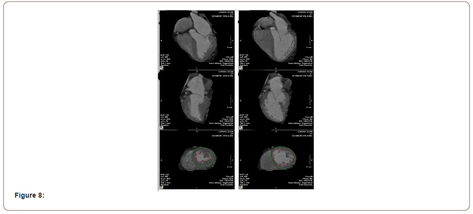
(Table 3) Normal left ventricular systolic function with no resting SWMA. There are no calcifications within mitral and aortic annuli, including valves. There is no pericardial or pleural effusion. There is no aneurysm or dissection of the thoracic aorta [3,4]. Impression: Atherosclerotic CAD. Critical ostial LM stenosis with no other significant lesions.
• Calcium Score is calculated to be 53 by Agatston Score. Normal LV EF (56%) with no resting SWMA’s (Figures 2-8).
Conclusion
C.T angiography is having a good diagnostic value in detection of the left Main coronary artery disease.
To read more about this article....Open access Journal of Biomedical Engineering & Biotechnology
Please follow the URL to access more information about this article
To know more about our Journals...Iris Publishers


No comments:
Post a Comment