Friday, May 31, 2024

Iris Publishers-Open access Journal of Anaesthesia & Surgery | Breaking Bad News: Addressing Hope, Spiritual Needs, And Denial to Achieve High Value Care


Authored by Kushinga Bvute*,

Introduction

Breaking bad news to a patient in the hospital is a powerful experience for doctors in training [1] and is widely regarded as a demanding task for physicians [2]. One challenge for the admitted patient is the frequent handoffs; one provider initiates the conversation about the disease and prognosis while a different physician may provide the follow-up. Additionally, inpatient providers lack pre-existing longitudinal relationships with their patients and insight into their values, cultural, spiritual, and social issues, or the family support system. Thus, preparation for these conversations inside the hospital is more complex [3].

When disclosing bad news, the quality of the communication significantly influences the patients’ emotional adjustment and compliance with recommendations [4]. In addition, patients’ belief in miracles or denial may hinder prognostic discussions [5]. With the high cost of healthcare, providers are expected to practice high value care by improving communication and incorporating patient concerns and values into care plans. This case illustrates the challenges of revealing bad news, examines the patient’s responses, and provides recommendations to better prepare physicians for disclosing bad news.

Case Presentation

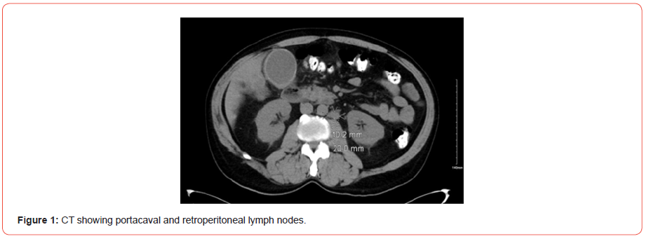
A 59-year-old Brazilian male presented to his primary care physician (PCP) with chronic epigastric pain. He was worked up for dyspepsia but continued to endorse intermittent diffuse abdominal pain as well as nausea and vomiting after dinner, so a right upper quadrant ultrasound was ordered, which showed multiple hypoechoic ill-defined mass lesions throughout the liver suspicious for metastatic disease and a gallbladder filled with sludge and stones. This prompted his PCP to send him to the hospital for a CT scan which showed multiple hepatic hypodense lesions, likely reflecting metastasis [Figure 1].
irispublishers-openaccess-anaesthesia-surgery

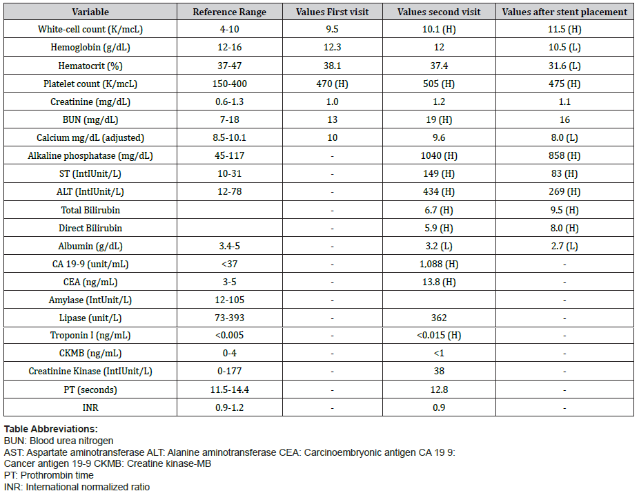
When he was informed of the findings suggestive of metastatic disease, he opted to leave without the full evaluation to identify the primary source of the malignancy, but then returned to the hospital days later for worsening symptoms. The patient was jaundiced, afebrile, and hemodynamically stable. Labs were significant for elevated alkaline phosphatase, AST, ALT, and total and direct bilirubin [Table 1].

Table 1:Blood work on first and second admissions, and after stent placement.
irispublishers-openaccess-anaesthesia-surgery


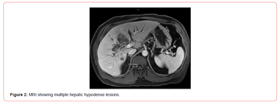
An endoscopic ultrasound and endoscopic retrograde cholangiopancreatography with biliary sphincterotomy and liver biopsy was performed, which unfortunately confirmed moderately differentiated cholangiocarcinoma. MRI showed a large infiltrative mass encasing the common hepatic and proximal common bile ducts with metastatic lesion within the right and left liver lobes. He also had metastatic nodal involvement along the periportal portacaval and retroperitoneal regions [Figure 2].
irispublishers-openaccess-anaesthesia-surgery

The patient was informed of his diagnosis during the hospital stay and the gastroenterologist placed a common bile duct stent. A consultation was placed with an oncologist; however, the patient preferred to follow up with an unspecified oncologist closer to home. Notes from the hospitalist suggested he was concerned the patient had an element of denial at the time of discharge. During hospitalization, he had made numerous comments in response to being told he has a malignancy, such as “I’m fine” and “I really think I’m OK.” When asked if he understood his diagnosis, he responded that he did and would indeed be seeing a specialist.

Days later, however, he returned to his PCP and stated he was not aware of the outcome of the biopsy or imaging, nor had he followed up with oncology. He endorsed itchy skin, weight loss, and poor sleep. He also noted that he changed his diet and was now drinking pureed vegetables in an effort to “cleanse his liver.” His wife was in the room with him and did not appear to understand the severity of her husband’s condition. The PCP created a safe and intimate atmosphere for the patient and his wife and asked them exactly how much they knew, how much they wanted to know, and how they would want that information conveyed. The patient responded that he wanted her to explain the biopsy and imaging results without foregoing any detail. The physician displayed the results on her computer and explained everything to him. After answering his questions, she calmly—but clearly—informed him and his wife that he had metastatic cancer.

The patient subsequently displayed a clear understanding of the severity of his condition and informed us that although he is very religious and believes in miracles, if God decides it is his time to go, then that is something he can accept. The PCP discussed the unlikelihood of a cure given how advanced his cancer already was but did not trivialize the power of hope. After the visit, the patient requested a referral to an oncologist for a visit that week. However, upon discussing the long-anticipated wait time and cost of treatment, he decided it would be best to seek further evaluation with an oncologist in Brazil, and by the end of that week he and his wife moved back to Brazil.

Discussion

The skill of giving patients bad news, as with other aspects of clinical medicine, is of great importance [1]. The quality of the provider’s communication and the patient’s initial response, such as denial or hope for a miracle, makes delivering bad news an essential skill not frequently found in the medical curriculum [6]. Denial is a basic mechanism for coping with stressful situations, especially in patients facing the challenge of cancer, as it reduces anxiety and allows them time to process the distressing information at a manageable rate [7]. However, denial may also interfere with getting treatment, for instance, leading patients to delay appointments with their oncologists, miss follow-ups, and exhibit non-compliance [8].

Healthcare providers commonly encounter patients hoping for a miracle stemming from a religious worldview and neglecting the patient’s spiritual needs leads to the patient disengaging in healthcare discussions. The way patients communicate hope for miraculous recoveries requires tailored responses by the clinician [9]. Providers who invite the patient to talk about miracles not only communicate acceptance and willingness to listen, but the patients are more likely to accept a diagnosis. A study at the University of Pennsylvania found that 66% of patients agreed that physicians who inquire about spiritual beliefs would strengthen their trust [10]. In comparison, 94% of patients for whom spirituality was meaningful wanted their doctors to address their spiritual sentiments and be sensitive to their values framework.

Since patients may be more likely to accept a diagnosis and comply with recommendations if they are informed of their condition by their previously established provider, a lack of a longitudinal relationship may increase communication complexity, leading to misunderstanding of prognosis or the purpose of care. Our patient’s hospital providers might not have had enough knowledge of his values and cultural, social, and spiritual nuances. The lack of such insight could explain why our patient denied understanding his diagnosis and its implications upon follow-up with his PCP despite being directly informed of his diagnosis in a hospital setting. The PCP was able to foster an understanding of the diagnosis and better convey the prognosis to him. Subsequently, the patient opted to seek affordable care in Brazil as opposed to not pursuing care after hospital discharge.

Despite the importance of the skill of breaking bad news in clinical practice, formal education for medical students and residents to communicate difficult information is limited [6]. Although denial may be a component in the early phases of coping, this thought process can become dysfunctional if it interferes with the patient seeking treatment [7]. The PCP with an established relationship can improve patient care through better communication. The PCP can incorporate the patient’s values into the care plan and address denial and spiritual needs leading to high-value care. Therefore, hospital providers should encourage a prompt office visit after discharge for the PCP to aid in prognostic discussions.

To read more about this article...Open access Journal of Anaesthesia & Surgery

Please follow the URL to access more information about this article

https://irispublishers.com/asoaj/fulltext/breaking-bad-news-addressing-hope-spiritual-needs-and-denial-to-achieve-high-value-care.ID.000566.php

To know more about our Journals...Iris Publishers

To know about Open Access Publishers

Iris Publishers-Open access Journal of Cardiology Research & Reports | High-Sensitive Cardiac Troponin I - An Important Biomarker in the Course of COVID-19 Disease in Adult Patients


Authored by Mariana Georgieva Yordanova*,

Abstract

Introduction: SARS-CoV-2 virus, in addition to causing pneumonia, can cause direct damage to the heart, inducing myocarditis, pericarditis or AMI with significant impairment of cardiac contractility. Adult patients with hypertension, diabetes mellitus and obesity are at increased cardiovascular risk and complications from COVID-19.

Aim: To assess high-sensitivity troponin I (hs-TnI) levels among hospitalized patients with COVID-19 as a biomarker associated with myocardial damage and determine the correlation with disease severity and outcome.

Material and Method: A retrospective analysis of hospitalized patients was performed between January 1 and May 1, 2021, with clinical and laboratory evidence of COVID-19. Monitored were the laboratory parameters of myocardial damage with baseline and peak hs-TnI values above the median (≥19.8 ng/L) in 110 patients. The extent of the inflammatory response (СRP), coagulation status (DDimer), acid-base balance parameters, hypoxemia abnormalities and LDH levels according to disease severity and outcome, and length of hospital stay was assessed. Data were evaluated using descriptive, correlation and multivariate analysis.

Results: The present study showed a statistically significant relationship between disease severity and heart damage. The hs-TnI values in patients with COVID-19 and CVD were statistically significantly higher than those with COVID-19 but without CVD. The most significant levels of the cardiac biomarker are observed in deceased patients.

Conclusion: High-sensitive troponin is a valuable biomarker for early diagnosis and assessment of disease progression with a worse prognosis in patients with COVID-19. Elevated hs-TnI in patients with SARS-CoV-2 infection who have concomitant heart disease may suggest the severity of the clinical picture and the course of the disease.

Introduction

From early 2020, the world faces a new medical challenge, a pandemic caused by an unknown SARS-CoV-2 coronavirus, requiring new knowledge and approaches. Today, more than 250 million people worldwide are affected by the covid infection, of whom 2.1% have lost their lives. The most common pathological manifestation of SARS-CoV-2 is respiratory distress syndrome, considered the leading cause of death. However, with each new epidemic wave, more evidence also accumulates of direct damage to the heart, causing myocarditis, arrhythmia, and acute coronary syndrome as likely concomitant damage. The presence of previous cardiovascular disease or cardiovascular risk factors leads to a higher mortality rate than patients without prior cardiovascular disease [1]. In addition, several studies have shown that COVID-19 patients who did not have heart disease before infection may also develop cardiac complications [2].

Cardiac troponins are biomarkers that have gained a reputation as the gold standard in diagnosing myocardial damage. The troponin complex consists of three subunits (troponin C, troponin T and troponin I). Together with calcium ions, troponin proteins regulate and facilitate the interaction between actin and myosin fibres during muscle contraction, including the myocardium. Troponin I is particular for the heart muscle and is not isolated from skeletal muscle. This absolute specificity makes it an ideal marker for myocardial damage [3].

Cardiac isoforms of troponins are quantitative markers of cardiomyocyte damage, and the likelihood of AMI increases with increasing levels in the blood. Their increase is observed in the interval 4-8 hours after myocardial injury, with a peak value of 12-24 hours and retention in the levels for 7-10 days [4]. A Contemporary high-sensitivity cardiac troponin (hs-cTn) assays can detect concentrations 10 to 100 times lower than conventional assays, increasing their diagnostic accuracy in patients with acute chest pain [5]. In this aspect, it is reasonable to investigate hs-TnI in patients with COVID-19 as an essential biomarker for both an early diagnosis of myocardial injury and prognosis.

Aim

To analyze the levels of high-sensitivity troponin I among hospitalized patients with COVID-19 as a biomarker associated with myocardial injury and to determine the correlation with disease severity and outcome.

Material and Method

112 patients from 320 hospitalized patients with proven SARSCoV- 2 infection in the period 1.02. to 1.05.2021 were found to have cardiac lesions with initial and peak hs-TnI values above the median (URL of 99th percentile ≥ 19.8 ng/L). The review included 82 men and 30 women aged 67.7±15.3 years and 71.6±18.8 years, respectively. Levels of hs-TnI were determined by chemiluminescent immunoassay on an Assay 2 analyzer (B. Coulter - USA). The extent of inflammatory response was assessed by CRP values (Olympus AU640 B. Coulter - USA), change in coagulation status and tendency to thrombogenicity (DDimer - Stago Satelite), disturbances in AB balance and hypoxemia by KGA Modular Pro (ESCHWEILER - Germany), LDH levels (Olympus AU640 B. Coulter - USA), length of stay and disease outcome. The severity of SARS-CoV-2 infection was assessed according to WHO criteria from January 2021.

Data analysis was done using GraphPad Prism v. 6.0 software by standard statistical methods (descriptive statistics, Anova with Bonferroni correction for median comparison, and Spearman correlation analysis). Biochemical parametric data were presented as mean ± standard deviation. Statistical significance was indicated at p<0.05.

Result

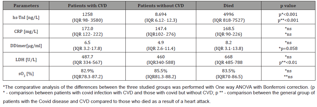
According to the WHO criteria for the clinical course, patients were divided into groups - moderate grade (22%), severe grade (45%) and critical grade (33%). Eighteen deaths with critical grade disease, representing 16.1% (12 males and six females) were. Of these, only two men in young age (45-50 years) had no evidence of previous and concomitant diseases. All other patients had comorbidities, most commonly AH alone or combined with CVD and/or diabetes and obesity. Furthermore, COVID-19 patients according to clinical presentation (subjective and objective criteria) of acute cardiac injury were divided into three groups (Table 1).

Table 1: Parameters were studied in COVID-19 patients in the three groups. Patients with CVD events, those who died as a result of them and patients with COVID-19 without CVD disorders.

irispublishers-openaccess-cardiovascular-research

The analysis performed found that patients with COVID-19 and cardiovascular symptoms or complications had significantly higher hs-TnI values than other patients with COVID-19 but without cardiac problems. The highest myocardial necrosis marker values were found in those who died due to AMI, myocarditis, rhythm and conduction disturbances, or PTE. In the group with lethal outcome, hs-TnI values showed a statistically significant difference compared to the entire CVD group and those without such complications (p<0.001). Regarding the other studied parameters characterizing the severity of pulmonary perfusion disturbance, haemostasis and hypoxaemia, no such differences were found between groups. There was a tendency for a difference in DDimer values in the deceased patients compared to the general CVD group. Only LDH as an enzyme reflecting both necroses and activated anaerobic glycolysis showed significantly higher levels in the deceased than the total patient group with CVD complications. This explains the moderate statistically significant correlation between hs-TnI and LDH values in the overall COVID-19 group with CVD (r=0.438 p=0.0008).

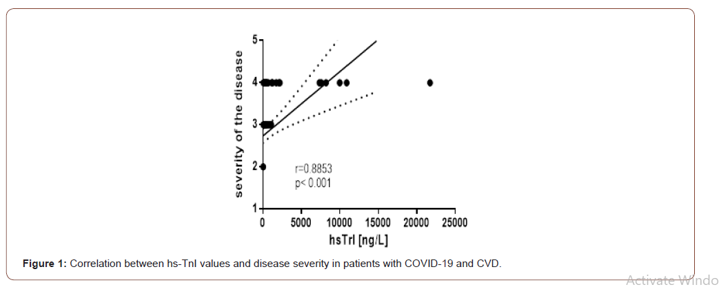
We found that hs-TnI levels showed a high correlation with disease severity (Figure 1). A significant degree of correlation was observed between the severity of the disease and the length of hospital stay (r = 0.635; p <0.001). The mean length of stay in patients with CVD was 16 days while in the critical course of the disease and lethal outcome is 14 days (range 8-30 days). An analysis of the correlations between hs-TnI and other indicators for monitoring vital and metabolic disorders in patients from the group of the deceased revealed the following data (Table 2).

Table 2: Correlation between the values of hsTrI with the other studied indicators in deceased patients with Covid 19 and CVD incidents.

irispublishers-openaccess-cardiovascular-research

irispublishers-openaccess-cardiovascular-research

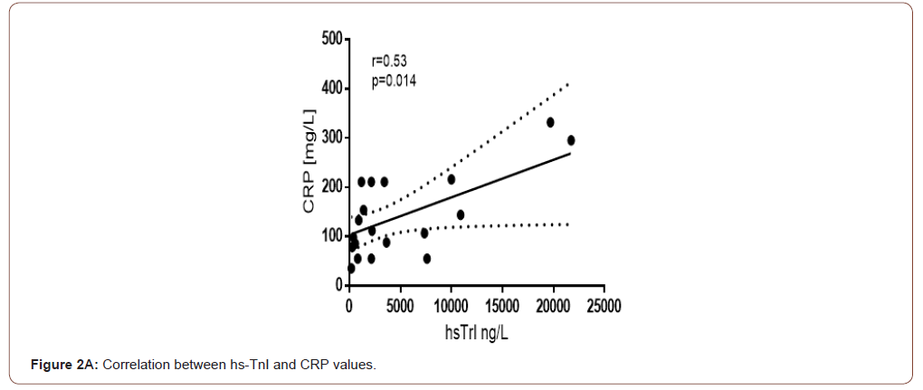
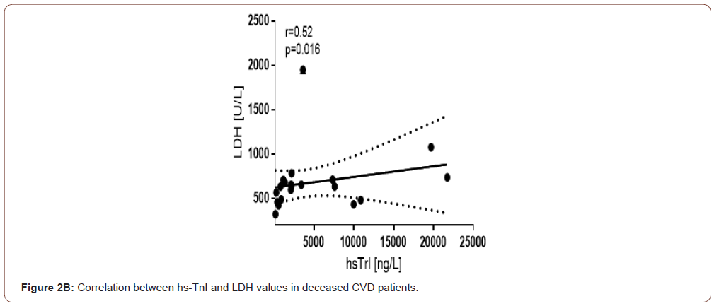
A moderate correlation between hs-TnI and LDH is currently established in the general group of COVID-19 and GCC. In the deceased group, this correlation is preserved and is significant in size (Figure 2B). We observed a similar correlation force with CRP, a biomarker of inflammation (Figure 2A). This is probably because CRP, in addition to being a well-known acute phase protein, is a marker that reflects and correlates with the size of the necrotic myocardial lesion.

irispublishers-openaccess-cardiovascular-research

irispublishers-openaccess-cardiovascular-research

Discussion

Shi et al. showed as early as 2020 that myocardial injury is a common condition among COVID-19 patients in Wuhan [6]. The authors reported that more than half of these patients died. A recent report suggests initial and follow-up testing during hospital stay of high-sensitivity troponin in patients with SARS-CoV-2 infection as a way to help clinicians identify individuals with possible cardiac damage and predict progression of COVID-19 to a fatal outcome [7]. The aetiology of acute myocardial injury in response to SARS-CoV-2 infection is still poorly understood. Possible mechanisms include cytokine storm resulting from a dysfunctional and uncontrolled immunologic response evidenced by elevated levels of d-dimer, IL- 6, CRP, and LDH and primary cardiac injury caused by SARS-CoV-2 [8]. One of the hypotheses is a direct attack on myocytes in the viraemic phase because of the affinity between the SARS-CoV Spike protein and angiotensin-converting enzyme type 2 (ACE-2), which is well represented in myocardial cells [9]. ACE-2 is also present in the vascular endothelium, so vasculitis and endothelial dysfunction are possible, further activating haemostasis with the occurrence of thromboembolic events and myocardial ischemic injury [10,11].

The amount of ACE2 may vary between individuals as well as in different tissues and cells. Some evidence suggests that ACE2 may be higher in patients with hypertension, diabetes, and coronary artery disease, explaining their greater vulnerability to SARSCoV- 2 infection [12]. The primary role of ACE2 is that of a negative regulator of the renin-angiotensin system (RAS) by degrading angiotensin II to angiotensin. With this, it exerts vasodilating, anti-inflammatory and antifibrotic effects by binding to the Mas receptor. The RAS plays a critical role in the maintenance of blood pressure homeostasis as well as water-electrolyte balance and is closely related to the pathophysiology of heart disease [13].

Our data show an increase in hs-TnI in patients with myocardial injury, consistent with the findings of the meta-analysis by Dawson Det al. [14]. There was also an increased risk of death with increasing cardiac marker levels found in other studies [15]. Patients with COVID-19 have reduced oxygen supply due to hypoxic respiratory failure and concomitant increased oxygen demand due to tachycardia, febrility and endocrine dysregulation, which enhances myocardial hypoxia [16]. Patients who passed through the Covid unit during the relevant period were predominantly elderly (over 65 years of age and 29.5% of them are over 80 years of age) with concomitant cardiovascular disease. This predisposed to SARS-CoV-2 induced myocardial injury and increased mortality associated with COVID-19. Ample evidence has now accumulated to explain the increased susceptibility and severity in the course of the disease, resulting from multiple factors, most commonly dependent on altered ACE2 expression, age, sex, medications and comorbidities s.

A clear picture of hypercoagulability with elevated D-dimer values is observed in the course of the disease. The most significantly increased levels of the parameter are seen in patients with critical stage disease. D-dimer is a fibrinolytic degradation product of pathologically increased blood clot formation. Some studies found that D-dimer levels at hospital admission predicted worse clinical outcomes [18]. In contrast to the meta-analysis by Dawson D, et al. in the present study, we found no statistically significant difference in DDimer between the group with and without CVD versus deceased [14]. COVID-19 infection likely causes a range of multidirectional pathological conditions in different patient groups, whereby differences between groups are blurred. However, the D-dimer is also an inflammatory marker of the acute phase, which may explain the non-statistical differences in the three groups of patients.

Conclusion

cTnI appears to be a beneficial biomarker, the gold standard for early diagnosis of cardiac complications and myocardial damage in the course of COVID-19 infection. Serum cTnI testing is an independent predictor of disease severity and mortality, especially in elderly patients with comorbidities.

To read more about this article...Open access Journal of Cardiology Research & Reports

Please follow the URL to access more information about this article


To know more about our Journals...Iris Publishers

To know about Open Access Publishers

Thursday, May 30, 2024

Iris Publishers- Open access Journal of Gastroenterology & Hepatology | Rapid Review on Inflammatory Bowel Disease: Current Management and Future Research


Authored by Sepideh Khazeni*,

Mini Review

Inflammatory bowel disease (IBD) is a chronic and progressive inflammatory condition involving the gastrointestinal (GI) tract with an increased risk for colon cancer. Two main IBD are affecting the GI tract: Crohn’s disease (CD) that the inflammatory conditions that can affect the entire GI tract, and ulcerative colitis (UC), in which the inflammatory conditions only affect colonic mucosa [1]. Since 1990, the prevalence of IBD has been stabilizing in western countries, but the incident has been globally accelerating in newly industrialized countries in Africa, Asia, and South America [3]. The exacerbation of IBD forces an excessive economic burden on the healthcare system [4] and impairs patients’ quality of life [4, 5].

The systematic reviews highlighted the role of genetic and environmental factors as the main risk factors associated with the IBD, but the leading cause of IBD has not yet been fully understood [2]. Studies showed that patients with a family history of IBD have four times the risk of developing the disease [6]. Over 200 risk loci have been identified for IBD. Many loci overlap between IBD and some autoimmune disorders, particularly with psoriasis and ankylosing spondylitis [7]. In addition, some medications, such as oral contraceptives [8] and non-steroidal anti-inflammatory drugs [9], are associated with an increased risk of IBD, while Breastfeeding appears to decrease the risk of IBD [10]. Other than genetics, dietary intake might affect the risk of GI tract inflammation in CD and UC. For example, it has been reported that western diets with a high amount of total fats, total polyunsaturated fatty acids (PUFAs), omega-6 fatty acids, and meat might increase the inflammation in the GI tract and subsequently increase the risk of CD and UC. On the contrary, diets with an abundance of vegetable intake might decrease UC risk, and those richer in fibre and fruit might reduce the risk of CD [11].

Theoretically, the Mediterranean or vegetarian diet is more beneficial for IBD prevention and management than the western diet. Because the Mediterranean and vegetarian diets provide plentiful anti-inflammatory micronutrients, essential fatty acids, and fibres [12]. Further clinical studies can demonstrate the benefit of Mediterranean or vegetarian diets in the prevention, treatment, and management of IBD. Meta-analysis showed that Mediterranean Diet might prevent the development of IBD through gut microbiota [13]. Shifts in microbial composition in IBD patients between remission and active disease might contribute to IBD flare-up [14]. It is known that an increase in Fusobacterium bacterium is associated with IBD [13]. Limited clinical studies showed that probiotics might be as effective as conventional therapies in preventing IBD relapses [12]. However, the solid preclinical data suggested that probiotics can aid patients with IBD management [15]. Further clinical studies can demonstrate the benefit of the probiotics in prevention, treatment, and management of IBD.

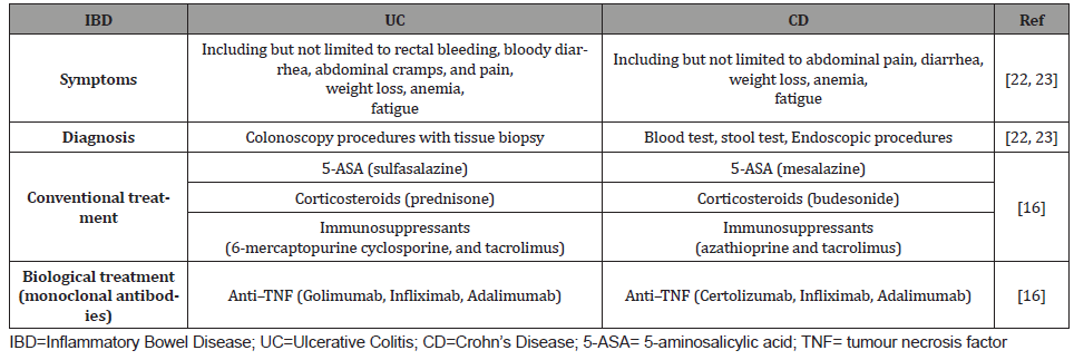
The IBDs are mainly managed with conventional therapy through groups of medications such as 5-aminosalicylic acid (5-ASA) derivatives, Corticosteroids, and immunosuppressants [16]. However, meta-analyses showed that in IBD patients, immunosuppressants such as methotrexate, azathioprine, and 6-mercaptopurine have no statistically significant advantage over placebo for induction of clinical response and remission [17]. Furthermore, meta-analyses showed superior clinical response induced by tacrolimus compared to placebo in patients suffering from UC. Tacrolimus also induced mucosal healing in UC patients [17]. The second-line therapy for IBD patients not responding to conventional treatment is biologic therapy by administrating monoclonal antibodies as anti–tumour necrosis factor [TNF], anti– integrins, and anti–cytokines [18]. The anti–TNF such as Golimumab and Certolizumab is prescribed for treatment or management of UC and CD, respectively. Moreover, other anti–TNFs such as Infliximab (IFX) and Adalimumab (ADA) are approved to manage both UC and CD. However, in severe IBDs, the increased levels of proteases, including matrix metalloproteinases (MMPs) in inflamed bowel, might fail the response to anti–TNFs [19].

(Table 1) summarised the correct IBD management. IBDs might increase the risk of developing extra-intestinal cancers. Meta-analyses demonstrated an increased risk of skin cancer by 2.2 fold and hepatobiliary malignancies by 2.3 fold in IBD patients. Furthermore, the risk of hematologic malignancies increased for CD patients by 2.4 fold and lung cancer by 1.53 fold [20]. The recent systematic review showed that in patients with IBDs, there is no risk associated with SARS-COV-2 infection. Moreover, IBD management by immunosuppressant or biologics might not affect the SARS-COV-2 infection prognosis. In contrast, IBD management by Corticosteroids might increase the risk associated with a worse prognosis of SARS-COV-2 infection [21-23].

Table 1:Summary of current IBD management.

irispublishers-openaccess-gastroenterology-hepatology


As the current treatment and maintenance of IBD are costly for patients and the healthcare systems, herbal treatments might represent an effective complementary and alternative medicine [24]. In addition, several herbal medications might potentially be effective for the treatment of IBD as they are known to have antiinflammatory effects such as immunomodulatory properties, antimicrobial activities, antioxidant activities, and antiulcer [25]. Clinical studies showed that using herbal medicines such as curcumin, Aloe vera and Boswellia serrata could induce clinical remission or improvement in patients with mild or moderate IBD as effective as conventional therapy [26]. However, randomized controlled trials of herbal therapy for IBD treatment remain limited and heterogeneous [27]. Therefore, we suggest further clinical studies to better demonstrate the benefit of herbal medicines in the prevention, treatment, and management of IBD.

To read more about this article...Open access Journal of Gastroenterology & Hepatology

Please follow the URL to access more information about this article

https://irispublishers.com/ajgh/fulltext/rapid-review-on-inflammatory-bowel-disease-current-management-and-future-research.ID.000555.php


To know more about our Journals...Iris Publishers

Iris Publishers-Open access Journal of Otolaryngology and Rhinology | Ectopic Thyroid Mimicking A Thyroglossal Duct Cyst: A Case Report


Authored by W Lendoye*,

Abstract

Thyroid ectopy is a rare condition, caused by dysfunction during migration of the thyroid gland during its embryogenesis. It is generally associated with subclinical hypothyroidism. Patients with this pathology are mostly asymptomatic. It is seen in childhood by an anterior cervical mass which confuses it with a cyst of the thyroglossal tract. The diagnosis is made by ultrasound imaging, scintigraphy and histological examination. Management is essentially hormonal. Surgery is considered in case of complication.

Keywords: Ectopic thyroid; Thyroglossal Duct Cyst; Fine needle aspiration cytopunction

Abbreviations: The thyroglossal duct cyst (TGDC); Fine needle aspiration cytopunction (FNAC)

Introduction

Ectopic thyroid refers to the presence of thyroid tissue in locations other than the normal anterior neck region between the second and fourth tracheal cartilages1. It is the most frequent form of thyroid dysgenesis, accounting for 48-61% of the cases. Prevalence of this condition is reported to be between 1 per 100,000-300,000 persons and occur one in 4,000-8,000 patients with thyroid disease [1]. The thyroglossal duct cyst (TGDC) is the most common congenital malformation in the neck, accounting for 70%of all congenital neck lesions. It may occur at any age. Most are detected in the first 2 decades of life [2]. Ectopic thyroid can be found along the way of thyroid descent, in the midline, or laterally in the neck or even in the mediastinum or under the diaphragm or in other different site.

We report the course and management of a child with ectopic thyroid which mimicked as thyroglossal duct cyst.

Case Presentation

This case is about a 7 years-old female child who presented in ENT outpatient department for midline neck mass, evolving for one year, and increasing the size slowly. There was no history of dysphagia, dyspnea, dysphonia, recurrent respiratory tract infections or dysthyroidism.

The clinical examination midline neck mass, firm, swelling, measuring 2×1cm in size in the hyoid region moving with deglutition and protrusion of tongue, on good general condition. Examination of cardiovascular system and abdominal region was normal. Patient was at first diagnosed to have thyroglossal duct cyst.

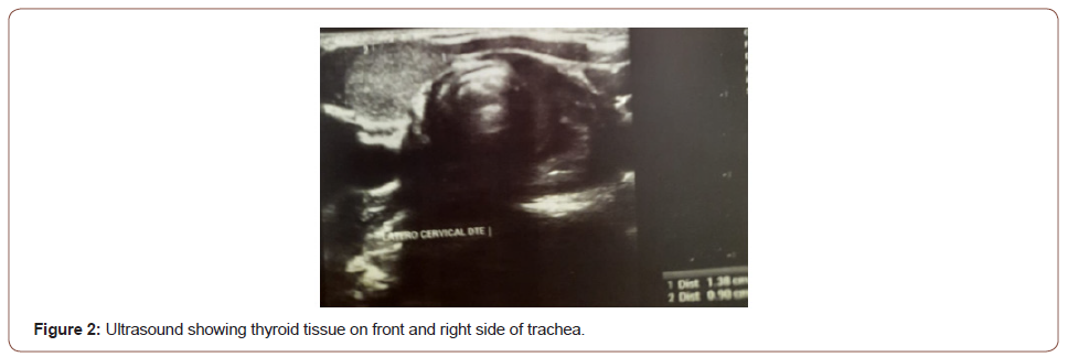
A neck ultrasound was peformed and revealed that the midline and lateral trachea mass was the only functioning thyroid tissue.

Thyroid function tests revealed a high TSH: 15,3 mIU/mL (0.3- 5.5 mIU/mL),

Fine needle aspiration cytology-thyroglossal cyst was performed, thyroid peroxidase antibody 26 U/mL (1-34U/mL).99 TC.

The patient was finally diagnosed with ectopic thyroid with subclinical hypothyroidism, and received L-thyroxine 50 mcg/day. Surgery was not necessary on this case. After 6 months follow-up, the patient was doing well, with normal TSH at 1,3 mIU/mL. There was no evolution of the mass size (Figures 1, 2).

irispublishers-openaccess-textile-science-fashion

Discussion

Ectopic thyroid tissue is defined as any tissue not located anterior and lateral to the second, third and fourth tracheal rings. This can occur anywhere along the path of descent of the normal thyroid gland [3]. Ectopic thyroid is a rare entity with a prevalence of approximately 1 per 100 000 to 300 000 persons and is reported to occur in 1 of every 4000 to 8000 patients who have thyroid disease [4].

The most common site of ectopic thyroid is a lingual thyroid. The wall of a thyroglossal duct cyst is the second most common site for ectopic thyroid tissue [5]. The thyroglossal duct cyst (TGDC) is the most common congenital malformation in the neck, accounting for 70%of all congenital neck lesions. It occurs as a result of failure of the thyroglossal duct to obliterate during embryonic development.

Ectopic thyroid is essentially asymptomatic, but 30% of patients present with hypothyroidism. The pathophysiology of hypothyroidism in ectopic thyroid is explained by an aberration in the migratory pathways of the rudimentary thyroid that can lead to ectopy, which almost certainly results in inadequate blood supply to support normal thyroid function Genetic factors leading to abnormal function and morphology may also contribute to hypothyroidism in these patients [4]. To diagnosticate an ectopic gland imaging methods such as scintigraphy, fine needle aspiration cytopunction (FNAC) employ technetium-99m pertechnetate which differs to other isotopes because of the better quality of imaging and the less radiation dose, so it might be used in children [6].

The treatment of the ectopic thyroid depends on the functional status of the thyroid, and the presence of compressive symptoms. Levothyroxine treatment is recommended for patients with hypothyroidism. Surgery is recommended for patients with a large thyroid gland causing pressure symptoms or thyroid showing malignancy signs.

Conclusion

Ectopic thyroid remains a rare disease. When it presents as a midline neck mass, it can be easily confused with a Thyroglossal Duct Cyst. Imagery and fine needle aspiration cytopunction (FNAC) using technetium-99 m pertechnetate is necessary for diagnosis, leading to an adequate treatment.

To read more about this article....Open access Journal of Otolaryngology and Rhinology

Please follow the URL to access more information about this article

To know more about our Journals...Iris Publishers

To know about Open Access Publishers

Wednesday, May 29, 2024

Iris Publishers-Open access Journal of Dentistry & Oral Health | Surface Treatments of Titanium and Zirconia Implants: Impact on Bacterial Adhesion (Systematic Revue of Literature)


Authored by Moudni S*,

Introduction

The use of dental implants to restore the loss of one or many teeth has become a widely used treatment option in daily practice. However, as soon as implant surfaces or their components are exposed to human oral cavity, they are immediately covered by an acquired film and instantly subjected to bacterial colonization. This is directly influenced by the surface properties of the materials, including chemical composition, surface roughness, surface energy... [1]. In modern biomaterial research, implant surfaces are primarily modified to increase bone integration into the alveolar bone. Recently, implant surfaces are also modified to reduce biofilm formation after exposure to the oral cavity [2]. Currently, many implant systems with different surface treatments are available on the market, which makes it difficult for the practitioner to choose. There are two main categories of implant surface treatments [3]:

-Either by adding substance: this is the addition treatment.

-or by altering the smooth surface: this is the subtraction treatment.

The main objective of this systematic review was to evaluate the impact of different surface treatments of titanium and zirconium implants on bacterial adhesion. The secondary objective was to compare bacterial adhesion on titanium and on zirconium implants.

Material and Methods

Research strategy

Three computer databases were used for the literature search: Pubmed, Cochrane, and Science Direct.

The search was conducted between 01/ 01/ 2011 and 01/ 02/ 2022. The review included well-conducted in vivo and/or in vitro trials and randomized trials written in English, evaluating the surface condition of titanium and zirconium dental implants and its relationship with bacterial adhesion. Exclusion criteria were case reports, systematic reviews of the literature, meta-analyses, literature reviews, animal and cadaver studies, and articles dealing with bacterial adhesion on supra-implant prosthesis.

The table below represents the keywords used in the different boolean equations and for each database (Table 1).

Table 1: keywords and boolean equations.

irispublishers-openaccess-dentistry-oral-health


Assessment of methodological quality

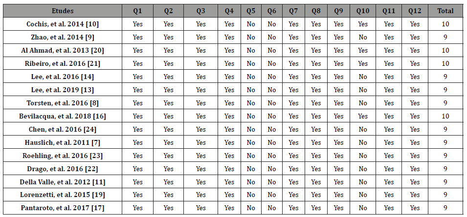
Two reviewers (M.S. & B.I.) assessed the methodological quality of the studies selected for analysis. This assessment was based on the tool “The Critical Skills Appraisal Program” (CASP) which was created from guides produced by the Evidence Based Medicine Working Group and published in the Journal of the American Medical Association [4].

Data extraction

To prepare and structure our systematic review, the focused question was developed using the PICO criteria:
P: Participants => Titanium and zirconium dental implants
I: Intervention => Implants with different surface treatments (by addition/subtraction) placed in contact with different bacteria.
C: Comparison => Control group (titanium or zirconium discs without surface treatment)
O: Outcomes => Relationship between the surface characteristics/the material of the implants and bacterial adhesion.
Data extraction was completed by 2 readers independently, with formal processes for discussion and consensus building in case of disagreement to minimize subjectivity during the multiple stages of completion.
The writing of this systematic review followed the PRISMA Statement “Preferred Reporting Items for Systematic reviews and Meta-Analyses” Moher, et al. [5].

Results

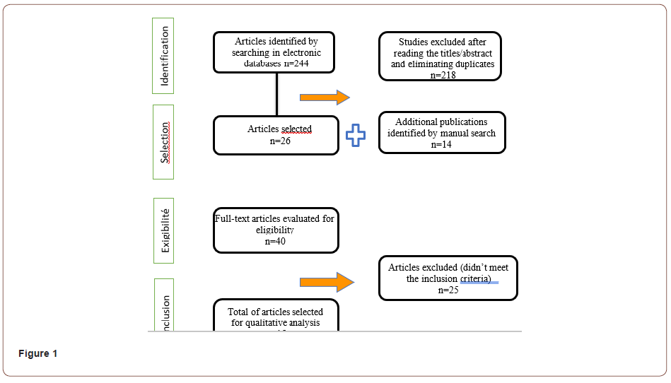
The search of the three databases identified a total of 244 studies from the automated search, from which we retained 26 studies after the first selection based on the reading of titles and abstracts and after eliminating duplicates. Through the manual search, we were able to group 14 articles. Thus, we had a total of 40 articles from which 25 articles were eliminated after applying the inclusion and exclusion criteria, resulting in the selection of 15 articles included in our review (Figure 1).

irispublishers-openaccess-dentistry-oral-health

The 15 selected studies include 11 in vitro studies, 2 studies conducted in vivo and 2 studies conducted in vitro and in vivo. They were all performed using titanium or zirconium discs with different surface characteristics. The control group was constituted of titanium or zirconium discs without surface treatment. In vivo studies were performed with intra-oral splints with different discs for each tested sample.

Of the 15 studies included in our review, 10 investigated bacterial adhesion only on titanium discs. The other 5 studies included, in addition to titanium discs, zirconium discs with different surface treatments.

The different parameters measured were essentially surface roughness of the material, its wettability through the measurement of the contact angle, chemical composition of the surfaces and analysis and quantification of the bacteria adhesion to the different surfaces after bacterial culture.

The different surface treatments used were multiple and heterogeneous and were classified into implant surface modification by addition or subtraction (Table 1) (Table 2).

Table 2: Classification of surface implant modifications used in the included studies.

irispublishers-openaccess-dentistry-oral-health


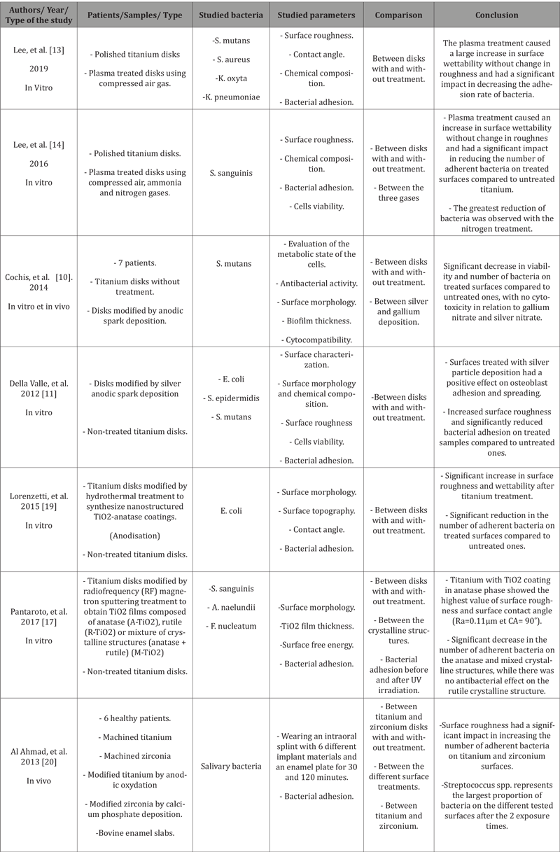
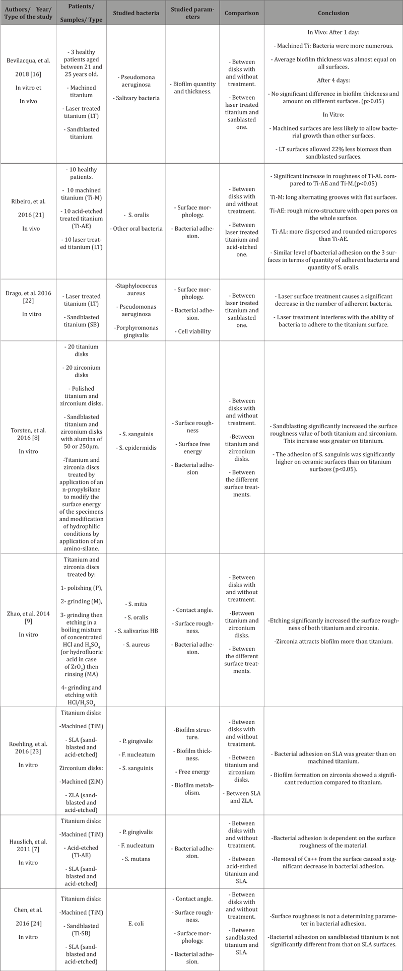
Informations about the characteristics of these studies are presented in (Tables 3, 4), including a brief description of the studies and their conclusions and the quality assessment of included studies is summarized in (Table 5).

Table 3: Bacterial adhesion and implant surface modification by addition.

irispublishers-openaccess-dentistry-oral-health


Table 4: Bacterial adhesion and implant surface modification by subtraction.

irispublishers-openaccess-dentistry-oral-health


Table 5: Assessment of methodological quality of the included studies.

irispublishers-openaccess-dentistry-oral-health


Discussion

This work focused on the effect of different surface treatments of titanium and zirconium dental implants on bacterial adhesion. This question was addressed by in vitro and/or in vivo studies. Several parameters are involved in the phenomenon of bacterial adhesion on dental implants. Surface roughness is a widely evaluated parameter. According to Wennerberg, et al. [6], dental implant surfaces are classified into four different groups based on their roughness: smooth surfaces (Ra < 0.5 μm), minimally rough surfaces (0.5 < Ra < 1.0 μm), moderately rough surfaces (1.0 < Ra < 2.0 μm), and rough surfaces (Ra > 2.0 μm).

According to Hauslich, et al. [7], increasing surface roughness increases bacterial adhesion to implant surfaces. However, other studies (12-20) have shown that surface roughness only affects the early stage of biofilm development. Torsten and Zhao [8, 9], on the other hand, have shown that roughness is not the only determinant of bacterial adhesion. Three-dimensional analyses of the microstructure are necessary [8]. Considering the studies carried out in vivo and in vitro using complex bacterial communities close to the oral context where the bacterial flora is rich and varied, it was concluded that the surface roughness intervenes only during the initial bacterial attachment.

Furthermore, the use of surface treatment by electro-deposition of silver or gallium showed a great decrease in bacterial adhesion on the surfaces despite the increase in surface roughness [10, 11]. This was explained by the fact that silver nanoparticles in the microporous titanium oxide layer are readily available to react with water and release silver ions. These later are known to bind strongly to electron donor groups in biological molecules containing sulfur, oxygen or nitrogen causing defects in the bacteria cell wall so that cell contents are lost. In addition, silver ions bound to proteins can alter bacterial cells metabolism and change membrane permeability and respiration. Both of these effects lead to bacterial cell death [9]. According to Wennerberg and Albrektsson [12], there is an optimal surface roughness window of 1 to 1.5μm. They consider that a higher value leads to a loss of bone anchorage. They add that it is very difficult to compare different studies, especially because the techniques used for characterization of surface topography vary considerably.

The surface energy of implant materials is also an important parameter influencing bacterial adhesion. Hydrophilic surfaces prevent bacterial attachment and can be achieved by increasing the wettability. The treatment of implant surfaces with plasma had led to significant increase in surface energy of titanium (4x more than untreated one), and consequently, to a significant reduction in the number of adherent bacteria [13, 14]. This was explained by the fact that bacteria with hydrophobic properties prefer hydrophobic surfaces and tend not to attach to hydrophilic surfaces and vice versa. However, the majority of bacteria involved in peri-implantitis are hydrophobic species. These results are in agreement with a review of literature about the wettability of dental implant surfaces, which concluded that among the benefits of hydrophilic surfaces is the reduction of adhesion of bacteria such as Staphylococcus aureus, Streptococcus sanguinis, and Staphylococcus epidermidis [15].

Concerning the crystalline phase of the surface, titanium dioxide can be found in three crystalline structures: anatase, rutile and brookite. These have a photocatalytic activity responsible for their anti-bacterial properties. The different structures can be generated by different methods like anodic oxidation, hydrothermal treatment and plasma treatment. TiO2-anatase coatings, which were previously proven to improve corrosion resistance, affect the plasma protein adsorption and enhance osteogenesis, have also shown strong antibacterial activity on titanium surfaces [16]. This can be attributed to the presence of anatase phase and its large band gap (3.2 eV) promoting high energy to create electrons and holes, and consequently to form more reactive oxygen species, when compared to rutile [17].

With regard to the implant material, titanium is the most frequently used reference material because of its biocompatibility and excellent mechanical properties. Recently, high-strength zirconia (ZrO2) implants have been invented as an alternative to titanium implants because of their resistance to corrosion and their enhanced aesthetics in case of exposure. Bacterial adhesion on titanium and zirconia does not seem to show any significant difference. Titanium is coated by a layer of surface oxide, which physical and mechanical characteristics are more closely related to ceramic than to metal. This phenomenon may explain why similar protein-binding properties on titanium and zirconium oxide have been reported and why zirconia did not show any reduced bacterial adhesion [8]. A randomized clinical trial, performed in vivo and in vitro, revealed no significant difference in the colonized surface area in the different discs (p=0.0730) as well as a high percentage of coverage by biofilm on all materials tested (90.9% of the total surface area of zirconia and 84.14% on machined titanium) [18]. Nevertheless, the number of studies on bacterial adhesion on zirconia remains very low, and other studies should be carried out on a larger number of patients to confirm the experimental results found [19-24].

Conclusion

Based on the results of this systematic review, the following conclusions can be drawn:

-Roughness is only involved in the early stage of biofilm development and not in its maturation.

-Increasing the wettability of materials by creating hydrophilic surfaces has shown very good results in reducing the number of bacteria adhering to them.

-The use of materials in anatase crystalline structure has proven to be an important factor in the development of an antimicrobial surface.

-The deposition of a thin layer of silver nanoparticles may be an option to provide the implant with anti-bacterial characteristics on its surface and contribute to the prevention of peri-implant inflammatory processes. However, it is essential that the particles are securely fixed to prevent their entrance to the circulation.

-As for adhesion on zirconia and titanium, the results of the majority of studies have shown no difference in bacterial colonization between the two. However, further clinical investigations are still needed.

To read more about this article...Open access Journal of Dentistry & Oral Health

Please follow the URL to access more information about this article
https://irispublishers.com/ojdoh/fulltext/surface-treatments-of-titanium-and-zirconia-implants-impact-on-bacterial-adhesion.ID.000620.php

To know more about our Journals...
Iris Publishers

To know about 
Open Access Publishers
Iris Publishers-Open access Journal of Hydrology & Meteorology | Influence of Community Resilience to Flood Risk and Coping Strategies in Bayelsa State, Southern Nigeria

  Authored by  Nwankwoala HO *, Abstract This study is aimed at assessing the influence of community resilience to flood risk and coping str...