Friday, June 28, 2024

Iris Publishers-Open access Journal of Anaesthesia & Surgery | In Shortly About Spinal Anesthesia


Authored by Siniša Franjić*,

Abstract

Spinal anesthesia is a method of neuroaxial anesthesia in which a local anesthetic is applied to the subarachnoid space of the spinal cord leading to blocking the conduction of painful stimuli in the area innervated by anesthetized nerves. This form of anesthesia is particularly suitable for surgeries performed below the level of the navel. When performing this form of anesthesia, the local anesthetic is applied to the space between the two vertebrae with a thin needle. During this procedure, the patient sits or lies on his side, and the speed of drug administration depends on the patient’s cooperation and the anatomy of the spine. Spinal anesthesia typically acts for about 2-3 hours, after which the patient begins to regain sensation in the anesthetized area.

Keywords:Spinal Anesthesia; Spinal Cord; Patient; Health Care

Introduction

Spinal anaesthesia (Subarachnoid/intrathecal anaesthesia) likely first done via way of means of Corning in 1885, however first done for surgical procedure via way of means of Bier in 1898. Initial use of cocaine changed into related to tremor, headache and muscle spasms [1]. The much less poisonous procaine changed into first utilized by Braun in 1905 and changed into quickly used widely. Hyperbaric answers had been brought via way of means of Barker in 1907. Further refinements had been associated with new nearby anaesthetic agents. Continuous spinal strategies had been defined withinside the 1940s, to begin with thru rubber tubing related to the needle left in situ.

Popularity waned withinside the overdue 1940s following reviews of neurological harm and the creation of neuromuscular blockading capsules for general anaesthesia (GA). In the conventional Woolley and Roe case withinside the UK in 1947, instances of paraplegia throughout the equal running listing observed spinal anaesthesia. Phenol infection thru cracks withinside the cinchocaine ampoules changed into blamed on the time, even though infection of the syringes and needles with acidic descaler answer from the steriliser has on account that been recommended as being greater possibly.

Erector spinae plane (ESP) block is an interfascial plane block which has grown in reputation as an powerful and secure analgesic nearby method [2]. It has extensive style of packages starting from manipulate of acute postoperative ache to persistent ache. Anatomical and radiological research in sparkling cadavers shows that its possibly webweb page of movement is on the dorsal and ventral rami of the thoracic spinal nerves. This block is generally done beneathneath ultrasound guidance, and includes figuring out the transverse system of a decrease thoracic vertebrae. The probe is located in a vertical alignment and the erector spinae muscle groups are visualised, mendacity below the trapezius muscle. A needle is located beneathneath ultrasound visualisation, below the fascia of the erector spinae muscle. Local anaesthetic answer may be given both as a bolus or with an indwelling cathether method. A latest overview observed that it’s miles typically used as a part of multimodal analgesia, and that round a third of research confirmed a discount in opioid requirements. There is presently little proof in hepatic resection surgical procedure, however it could be beneficial as a part of a multimodal method.

Spinal Cord

As a fetus, the spinal cord spans the whole duration of the spine [3]. As the little one ages, the spine lengthens at a quicker charge as compared to the boom of the spinal cord. As a result, the spinal cord ends on the L3 stage in early childhood, and in maturity the spinal cord extends from the foramen magnum to the L1-2 level.

As the spinal cord procedures L1 it tapers off into the conus medularis and in the end turns into the cauda equina, or “horse’s tail,” a package of nerve roots that glide highly freely withinside the cerebrospinal fluid (CSF) of the spinal canal. A spinal anesthetic done underneath L1 might be withinside the territory of the cauda equina and reduce the capacity for spinal cord trauma because the needle theoretically displaces the nerve roots to the side.

There are 31 pairs of spinal nerves (8 cervical, 12 thoracic, 5 lumbar, and 5 sacral). The nerve roots go out the spinal column via bilateral intervertebral foramen. In the cervical spine the nerve roots go out above their corresponding vertebral frame. At the C7 frame, the C7 root exits above and the C8 root exits underneath (among C7 and T1). Starting at T1, the nerve roots then go out underneath their respective vertebral bodies.

Anesthesiologists are reluctant to apply spinal anesthesia in persistent spinal harm sufferers, even though there may be no proof suggesting worsening of neurological final results with spinal anesthesia [4]. Spinal anesthesia may be given for decrease belly and decrease limb surgical procedure. Avoidance of complications of popular anesthesia and dependable suppression of the autonomic hyperreflexia are the blessings of spinal anesthesia. However, the hazards are that standard dose/reaction relation might not be visible in those sufferers and it’s miles nearly not possible to decide the extent of block, if the extent of spinal anesthesia has now no longer unfold above the extent of spinal harm. Spastic paresis turning into flaccid might also additionally assist in figuring out the extent of block in few sufferers. Spinal anesthesia is related to exaggerated hypotension because of low sympathetic tone in sufferers with spinal twine harm. Hence, good enough preloading with fluids is suggested earlier than giving spinal anesthesia. Epidural anesthesia is much less excellent than spinal anesthesia because of distortion of the epidural area and possibilities of ignored segments. Epidural pethidine and fentanyl were used to govern autonomic hyperreflexia.

CSE

An epidural catheter gives extended anesthesia and an choice to use for postoperative analgesia [3]. A single-shot spinal is constrained to the period of movement of the injected drug (60–90 minutes). The combined spinal–epidural (CSE) method is the overall performance of a unmarried shot spinal block and the location of an epidural catheter withinside the equal setting. A CSE combines the gain of a unmarried shot spinal (rapidity and predictability of block) with epidural anesthesia and analgesia (alter and amplify the block as needed).

Epidural extent extension: a low subarachnoid block can amplify cephalad via way of means of an epidural ‘top-up’ of 10 ml of everyday saline given inside 5 minutes of the spinal block. Compression of the subarachnoid area via way of means of the saline withinside the epidural area probably effects in cephalad unfold of nearby anesthetic in the subarachnoid area. In morbidly overweight sufferers, the desired anesthesia method for a cesarean phase is a CSE on account that duration of surgical procedure is unpredictable and popular anesthesia is related to elevated complication hazard. Disadvantages consist of highly longer period of anesthetic method and not on time confirmation of practical epidural catheter.

Injection

The biggest determinants of anesthetic unfold consist of affected person role throughout and right away after injection and the dosage and baricity of the nearby anesthetic injected [5]. A large injected overall dose (now no longer extent or awareness on my own) of nearby anesthetic will bring about in addition unfold in both route. The baricity of a nearby anesthetic essentially refers back to the relative density of the drug in terms of that of CSF (cerebrospinal fluid). A hyperbaric drug is denser, and accordingly heavier, than CSF and could migrate in a route steady with gravity (ie, it’ll sink with gravity). A hypobaric answer is lighter than CSF and could unfold in a route contrary to gravity (upward thrust towards gravity). An isobaric anesthetic has the equal density as CSF and could possibly continue to be wherein it’s miles injected. For example, if a affected person is injected with an answer whilst seated in an upright role, a hyperbaric answer will unfold caudally, a hypobaric answer will unfold cephalad, and an isobaric answer will continue to be on the site of injection. Dextrose is commonly delivered to a method to make it hyperbaric; whereas, fentanyl or sterile water delivered to an answer will make it hypobaric.

Patient positioning after injection will even decide unfold. If a affected person is located in Trendelenburg position, the anesthetic will tour cephalad, and if the affected person is in reverse-Trendelenburg, the answer will tour caudally. This isn’t always proper for isobaric answers, however, as gravity does now no longer play a function in those instances.

Patient

When a hyperbaric answer is used for a thoracic neuraxial in a affected person withinside the supine role, the injected drug will migrate to the maximum based area of the spine [5]. The herbal curvature of the backbone dictates that the maximum posterior and based curvature of the thoracic spine takes place on the T4-T8 stage, consequently generating an anesthetic stage closer to T4.

The unfold and distribution of any spinal anesthetic is likewise extraordinarily depending on the extent of CSF withinside the spinal column. In popular, elevated CSF extent is related to reduced unfold of nearby anesthesia, and reduced extent effects in elevated unfold. Patient elements affecting CSF extent consist of affected person top (taller sufferers require greater nearby anesthetic), intra-belly strain together with being pregnant or big intra-belly tumors, and man or woman anatomic versions of the spinal column. In the case of being pregnant, the idea is that the elevated intra-belly strain ends in engorgement of epidural veins, thereby lowering CSF extent. Thus, parturients might require much less anesthetic method to reap the equal unfold.

Age additionally can be an unbiased determinant in that the aged have reduced CSF extent, in all likelihood because of extreme kyphosis, and require smaller doses of nearby anesthetic. The top of the injection webweb page and the route of injection via the needle also are elements that can have an effect on the extent of anesthesia obtained.

Caesarean Section

In skilled hands, spinal anaesthesia may be nearly as speedy as popular anaesthesia, and there are few events whilst the urgency of the state of affairs method that there may be no time for this method [6]. If the mother already has an powerful epidural in situ then, time permitting, this must be crowned up in desire to organising a brand new block. If time is short, a single-shot spinal has been recommended as an opportunity to popular anaesthesia in a mother with an epidural in situ. If spinal supplementation of an present epidural block is idea appropriate, it could be important to apply a discounted dose if an epidural top-up has currently been given, as there were case reviews of very excessive blocks in those circumstances.

Spinal anaesthesia is contraindicated in sufferers with hypovolaemia, coagulation disorders (whether or not iatrogenic or pathological) and systemic sepsis. Although nearby anaesthesia changed into historically prevented formerly if big blood loss changed into expected, consisting of in placenta praevia, many anaesthetists might now use a spinal block on this state of affairs. There is a few proof to signify that blood loss and the want for blood transfusion is decreased if nearby anaesthesia is utilized in those circumstances.

Although historically favoured as being higher for the toddler than popular anaesthesia, there may be proof that spinal anaesthesia can be related to more neonatal acidosis than after epidural or popular anaesthesia, in all likelihood associated with the rapidity of onset and cardiovascular changes. However, the fast onset and greater profound block as compared with epidural anaesthesia, and the more maternal protection profile as compared with popular anaesthesia, make spinal anaesthesia the method desired via way of means of maximum obstetric anaesthetists for CS (caesarean phase).

Standard tracking is mandatory, and appropriate intravenous get admission to is essential. Most anaesthetists choose to carry out spinal anaesthesia with the affected person at the running table, on account that this minimises the want for motion after the nearby anaesthetic has been administered. Sitting and lateral positions are each acceptable, even though there may be proof that the previous can be simpler if the bony landmarks are hard to palpate. The block has a tendency to expand greater swiftly withinside the lateral role, likely thanks to the slope of the vertebral canal on this role in women (the hips are wider than the shoulders, inflicting a downward slope closer to the head, in contrast to in men, wherein the other has a tendency to occur).

In hard work and delivery, the ache of uterine contractions may be a stimulant for AD, which may be attenuated via way of means of management of spinal, or epidural anesthesia [7]. Further blood strain control may be completed with sodium nitroprusside or trinitroglycerin as needed. In maximum sufferers, affirmation of spinal anesthesia may be showed via way of means of the absence of a Babinski sign and the patellar tendon reflex and the lack of spasticity, even though figuring out the precise stage of block may be hard. Finally, extra care should be taken to save you ascending urinary tract infections and thromboembolic occasions in pregnant women with spinal cord injuries.

Technique

The anesthetic company must recall the form of fracture whilst getting ready for surgical procedure [8]. Intertrochanteric fractures are related to large blood losses and longer operations, due to the fact a plate and screw are inserted, than intracapsular fractures that can be repaired with cannulated screws or a hemiarthroplasty relying at the viability of the femoral head.

Advantages of nearby anesthesia, consisting of supplied via way of means of a spinal anesthetic, are that (1) it avoids endotracheal intubation and airway manipulation and the medications that want to be administered to perform this, (2) it decreases the full quantity of systemic remedy the affected person gets at some point of the process, and (3) it could play a function in lowering the hazard of thromboembolism. The vasodilatory impact of the spinal anesthetic might also additionally assist the affected person with CHF (congestive heart failure). However, intravascular fluid nevertheless must receive carefully due to the fact CHF might also additionally get worse because the intravascular vasodilatory impact of the spinal recedes.

Preoperatively, intravascular extent fame is a subject as fractures can bring about extensive blood loss, and a spinal anesthetic withinside the presence of hypovolemia can bring about profound hypotension. An extra subject is the quantity of time the affected person should lay at the fracture table, specially withinside the aged, as even small quantities of sedation can bring about extensive respiration depression.

Peripheral nerve blocks together with lumbar plexus, femoral, and lateral femoral cutaneous nerve (LFCN) blocks can also be utilized in decided on situations. Chayen and coworkers proven the effectiveness of lumbar plexus blocks in fractured hip sufferers. This block may be done with the nerve stimulator method or the usage of ultrasound guidance. Fracture restore requiring best cannulated pins can be done with blended femoral and LFCN blocks. The femoral nerve block gives analgesia withinside the area of the hip, and the LFCN block will anesthetize the area of cannulated pin insertion placed at the lateral issue of the thigh. An LFCN block is done via way of means of administering partial to nearby anesthetic in a cephalad route from a factor 1 cm medial and not as good as the anterior advanced iliac spine. The LFCN is a sensory nerve and consequently now no longer amenable to region with a nerve stimulator. Alternatively, the nerve may be blocked the use of ultrasound guidance.

Intraoperative concerns for sufferers present process fractured hip restore consist of right positioning and padding at the fracture table, retaining good enough intravascular extent fame as blood is lost, and correctly retaining frame temperature. Observation for hemodynamic alterations, and different unanticipated responses withinside the aged affected person, is specially essential because the process progresses.

4 Ms

All sufferers with malignancy must be assessed with the 4 Ms in mind: mass impact of the tumor specially on airlines and wonderful vessels; the facet outcomes of chemotherapeutic medications, together with respiration toxicity from bleomycin or cardiac or renal toxicity from different agents; metabolic outcomes associated with paraneoplastic disorders; and different organ disorder associated with metastatic disorder [9]. Finally, placement of neuraxial or nearby nerve block catheters calls for exclusion of pre-present coagulopathy and discontinuation of anticoagulant remedy according with the American Society of Regional Anesthesia recommendations. Anticoagulant medicinal drugs must consequently be reviewed and held for the right preoperative interval.

An essential a part of the preoperative assessment is the overview of the preoperative imaging, specially the computed chest tomography (CT) test. The preoperative CT gives precious facts that may be used to cautiously plan airway control and assume problems with endobronchial tube placement, oxygenation, or ventilation throughout one-lung ventilation (OLV). Anesthesiologists must discover ways to compare the snap shots themselves, because the radiologist’s document might not touch upon unique elements applicable to lung isolation. For example, if a left-sided resection or pneumonectomy is deliberate or the affected person affords with a left mainstem bronchus tumor, a left-sided double lumen tube might be contraindicated. The assessment of primary airway anatomy is likewise essential in sufferers offering for a next lung resection. For left-sided double lumen tube placement, the left mainstem bronchus diameter may be measured at the CT test and be used to manual the choice of double-tumen tube length. Radiologic lung pathology may be diagnosed on chest CT and might suggest the presence of bullous lung disorder or blebs, fibrotic changes, or interstitial patterns, together with pulmonary edema, atelectasis, consolidation, or pleural disorder together with effusions. The length and particular region of any lung mass, specially whilst centrally placed, might also additionally yield beneficial clues as to the surgical problem with resection and the probability of conversion from thoracoscopic to open procedures. Mass impact on wonderful vessels, together with the advanced vena cava, brachiocephalic veins, or pulmonary arteries, can regulate the plan for venous get admission to, consisting of placement of extra get admission to withinside the decrease extremities. Extrapulmonary pathologies, together with coronary artery calcification, extreme cardiac chamber enlargement, pulmonary artery enlargement, or pericardial effusion, may be visible. Finally, overview of the imaging will facilitate neuraxial catheter placement, each the intensity of laminae for paramedian epidural placement and the absence of spinal deformities.

The gain of the excessive spinal method is possibly associated with attenuation of the strain reaction to cardiac surgical procedure and the related cardiovascular balance with stepped forward cardiac indices and decrease pulmonary vascular resistance [10]. In addition, a few reviews have recommended that excessive spinal anesthesia led to much less beta-receptor disorder, decrease strain reaction, and changed anti inflammatory and immune structures whilst as compared to popular anesthesia on my own withinside the cardiac surgical setting. Careful affected person choice is crucial, and Trendelenburg positioning with extensive vasoactive assist is regularly required to hold good enough imply arterial strain. Commonly, hyperbaric nearby anesthetic answers are administered blended with opioids for stepped forward analgesic impact postoperatively, with maximum of the posted facts the use of a mixture of hyperbaric nearby anesthetic answer and intrathecal morphine (e.g. 20–40 mg of 0.75% hyperbaric bupivacaine with 0.3–0.4 mg morphine, and regularly sufentanil 10–20 mcg).

Conclusion

Spinal anesthesia occurs when drugs are given into a fluid that surrounds the spinal cord. Pain signals from the spinal nerves are blocked before they reach the brain so surgery can be performed painlessly and the patient is awake. Depending on the position of the body after the injection, the entire lower half of the body or only one side of the lower half of the body is blocked. After the anesthetic stops working, sensation and motor skills are restored. Two to four percent of patients develop headaches after spinal anesthesia.

To read more about this article...Open access Journal of Anaesthesia & Surgery

Please follow the URL to access more information about this article

https://irispublishers.com/asoaj/fulltext/in-shortly-about-spinal-anesthesia.ID.000568.php

To know more about our Journals...Iris Publishers

To know about Open Access Publishers

Iris Publishers-Open access Journal of Cardiology Research & Reports | Contribution of Anomalous Magnetic Fields to Diabetes Mellitus Etiopathogenesis


Authored by Nikola Trifunović*,

Introduction

Man is created, lives and dies in the ENMF. However, the enormous intensity - flux ENMF can cause harmful effects on the human body, i.e. create pathological conditions that can cause a variety of diseases. Although official medical science has not proven the causes for the most common diseases of this century, our previous studies have shown association of unnatural magnetic and variable magnetic fields and explained the development of cancer [1, 2], atherosclerosis [3], and mental disorders [4]. Knowing that the cause of diabetes is mainly unknown previous papers indicated that the effect of external influence, particularly, anomalous magnetic fields, on the etiopathogenesis of diabetes should be investigated.

General data

The Earth as a planet is magnetized sphere which is surrounded and penetrated through by the magnetic field. It means that the components of earth (lithosphere, hydrosphere, atmosphere, and biosphere) are characterized by the existence of macro earth’s magnetic forces. ENMF magnetizes everything in the biosphere that has paramagnetic properties. The intensity of the magnetization depends on the intensity of ENMF, magnetic characteristics (MC) of building blocks of substances, ambient temperature, and molecular density substances, taking into account that MC are additive and that in the micro world there are micro-MC. Elementary magnetic particles have two poles, north and south, called micro magnetic dipoles or domains. By stacking several magnetic dipoles macro-M fields that are also dipole are formed. It should be noted that the MC of any substance affect the concentrations of substance magnetization. If with reduced MC, they reduce the overall magnetic effect of the substance they are made up of, and vice versa. It also should be noted that the substance with increased MC perform constriction to parts of vascular system while with reduced MC they cause dilation.

It should be emphasized that our body contains micro-currents generated in the brain in higher potential up to lower potential through autonomic and peripheral nervous system [4]. ENMF flux affects these micro-currents, particularly anomalous ENMF, which are present in many living spaces. It is well known that if an organ, and the whole organism, is for a long time exposed to enormous (unnatural) MF, circulation, and metabolism of substances in the cells, organs and the whole organism is disturbed leading to the development of diabetes (D). D is divided into four groups.: D type 1 (frequency of 5-10%), D type 2 (frequency of 90-95%), gestational - D and a secondary D. In all types of diseases D metabolism of glucose is disturbed, It is not known that both glucose and insulin are paramagnetic [3]. As the MC are additive, this means that paramagnetic properties will be increased when merged and therefore glucose and insulin will enter the cells of tissues and organs except for the cells of the CNS. Because neurons have the strongest MC among the cells of the body [4], insulin is not needed to amplify the MC of glucose. Glucose itself penetrates neurons, MC of which are stronger than other cells, and glucose is the main supplier of CNS with energy.

Experimental Setup and Results

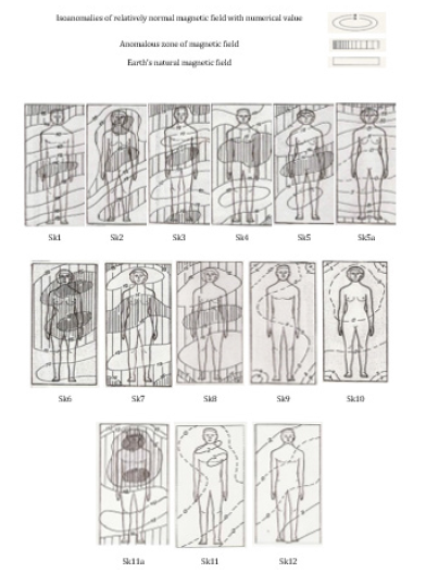
Overall intensity of the resulting total magnetic field was measured by proton magnetometer made in USA. Geology Brunton compass was also used. The results are shown in sketch consisting of Isonomies, and pictures of human body. Isonomies are lines with the same values of growth of anomalous magnetic fields (AMF), and they are the borderlines that separate distinct growth of AMF. Surfaces represent anomalous zones (AZ). High variability of magnetic field was observed on bed surfaces of examinees diagnosed with D. It is obvious that AZ covers the area where the head and torso of examinees are placed. These body parts contain organs that are regulators of glucose in the body. Sketches show the measurement results of: five patients with type 2 D, three patients with type 1D, and two people completely healthy.

There were two examined groups, group A and group B and their health status was observed. Examinees in group A were living in normal ENMF and examinees in group B were living or sleep in beds in anomalous magnetic fields. Results show statically significant deference in health status in these groups. In group A there was health improvement and tendency to normal blood glucose level. Health deterioration was observed in examinees of group B for a longer period of time. Sketch of healthy or sick examinees represents 12 type of health problems. Legends which represent measurement results of magnetic field are used.

Sketch 1: The present example clearly shows that the AZ engulfed the head and abdomen of examinee. It is characteristic that the enormously high value of AMF swept the whole bed. The user was first diagnosed with D and after a few years of malignant disease of the colon. Then we set up living area for patient to spend DaNR (day and night rest) in ENMF. Condition glucose very quickly normalized and reached key values.

Sketch 2: The whole surface of bed was in AZ. First, the patient got an elevated blood glucose, and type 2 D is the diagnosed, and after several years appears distinct angina pectoris, thyroid tumor, and cataracts. After all diseases mentioned above, the examinee was moved to living space with normal ENMF in DaNR. Shortly thereafter glucose was reduced to a tolerable level. Medicines for lowering blood glucose were discarded. After a while the thyroid tumor disappeared, and the angina pectoris appeared no longer. Surgery was performed on one eye because of cataracts and second surgery was scheduled on another eye. Another surgery was to be carried out a few months after the patient started living in ENMF, but doctor stated that condition of another eye was excellent, and that surgery was not needed. Monitoring of her health lasted for 20 years.

Sketch 3: This patient had had D for 30 years. State of AZ in his bed was very disturbed and whole bed had very high values of AMF. The patient had been on insulin three times a day before he was moved to living space with normal earth magnetic field. After our intervention, he entered the ENMF and insulin requirements were reduced.

Sketch 4: A patient was diagnosed with hyperglycemia 15 years ago, his body mass was 150 kg with a height of 180 cm and a body mass index 46, which clearly indicates that the cause of the emergence of D - Obesity. This should give an explanation of formation of this type of 2D. Specifically, triglycerides (white and gray) are on the membrane of cells and gray triglycerides have more energy than white has and raise the temperature of the cell membrane where the insulin receptors are placed, and their MC are reduced and sensitivity to complex insulin and glucose is reduced and therefore leads to hyperglycemia. Our intervention brought the patient into the normal ENMF. The patient did not lead healthy life but he slept in normal magnetic field and used drug Metformine hydrochloride (Sijafor 850) twice per day. Blood glucose was 5-7 mmol/l and that status did not change for 5 years.

Sketch 5: This patient was diagnosed with Diabetes mellitus 15 years ago. You can see that she spent DaNR in AMF with very high levels with two distinct AZ. Lately she took insulin 4 times per day. In June 2015, the patient went to the place of her birth in Montenegro and rapidly decreased insulin dose to once daily. Measurements show (sketch 5a) that at night her body was placed in a normal ENMF. It is obvious that the stay in normal living magnetic field allowed a drastic reduction in intake of insulin. After the examinee had returned to permanent residence the insulin level was as previous.

Sketch 6, 7, 8: Patients aged 5-13 years. In these drawings are patients with type 1 D. Measurements showed the presence of very high values of AMF and almost all bearings are in AZ. Suggestion to parents, was to set up the living space for children to spend time in the normal ENMF. Whether it is done or not at this moment, we have no information.

Sketch 9: A healthy person in the bed is genetically predisposed to getting D. The mother of this person, then brother and uncle had D for decades. (We showed mother and brother in sketch number 1 i 2). It is characteristic for this person to use sweeteners in the form of crystalline sugar in enormous quantities. Condition of blood glucose in a person’s blood, 26 years ago, was elevated about 100-120 mg/dl by the old measures. Then the setup of his living space was performed and now he spends DaNR in ENMF. His check-ups of glucose in the blood are in the reference values for over 26 years.

Sketch 10: Intensities of value growth of AMF on this diagram are 0 such as on the sketch 9. It is normal ENMF or healthy condition for sleeping. Person is completely healthy, and balance of glucose was monitored over 30 years. This person’s mother and three sisters died early, because their four beds were placed in AZ. Her father lived 94 years and in his bed there was no AZ, as well as with the person from the sketch 10, who entered into the 90th year of age. This is a fine example of the necessity to live in the ENMF.

Sketch 11a: This picture show examinee on his bad and he had had diabetes, type 2. Therapy has been one dose of insulin per day. AZ was almost over the entire patient’s trunk. After some time, the person has got thrombocytopenia of idiopathic type. He was treated at the Clinical Center of Serbia with the therapy, which included blood plasm change. Results of the treatment were unfavorable. His spleen would have to be operated according to expert decision. On his request, he was released from hospital to spend a weekend at home. In this moment he moved away from the bed in the sketch 11a into the bed drown on the sketch 11. He spent in the normal ENMF four days and nights, and he ate better food, he come back to the Clinical Center where analyses show that thrombocytes increased almost to the referent level. Doctors understood that staying at home did good to him, postpone the operation, and suggest that he would wait for some time. The bed in the sketch 11 he has been using for more than 10 years and his health has been better. Insulin therapy for diabetes 2 he is using no more, he takes pills of Diprian (gliklazid) twice a day and is not on any diet. Glucose is around 5-7 mmol/l, and thrombocytes are within referent values.

Sketch 12: Patient spends his days and nights in ENMF for over 20 years. He has been pathologically fat for 11 years, body mass index was over 40 (188 cm height, 146 kg weight, born in 1939.). His blood sugar finds 5,4-6,5 mmol/l. He do not pay attention to food and don’t take medication. The normal ENMF condition is shown on the sketch 12, where there is not AZ.

There were a lot of processed cases (more than 100) by measuring during the period 1988. - 2016. Observation is that there is not D without striking AZ located in the area of the bearing, accept only the secondary D and with very obese patients. Therefore, authors conclude that the pathogen D is been created by AZ in the ENMF.

Immune System Functioning and Auto-Immune Diseases

To understand autoimmune diseases (AiD) appearance, we should briefly explain the functioning of the immune system (IS). An immune system function is due to the natural magnetization of leukocytes, tissue cells and extraneous agents (viruses, bacteria, fungi, parasites, toxins and extraneous tissues), received in ENMF. It is known that leukocytes are generated in bone marrow, it is processed in thymus, where T lymphocytes are processed and recognized by macrophage, and only 10% of started T lymphocytes have been approved for exit. The rest of T lymphocytes have been destroyed by magnetic forces. Therefore, only this 10% of T lymphocytes are in magnetic equilibrium with MC of other cells in an organism and do not attack their own cells. Extraneous agents enter tissue cells; have their own magnetization because they are generated in ENMF or AMF. The host cells and extraneous agents get stronger MC than the cells out of the diseased space, because of additive MC. Leukocytes are attracted by MC of infected cell, which in various ways, protect the tissue cells from an intruder.

The other way of getting stronger cells magnetization is when an organ or its part spends longer time in AMF, where these cells get an additional magnetization, which sums up with the cells MC equilibrium and cells get stronger Mf than the rest of them. Reinforced Mf of healthy cells attract leukocytes that attack their own cells. This additional magnetization only occurs in AMF and is the cause of AiD [6, 7]. It should be repeated that cells, as constructive elements of tissues and tissues of organs, are in the full equilibrium in organism, what is enabled by homeostatic internal somatic mechanism and there is no attacking of own cells. Cells magnetizations give two magnetic vectors, generated in the normal ENMF. It is the genetic remnant magnetization (inherited from parents and ancestors according to a magnetic code, i.e. genetic code) and induced, which depends on the current ENMF intensity from the external surrounding, representing epigenetics.

Figure 1:

irispublishers-openaccess-neurology-neuroscience


Note: Biology science should modify names to the existing MC. The fictional genetic code should be given an appropriate magnetic code name which, in fact, is remnant magnetization inherited from ancestors (parents and other ancestors) and it has stronger intermolecular Mf because it is historical, i.e. generated in the time of a strong ENMF and that is why it is hard to disturb. Epigenetics occurs from an induced ENMF, and it is currently MF, which is very variable, but if it is stable, it can be inherited.

Diabetes Mellitus

D Type 1 is characterized by loss of insulin production by the beta cells of the islets of Langerhans in the pancreas, leading to lack of insulin. This type can be further classified as idiopathic or immune mediated. [5] The majority of the D type 1 mediated immune nature, in which T-cell - mediated autoimmune leads to loss of beta cells, and thus insulin. Most people affected by the D-type 1 were healthy and of normal weight at the beginning of the occurrence of D. The sensitivity and responsibility to insulin are usually normal, especially in the early stages. Type 1 D can affect children or adults. It is often referred to as juvenile-D because most of these D cases are children. The cause of this type of D is very often called as labile (unstable) D... However, the D-type 1 may be accompanied by irregular and unpredictable hyperglycemia, often with ketosis, and sometimes with severe hypoglycemia [5]. All of these variations are due to patient’s habit at different intensities of AMF, which is determined by measurement.

D Type 1 is partially inherited, with multiple genes, including HLA-specific. it is known to affect the risk of D1. In genetically susceptible people, the beginning of D can be induced by one or more environmental factors, such as the nourishment, or viral infection. There is some evidence to suggest a link between type D 1 and Coxsackie B4 virus. In contrast to the D type 2, the beginning of D type 1 is brought into connection with lifestyle. It is known that genetic mutations in PVM (Physical viscous charge) are varied and very present [1-3]. However, they are a consequence of AMF and not the cause of D. Virus in the formation of D cannot be the cause, but it is true that it accelerates the formation of PVM, because each virus has its own magnetization and AMF as a result of a rapid proliferation. It also contributes to faster emergence of PVM [2, 3], that causes formation D [5].

In literature [5], cow’s milk diet, genetics and other environmental factors, are the main cause of occurrence of D type 1. We will explain how it came to such erroneous assertions that mixed cause and effect. The long stay of organs or organ in the AMF first occurs PVM of those parts of the body that have their natural magnetization developed in a normal ENMF, which is the magnetic equilibrium with the cells of organs of the whole body. It is logical that all cells located in the AMF gain enhanced MC because the accumulation of interphase of mitosis, substance with increased MC enter the cells and therefore occurs PVM in AMP. It is known that the young organism develops in normal ENMF. If it is fed with substances with paramagnetic properties, without there is no life, it is clear that these substances accumulate in the space of AMF action and accelerate the formation of PVM, then the cells form, and their antigens with increased MC. The natural response of the immune system is the formation of B and T lymphocyte clones to the antigen [6], and other leukocytes are involved in the attack on the antigen. Cow’s milk contains mostly sodium and calcium, which are paramagnetic and express a protein that is concentrated in PVM but it is a consequence of AMF as the sole cause of the emergence of D-type 1. Now it is clear that we should not look for the cause of D in milk and gene mutations, but the only cause is AMF that is located on a body part or an organ in the body. This statement is in full compliance with the advent of D on increasing latitudes of the Earth, temperatures from the sun, and the winter and summer days, and others [5]. All this just reinforces the AMF as the sole factor of D.

D Type 2 is characterized by insulin resistance, which can be combined with a decrease in the secretion of insulin [5], the defective response of body tissues to insulin is believed to involve insulin receptors. However, the specific defects are not known to science. In cases of known defects, D is separately classified. Type D 2 is the most common type. In the early stage of type 2, vulnerability on insulin was significantly reduced. A type 2 occurs primarily due to lifestyle factors and genetics, lifestyle numerous factors, it is known, that are important for the development of type 2 D including obesity (body mass index greater than 30). Lack of physical activity, poor diet, stress, and urbanization. An excess of body fat is associated with 30% of the cases of both Chinese and Japanese D, and 60-80% of the European and African D. Feeding as risk factors affecting the development of type 2 D consumption sugars, sweetened drinks are associated with higher risk too. Type of fat in food is very important as a risk factor, such as the saturated fatty acids which increase the risk, and unsaturated and polyunsaturated which reduce the risk. Lack of exercise is believed to cause 7% occurrence of type 2 D. All these factors in the literature, confirm the cause of D, which is AMF. If someone wants an explanation, we will gladly explain. Please note that we have obesity as a risk factor for D, explained by Sk. 4, where a body mass index is greater than 46. The most common cause of D type 2 in this type of disease is frequency of AMF in living area. As the pollution with magnetic and electromagnetic smog increases, which is a generator of origin of AMF, D is growing uncontrollably. The patients with type 2 D may produce insulin but the insulin does not fulfill its role as a resistance of the cells to insulin, i.e. body ignores insulin and glucose is not metabolized correctly, so hyperglycemia occurs. This is about the type of magnetization, if AMF is reversed, cells show resistance to glucose-insulin complex and hyperglycemia occurs (vagus nerve stimulation is missing, and islets of Langerhans normal function stops). Obesity as a risk factor for D is explained in Sk. 4 and 12 where body mass index is larger than 46.

Gestational D. Gestational D is similar to type D 2 in several respects, such as the combination of the relatively inadequate insulin secretion and the insulin response cells. This type D occurs with pregnant women with high blood pressure and obesity. D occurs in approximately 2-10% of all pregnancies and improves or disappears after delivery. Staying in the AMF is known to increase blood pressure. Obesity is a serious risk factor in the D. Adipocytes triglyceride fatty connective tissue is on the surface of cell membranes and there are white and grey. Grey ones release energy and raise the temperature which reduces the magnetic properties of the membrane insulin receptors and cannot be magnetically linked complexes of glucose and insulin to the cells and that results in hyperglycemia. It is known that after childbirth, 5-10% of women receive D type 2. This type D is also formed in the AMF as determined by measurement. Logical thinking gives all the answers to questions (such as why 5-10% of women after childbirth get D type 2, because they remain in AMP and so on).

Other types D. Some cases of D have been caused by a reduced magnetic sensitivity of cells to insulin, and do not react. This form is very rare. Genetic mutations can often lead to incorrect functioning of the beta cells where AMF has already been determined as a cause. In addition, abnormal insulin is often present, and may be genetically determined in some cases, which can only produce AMF with abnormal vagus nerve influence. Many diseases that cause damage to the pancreas may lead to D. Literary studying this disease, we concluded that they are all developed in the AMF. Diseases associated with high secretion of insulin can also cause diseases. Presence of AMF stimulates the vagus to amplify action potentials of beta cells that secrete insulin enormously. Some toxins, damaging the beta cells of the pancreas, which may cause D.

Pancreas

Arterial blood brings food and O2 in the pancreas and there are nerve fibers of the vagus and artesian blood with red blood cells, glucose and O2 are distinct paramagnetic. While it is still increased, glucose in the arterial blood passes by the vagal nerve fibers, pancreas, and increases the magnetic flux. Which generates stronger induction of action potentials that stimulate the secretion of insulin exocytosis from the pancreas into the blood. It binds to the insulin-glucose and glucose delivering helps cells. Please note that the glucose is connected with magnetic connections to insulin. Additive MC of the complex provides a stronger MC which cell inserts into the cytoplasm with its magnetic forces and use it for energy [6].

Operation of the beta cells of the islets of Langerhans - Beta cells obtained food and oxygen through artesian blood. In interphase the accumulation of beta cells by the genetic code (MC coded) take food by MC specific for beta cells [1]. In metabolism, the insulin is secreted from the beta cells, independent of the concentration of glucose in the blood, and that is normal operation of the beta cells of the islets of Langerhans. In pancreas, insulin is magnetically bound to calcium, which is always present in the cells and then with the help of the energy that increases the temperature of separating calcium from insulin secreted outside of beta cells to the pancreas. As noted, the movement of artesian blood with glucose next to the pancreas, glucose with its magnetic forces extracted insulin from the pancreas and with exocytosis comes to blood and magnetically binds to glucose and through the blood distributes all over the body. All that stimulate the vagus nerve action potentials.

Epidemiological Status

Apart from all the action of the World Health Organization and public health organizations the risk of D is booming, and it is expected that in 2030 the number of patients would be doubled. Serbia also has a tendency to rise. Diabetes is present everywhere in the world and it is particularly present in developed countries. D in Serbia is the fifth cause of death, the last twenty years there has been a significant increase in mortality from D, and it is expected that this trend will continue in the coming period. In 2013, it is recorded that 382 million people worldwide have diabetes- Type 2 accounts for about 90% of cases that is equal to about 8.3% of the adult population of both sexes. Rate increase follows the trend of urbanization including electrification, automation, and living space, manifested by pollution generated by the formation of AMF.

All medical literature in epidemiology is similar to the previously written one. It is also clear that everything described in pathogenesis is not even near to discovery of the cause of occurrence of D. In the etiopathogenesis, it is stated that the causes of D are genetics, obesity, pancreatic damage by infectious diseases (viruses, bacteria, mushrooms) and mentioned factors of external environment are obesity, training, movement, sitting. At the end, for all the above-mentioned risk factors of D, we can say that when they are present it is not certain that the D will occur. In addition, it is noted that environmental factors play a significant role in the D, but the literature does not know which factor is the cause of origin, that is why only consequential factors are stated. In this paper, it is clear that the cause of each D, except possibly secondary D, is AMF formed in the outside environment. Generators of AMF are all magnetic and electromagnetic in living space and movement of people. Here are some clear literature data that confirm that the cause of D is AMF in living space of people.

The biggest complications, which causes D. The biggest complications, which causes D- cardiovascular disease; stroke, myocardial infarction, constriction of blood vessels, hypertension, gangrene of the feet etc. For all these diseases and others, AMF [3] are responsible, what many of our publications have shown and proven. A fine example of the work of AMF is diabetic foot, because it is usually present in each AMF. AMF is present in every reinforced panel in houses where people live and work. Then with shoes (boots) with iron plate that gives shape and firmness to wear. The suggestion that you should not put protectors on shoes in order to prevent the diabetic foot, it is obvious evidence that the MC is additive, because the cleat increases the magnetic properties of wear. The cause of diabetic foot is AMF exclusively. The literature states that the glycosylated hemoglobin is better than morning glucose for determining risks of cardiovascular disease and death from any cause. This finding indicates that once again the only cause of D is AMF. It is known that hemoglobin (ferrous iron-ferromagnetic), and glucose, as distinct paramagnetic are bonded with magnetic connections. That would mean that magnetic connection is used in diagnostic purposes. D is most commonly encountered with dogs and cats, because they live in areas with people and everything that is the cause of D in humans is the same in dogs and cats, and that is AMF.

Conclusion

According to the described findings, it can be concluded that the cause of the emergence of diabetes is life in anomalous magnetic fields. In order to protect people from these physical agents, it is necessary to move them away from sources of magnetic and variable magnetic fields. Spending time in the Earth’s natural magnetic field is crucial for healthy life. Removal of patients from anomalous zone would be of huge help both for the patients and the physicians in the treatment of diabetes mellitus and in many cases it can lead toward to complete healing.

To read more about this article...Open access Journal of Cardiology Research & Reports

Please follow the URL to access more information about this article


To know more about our Journals...Iris Publishers

To know about Open Access Publishers

Thursday, June 27, 2024

Iris Publishers- Open access Journal of Gastroenterology & Hepatology | Esophageal Perforation with Successful Clinical Management: Case Report


Authored by Jarbas Faraco Maldonado Loureiro*,

Abstract

Esophageal perforation is a serious condition that has an unfavorable prognosis, especially when diagnosed late. The main causes are iatrogenics and foreign body ingestion. Clinical manifestations may be nonspecific, which delays their identification. When suspected, exams such as radiography and tomography should be requested to confirm the perforation. The conventional treatment is surgical, in addition to antibiotics and suspension of oral diet. Endoscopy has become an option in the therapeutic arsenal, being less invasive and showing good results. However, as seen in this case report, when the patient does not have systemic manifestations, expectant (non-surgical) management may be an appropriate option with a satisfactory outcome.

Keywords: Perforation; Esophagus; Endoscopy; Foreign body; Clinical management

Introduction

Esophageal perforation is considered a serious and rare medical condition that is associated with high rates of morbidity and mortality [1]. It has nonspecific clinical manifestations, which contribute to late diagnosis, causing worse outcomes. The main causes are: iatrogenics (after endoscopic procedures), trauma, ingestion of a foreign body or chemical substance. The perforation can also be spontaneous, which is known as Boerhaave syndrome. Regarding the ingestion of foreign bodies, in adults it usually occurs incidentally and in most cases the foreign body spontaneously progresses through the digestive tract and does not require additional conduct. Among the objects accidentally ingested, the bones of animals are considered the most prevalent, they can be sharp, generating a greater risk of perforation [2].

The clinical presentation of perforation is usually nonspecific, with pain being the most common symptom. The manifestations will depend on where the esophagus was perforated (cervical, thoracic or abdominal). When the involvement is cervical, symptoms can be dysphagia and neck pain. Perforation of the thoracic esophagus may present with pain in the back, chest or epigastric region. Distal lesions may cause abdominal pain and peritonitis. Other less frequent symptoms are vomiting, nausea and subcutaneous emphysema. Signs such as fever, hypotension and tachycardia may appear, characterizing systemic involvement. Early diagnosis is a challenge due to the nonspecific presentation of the condition. Imaging tests such as contrast radiography and tomography can characterize the perforation. Digestive endoscopy can be requested to elucidate the symptoms, it is able to identify the perforation and may also be a therapeutic option [3].

The management of patients with perforation is critical, because they can rapidly progress to septic shock, so they should be closely monitored. The main goals of treatment are infection control, maintenance of nutritional status and evaluating the therapeutic possibilities for repairing the perforation. Treatment is divided into surgical and non-surgical approaches. Non-surgical management can be performed in the portion of patients who do not show signs of septic shock. The main conducts are the initiation of antibiotic therapy and suspension of the oral diet using an enteral or parenteral diet until the resolution of the esophageal perforation is assured [1].

Digestive endoscopy has become more relevant in the treatment of esophageal perforation in the last decade. It can be used by itself, especially when the perforation is identified early (<24 hours) or associated with surgical treatment [4]. In cases where there is contrast extravasation, empyema or mediastinal contamination, drainage is mandatory. Interventional endoscopy techniques such as the application of clips, endoscopic stents and vacuum therapy have become less invasive options compared to conventional surgery techniques, therefore providing lower treatment-related morbidity and mortality.

The application of clips by endoscopy can be a treatment option, but they are used in very specific cases of early perforation, minimal inflammation, small holes (< 5 mm) and in the absence of fistulas. There is the option of over-the-scope (OTS) clips which are used in larger perforations. Endoscopic stents can also be used to treat perforation, combined or not with percutaneous drainage therapy, but the biggest complication is their migration, which limits their use. Predictors of good response to treatment are early identification of perforation and smaller luminal aperture. Vacuum therapy is a new treatment option, which has proven to be effective and its clinical use has become increasingly frequent. It is based on the application of a drainage system with negative pressure in intraluminal or intracavitary topography guided by endoscopy. Vacuum therapy results in decreased edema, removal of infected secretions, and gradual closure of the lesion [1]. Surgical treatment options includes draining the contaminated space, primary perforation repair, esophageal bypass, and esophagectomy. Surgical management is indicated for extensive lesions and in cases where there are severe complications, such as mediastinitis1.

Case Presentation

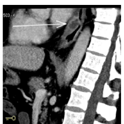
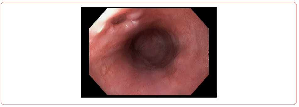
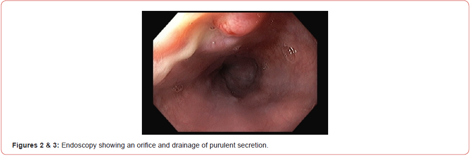
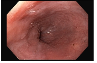
63-year-old male, with arterial hypertension and dyslipidemia, without other comorbidities. It started with pain in the lower thoracic region associated with a feeling of bloating. Symptoms started after participation in a family barbecue where fish and bone-in beef were served. The condition evolved for 5 days, when it became more intense and associated with difficulty of feeding, tolerating pasty foods. He sought medical care in the emergency, where he was evaluated and initially laboratory tests showed a significant increase in leukocytes and C-reactive protein. Abdominal ultrasound did not show relevant changes. It was decided to perform a tomography of the abdomen and chest, which showed thickening of the lower third of the esophagus, associated with parietal ulceration and reactive lymph node enlargement (Figure 1), and also upper digestive endoscopy which showed the presence of an 8 mm orifice in the distal esophagus draining purulent secretion (Figure 2 & 3). An evaluation was requested from the surgical team and as the patient remained in good general condition, with no signs of sepsis, a non-surgical approach was chosen. Antibiotic therapy was started with ceftriaxone and metronidazole, suspension of the diet oral and insertion of a nasoenteral tube to preserve the nutritional status. Patient evolved with progressive clinical and laboratory improvement. One week later, a new tomography was performed, which showed regression of the esophageal thickening and a new endoscopy (Figure 4) which showed complete closure of the orifice. The patient was discharged from hospital 10 days after admission, with good acceptance of the oral diet, without complaints and in excellent general condition.

irispublishers-openaccess-gastroenterology-hepatology

Discussion

Esophageal perforation, when diagnosed late, that is 24 hours after the injury, has worse morbidity and mortality rates. The fact of having non-specific symptoms contributes to delay the diagnosis, which will be identified only when the patient shows signs of systemic involvement, which demands more aggressive therapeutic approaches. For this reason, a detailed clinical history should be taken to increase diagnostic suspicion and early institution of treatment. To confirm the diagnosis, it is necessary to perform additional exams, such as tomography. Endoscopy, in addition to being a diagnostic option, is also considered a therapeutic option.

After diagnosing the perforation, the fundamental therapeutic approaches are infection control, maintenance of nutritional status and repair of the digestive tract. However, in cases where the patient is in a good clinical condition, without signals of systemic involvement or the presence of collections, management with antibiotic therapy and enteral nutrition can present a good clinical response and favorable evolution, without the need for surgical or endoscopic intervention, as it was described in this case report. In the case discussed, the diagnosis was made late with the patient presenting an insidious evolution and seeking medical care five days after the symptoms. When the perforation was diagnosed, the presence of a foreign body was not identified however due to the clinical history, the hypothesis was accidental ingestion of a foreign body. As the patient had no systemic signs, with a small perforation and drainage into the esophageal lumen, clinical treatment was chosen and showed an adequate response.

Conclusion

Therefore esophageal perforation is a serious medical condition that requires intensive patient care due to the high rates of complications. Early diagnosis is a challenge, but it is of paramount importance for a better prognosis. Conventional treatment for perforation is surgical, but in cases where the perforation is small, tamponade and the patient is stable with no evidence of sepsis, clinical management can be successfully performed. Surgical treatment is recommended in cases of extensive esophageal lesions and when there are signs of systemic involvement.

To read more about this article...Open access Journal of Gastroenterology & Hepatology

Please follow the URL to access more information about this article

https://irispublishers.com/ajgh/fulltext/Esophageal-Perforation-with-Successful-Clinical-Management-Case-Report.ID.000558.php


To know more about our Journals...Iris Publishers

Iris Publishers-Open access Journal of Hydrology & Meteorology | Influence of Community Resilience to Flood Risk and Coping Strategies in Bayelsa State, Southern Nigeria

  Authored by  Nwankwoala HO *, Abstract This study is aimed at assessing the influence of community resilience to flood risk and coping str...