Monday, September 30, 2024

Iris Publishers-Open access Journal of Civil & Structural Engineering | Installation of Pit Fencing in Cramped Conditions


Authored by Sokolov NS*,

Abstract

Construction of facilities in cramped conditions is always a complex geotechnical problem associated with ensuring the basal emergency operation of buildings and structures of the surrounding buildings. In the arsenal of geotechnicians, a great potential of geotechnical technologies for the installation of pit fences has been accumulated. Often, not everyone is suitable for the conditions of their use in cramped conditions. The most suitable technologies for such cases are the drilled wells used (wells for drill piles and soil anchors) with their subsequent filling with concrete and at the same time not disturbing the stress-strain state of the surrounding soil. The use of drilling piles and soil anchors arranged using electric discharge technology (ERT technology) in many cases successfully solves the problem of construction in cramped conditions. The article cites one of the geotechnical cases of fencing the pit in particularly cramped conditions. The work is an overview.

Keywords: Geotechnical construction; Electric discharge technology of ERT; Drill injection pile ERT; Soil anchors ERT

Introduction

The construction of buildings and structures in cramped conditions requires from builders and geotechnicians close attention [1-12] related to the need to preserve the objects of the surrounding buildings. In this article, a successful example of fencing the pit of a building under construction, located surrounded by existing structures, is given.
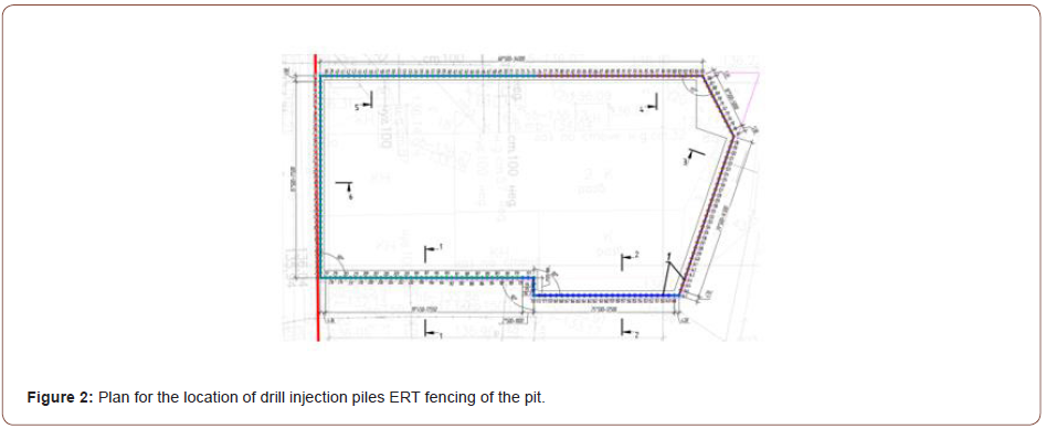
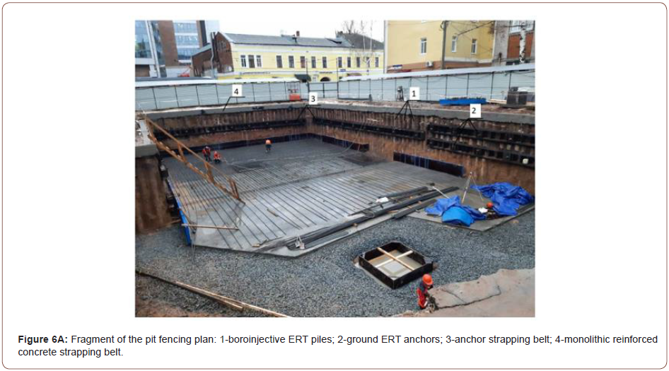
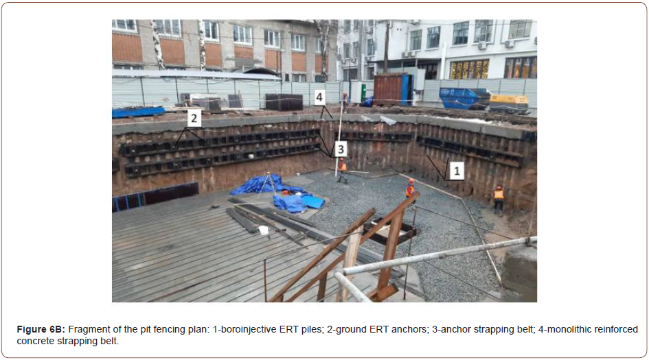
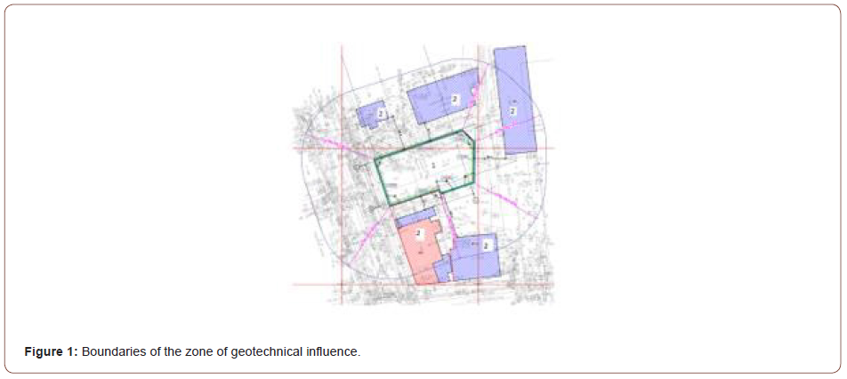
Administratively, the construction site of a six-storey residential building is located in Nizhny Novgorod in cramped conditions of urban development. It was built on a non-developmental territory. Surrounded by a house under construction in the zone of geotechnical influence, there are six objects of existing development (see Figure 1), including two objects of the OKN (objects of cultural heritage).
Geomorphologically, the survey site is located on the watershed plateau of the Oka and Volga rivers. Elevations of the surface of the earth within the site vary from 135.9 to 136.3 B.S. (at the mouths of engineering and geological workings). The terrain is planned. The engineering and geological structure of the site to a depth of 20.0 m is represented by undissected upper-middle quaternary loess loams (prQII-III), opened with a capacity of 1.7-2.5 m, undissected upper-middle quaternary loam loams (prQII-III), with a capacity of 14.5-16.6 m, from the surface of the sediments covered with bulk soil (tQIV), with a capacity of 1.7-3.0 m.
All engineering and geological workings are plotted on the layout plan of engineering and geological workings. The geological and lithological structure of the site is characterized by columns of wells and engineering-geological sections. The hydrogeological conditions of the site to a depth of 20.0 m at the time of the survey (July-August 2017) are characterized by the presence of an aquifer confined to Quaternary sediments. The groundwater level was recorded at depths of 4.5-6.4 m. The aquifer is non-pressure, the water-containing soils are loess loam. Water wells have not been opened. The aquifer is fed by atmospheric infiltration precipitation and leaks from water-carrying communication.
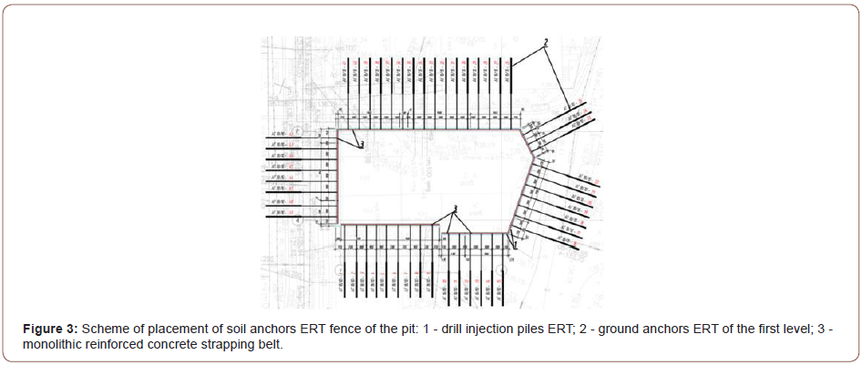
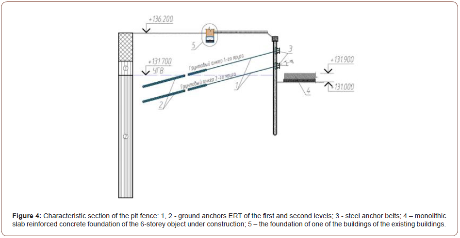
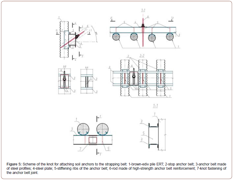
The geological and lithological structure of the site is presented in Table 1 (Table 1) (Figures 1-6).

irispublishers-openaccess-civil-structural-engineering

irispublishers-openaccess-civil-structural-engineering

irispublishers-openaccess-civil-structural-engineering

irispublishers-openaccess-civil-structural-engineering

irispublishers-openaccess-civil-structural-engineering

irispublishers-openaccess-civil-structural-engineering

irispublishers-openaccess-civil-structural-engineering

Table 1: Resource requirements by component.

irispublishers-openaccess-civil-structural-engineering

Table 2: Resource requirements by component.

irispublishers-openaccess-civil-structural-engineering

Below in Table Figure 2 shows the algorithm for the device of ERT drill injection anchors (Table 2).

Conclusion

1. Brown-injection piles and soil anchors ERT, being buried reinforced concrete structures of the pit fence during the construction of a six-story residential building, made it possible to build an object without negative consequences for buildings of the surrounding buildings.
2. The algorithm for the installation of soil anchors ERT has been tested on many objects of geotechnical construction. It is mandatory in the manufacture of brown injection ERT anchors.


To read more about this article...Open access Journal of Civil & Structural Engineering

Please follow the URL to access more information about this article
https://irispublishers.com/ctcse/fulltext/installation-of-pit-fencing-in-cramped-conditions.ID.000690.php

To know more about our Journals...Iris Publishers

To know about Open Access Publishers

Iris Publishers-Open access Journal of Biology & Life Sciences | Discovery of a New Kingdom of Life Without DNADNALESS Life Kingdom


Authored by James T Struck*,

Introduction

For some time, kingdoms of life have included Monera, Plants, Animals, Fungi, Protists, Archaea. Here I am suggesting the inclusion of a new kingdom of life without DNA.

Discussion

For Kingdoms of Life, we have had the Plants, Animals, Fungus, Protists, Monera and Archaea since 1977. Possibly we should have a new kingdom of life without DNA. This DNA less Kingdom would include.
• Red Blood cells
• Platelets
• Viroid’s

To be accurate describing life kingdoms, we have had the prokaryotes and eukaryotes for some time to describe life without a nucleus and with a nucleus. We have still had not had a biological scientists suggest the addition of life without DNA. Here I am suggesting we add a new Kingdom of Life to include life without DNA like the red blood cell, platelets and Viroid’s.

Suggestion- New Life Kingdom DNA Less Life Carl Linnaeus developed the binomial nomenclature classification system around 1700 with a genus and a species name.
The Kingdoms of life would change to 7 or 8 Life kingdoms.
We could have a new group of organisms to study.
We could see DNA less life in the Universe.

Conclusion

DNA less life is argued for as a new Kingdom of Life. Carl Linnaeus a Swedish scientist born in 1707 was associated with some of the classification of life. [1] Here we argue for the addition to the Kingdoms of Life of the DNA Less Life Kingdom.

To read more about this article...Open access Journal of Biology & Life Sciences

Please follow the URL to access more information about this article


To know more about our Journals...Iris Publishers

To know about Open Access Publishers

Friday, September 27, 2024

Iris Publishers-Open access Journal of Complementary & Alternative Medicine | The Myofascial System and Mind-Body Connections for Improving Health


Authored by Shawn M Drake*,

Myofascial System

Throughout the years, scientists dissected the human body to obtain knowledge of the body’s structure and functions. During dissection, fascia is cast aside in receptacles and rarely studied. Fascia is the connective tissue that forms a web-like structure that covers, and connects every aspect of the body including bones, organs, muscles, nerves and blood vessels [1]. Although this structure is found vastly throughout the entire body, all of the functions of this tissue are still being studied. The myofascial system acts as an “exoskeleton” for the body and its inherent architecture is described by principles of biotensegrity. Tensegrity principles describe architectural structures that have continuous tension with discontinuous compression providing stability and flexibility of structures. Within the human body, concepts of biotensegrity link organizational structures at the molecular, cellular, tissue, organ and organ system levels [2]. Biotensegrity refines the explanation of the body’s ability to adapt to change and the mechanics of movement by providing mobility, stability and function [3]. At the cellular level, microfilaments serve as the tension components (straightening) and microtubules function as compression (bending) components [4]. In addition, compression of the extracellular matrix transmits mechanical forces directly to the cell and nucleus through the architectural model. At the tissue level, integrins, extracellular matrix and the cytoskeleton, mediate mechanical signal transduction (mechano transduction) throughout the body [5].

Furthermore, the fascial system or “exoskeleton” provides compression-resistant or tension-generating components [2,6]. Fascia is composed of collagen fibers and ground substance, rich in proteoglycans, especially hyaluronic acid (HA). The HA acts as a lubricant so that fascia glides easily over muscle epimysium and acts as a shock absorber [7]. Fascia constantly transmits and receives mechano-metabolic information that can influence the shape and function of the entire body [3]. Following an injury or lack of exercise, the fascial system weakens, and HA becomes adhesive causing fascial restrictions. The fascial restrictions lead to changes in biotensegrity causing poor body alignment, decreased muscular strength and coordination. The compressive forces or lines of tension within the joint can lead to pain due to free nerve endings [7].

Mind-Body

Langevin HM [8] hypothesizes that unspecialized “loose” connective tissue, or fascia, functions as a body-wide signaling network. If the fascial network provides a communication system, could emotions use this same system to communicate? Pert [9] believed that the body is the unconscious mind and traumatic events are stored within the body. Emotions and bodily sensations are intricately intertwined through a bidirectional network. Memories are not only stored in the brain, but within the psychosomatic network of cells within the body. Pert [9], states, “The idea that the mind and body could be treated as a whole, that the emotions could be accessed through the body, not just the mind, for healing, and that the result could improve the health of the organism appealed to our deepest instincts.”

Additionally, the belief that tissues could possess the ability to store information or “memories” is still controversial in manual medicine. However, Tozzi P [10], explores the idea that every time a cell changes its shape, a vibratory message signals through the extracellular matrix forming a “body consciousness” connecting the nervous system. Furthermore, memories related to disease, physical or emotional trauma cause distorted vibrations, thereby altering the consciousness of memories stored in soft tissue [10].

Moreover, the idea of mind-body is not a new idea. Emerging disciplines such as psychoneuroimmunology (PNI) focus on the interactions between the endocrine, immune, and nervous systems and allow for a broader scope of how the mind and body interact [11]. Biopsychosocial factors, such as psychological stress, influence the immune system. Chronic stress leads to immune suppression, which is mediated by endocrine factors [12]. The connection of mind-body is even more apparent when studying Adverse Childhood Experiences (ACE). Research indicates that individuals with ACE’s have significantly poorer health outcomes, health risk behaviors and socioeconomic challenges [13]. Complementary and alternative medicine begins to bridge this gap in healthcare.

Myofascial Release

Uniquely, myofascial release (MFR) is a type of manual therapy that requires a low load, long-duration stretch to restore optimal length, decrease pain and improve function. The low load and long duration hold used in MFR allows for plasticity versus elasticity changes in the tissue. Myofascial release restores hydration of the tissue and physical changes to the body’s structure so that improved movement patterns can be appreciated. Changes to the biotensegrity of the body allow for decompression of structures in the body leading to reductions in pain. Structural changes that occur from MFR may provide an avenue for releasing such memories by unloading tissues that decrease neural input and the mechanical load of fascial structures [14]. Manual therapy may affect the changes in the viscosity of HA causing a reset of “dysfunctional memories” that were stored in the fascia [10]. Within the biotensegrity matrix system, the body’s information is communicated through a harmonic wave motion through vibrational chemo mechanical energy [15].

In a case report published by Goyal M, et al. [16], the use of MFR and myofascial unwinding improved depression and quality of life in a 39-year-old homemaker. Myofascial unwinding is a somatoemotional release technique that stimulates the parasympathetic nervous system that leads to the release of tissue emotions (Henley, et al, 2008). The authors contribute the improved quality of life and depression scores in the patient due to release of ascial restrictions at the physical and the emotional levels. Hence, MFR provides a conduit to the mind-body connection at the unconscious level in addition to structural changes to improve biotensegrity.

Recommendations

Undoubtedly, the mind-body relationship is a powerful connection. Traditional therapy typically works in silos for the treatment of either physical or emotional but not both. Western medicine often treats one system or aspect at a time. Myofascial treatment has the possibility to the change physical properties of the body (biotensegrity) and facilitate emotional release for a more holistic approach. Working interprofessionally should be encouraged to treat the biopsychosocial factors associated with improved quality of life.

To read more about this article..Open access Journal of Journal of Complementary & Alternative Medicine

Please follow the URL to access more information about this article


To know more about our Journals...Iris Publishers

To know about Open Access Publishers

Iris Publishers-Open access Journal of Textile Science & Fashion Technology | How Fashion Brands Can Benefit from the Power of Eroticism


Authored by Marc Paternot*,

Abstract

Eroticism has been affecting personal relationships for thousands of years and it still has a bad reputation. This article introduces eroticism to the field of management, in particular to brand management. In order to reveal what eroticism signifies a quantitative study using exploratory factor analysis was conducted. The findings have shown that eroticism is a multidimensional construct consisting of six distinct factors. It relies on a balanced attitudes model consisting equally of cognitive, affective and behavioral characteristics and enjoys significant differences with regards to gender and age. The developed erotic-based marketing strategy following a five-building block process should assist Fashion Marketing/General Managers in affirming their belief in the strength of turning their brand into an erotic brand.

Keywords:Fashion; Eroticism; Branding; Marketing mix; Construct measurement

Introduction

Eroticism enjoys a bad reputation. The aim of this article is to give this construct respectability within social sciences and to introduce it to the field of management. To consider eroticism as a new avenue for brands and for brand management. Much has been written on eroticism but mostly through definitions and typologies. Though, in the last decade there has been three attempts to consider this construct in a scientific manner. Hakim [1] published an article named “Erotic Capital” in which she highlights six elements of erotic capital: beauty, sexual attractiveness, social liveliness, social presentation and sexuality. Sarpila [2] developed a scale of erotic capital and extracted four factors: Identity, Fashion, Awareness and Vanity. Last, Requena [3] questioned eroticism’s impact on individuals’ well-being but restricted eroticism solely to beauty and attractiveness. The present paper’s research questions are twofold: How could eroticism be quantitatively measured ? How could eroticism transform its power into business proposals for the fashion industry? In terms of methology for the empirical study, an Exploratory Factor Analysis technique is employed to reveal the facets of the construct eroticism. This paper is divided in four sections. The first section presents the research design and the steps used to discover the facets of eroticism. The second section displays the empirical findings of the analysis. The third section highlights the recommendations for fashion brands to benefit from the power of eroticism. Finally, the last section critically analyses the limitations of the present paper and indicates directions for further research.

Research Design and Methods Leading to the Hexagonal Eroticism Scale

To answer the first research question, eroticism was taken without any prejudice from two main angles: a construct basedview and a theoretical-based view. Within a conceptual framework consisting of eleven dimensions (arousal, desire, sexual desire, sex, love, controversy, beauty, sensuality, mystery, pleasure and excess), apparently related to eroticism but still distinct, eroticism was put into context. A theoretical framework encompassing nine theories (sociology, sexology, religion, biology, psychology, anthropology, morality, philosophy and architecture) helped to assess eroticism’s interrelationships with these theories. Combined these two frameworks form the “Eroticism Framework” [4], which provides a simple and complete structure to understand the construct.

In order to quantitatively measure the construct, a seven-step method was employed.

Step 1: Based on an extensive literature review linked to the conceptual and theoretical frameworks 143 items were generated and then purified resulting in 116 items across the above-mentioned eleven dimensions to best describe eroticism.

Step 2: A survey about eroticism based on the 116 items was designed and pre-tested with 28 students from the University of Mainz. It was successfully assessed on five criteria: danger of social desirability, fear of extreme scale, primary-recency effect, danger of quick answer and fear of discontinuation rate. Corrective actions on the items were put in place.

Step 3: The final eroticism scale was reduced to 93 items (across the eleven dimensions from the conceptual framework) and was developed and translated into a questionnaire.

Step 4: 167 valid questionnaires were collected between March and June 2019 resulting in a mixed-sample (student and nonstudent). This sample covers age groups from 20 to 69 years old, has a balanced split between women and men (60% and 40% respectively) and participants are predominantly German. To the author, it was important to cover multiple age groups. It was expected that the engagement towards eroticism changes throughout life and thus impacts people’s understanding of the latent construct. To be noted: all questions could be answered regardless of any kind knowledge or experience in eroticism.

Step 5: Prior to the use of an exploratory factor analysis (EFA) technique, the results’ basic statistics were analyzed. The eroticism scale’s reliability based on Cronbach’s Alpha was computed and reached 0.929 (on standardized items), that greatly support the consistency of these 93 variables to reveal the latent construct. The item-to-total correlations were satisfactory as 88 variables out of 93 had a correlation between the optimal range of 0.1 and 0.6 supporting that they explain the latent variable well but without being redundant. Normality was assessed through two tests (Kolmogorov-Smirnov and Shapiro- Wilk) and both tests were significant for each variable. It means that the data diverts from normal distribution. This obstacle for running an EFA can be overcome by selecting a specific factors’ extraction method. Finally, multicollinearity was successfully assessed based on three indicators. Variance inflation factor, condition index and inter-item correlations showed moderate sign of multicollinearity. Overall, the data gathered displayed robust results and confirmed good psychometrics properties.

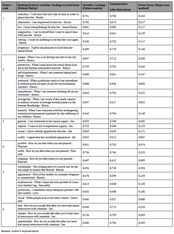
Step 6: An EFA has been conducted on the data gathered in step 4 using SPSS (version 25). Suitability tests (Kaiser-Meyer- Olkin and Bartlett) were incorporated and yielded satisfactory results to pursue an EFA. Following Fabrigar’s guidance [5], it is recommended in the case of data violating normal distribution to use a principal factor method (Principal Axis Factoring). In order to retain the appropriate number of factors, four methods were analyzed: Kaiser criterion, Scree test, Minimum Average Partial and Parallel analysis. As expected, the four methods do not lead to the retention of the same number of factors. Most probably, the final number lies between four and eight. It is assumed, as in most social sciences research, that the dimensions will be somehow correlated. Therefore, an oblique rotation (Promax) was chosen to maximize the number of variables loading onto each factor. The adequate rotation method should portray the cleanest factor structure or the “simple structure” as Thurstone LL [6] put it. The six final factors and associated 28 variables retained are displayed in Table 1. Three indicators (Variables’ loadings, communalities and factor scores) support the strongness of the data gathered. Each of the six subscales shows high internal consistency as their Cronbach’s Alpha are higher than the 0.7 threshold.

Step 7: convergent and discriminant validity were demonstrated. Convergent validity refers to what extent are the variables studied correlated and was supported by Average variance extracted (AVE) and Composite reliability. Discriminant validity finds out to what extent the factors retained are unique, as they should truly be distinct from other constructs. For each construct, the AVE was greater than the squared correlation of inter-construct showing that the factors obtained are somehow conceptually similar but still clearly distinct. Therefore, discriminant validity was also well supported.

Table 1:Characteristic of the patients with AED withdrawal (n=162).

irispublishers-openaccess-textile-science-fashion

Empirical Findings

Four main findings are revealed by the empirical study on eroticism.

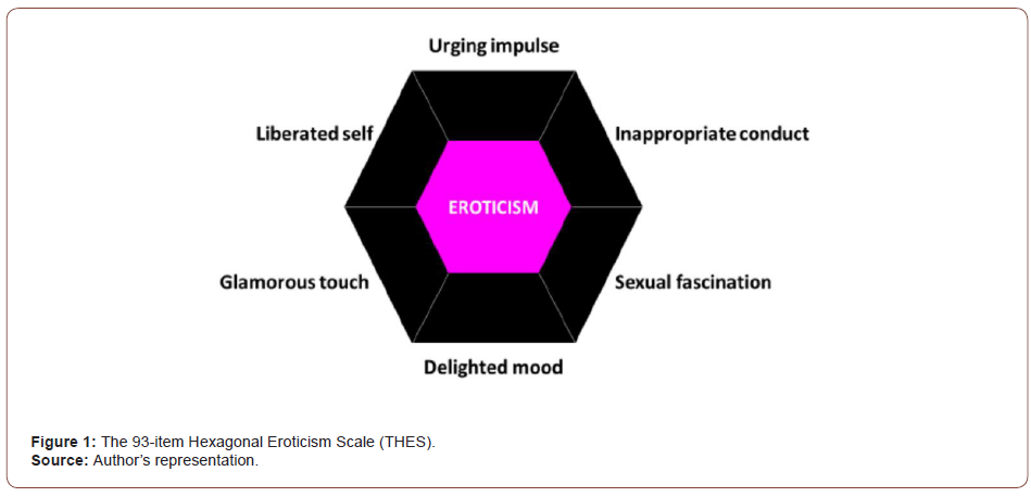
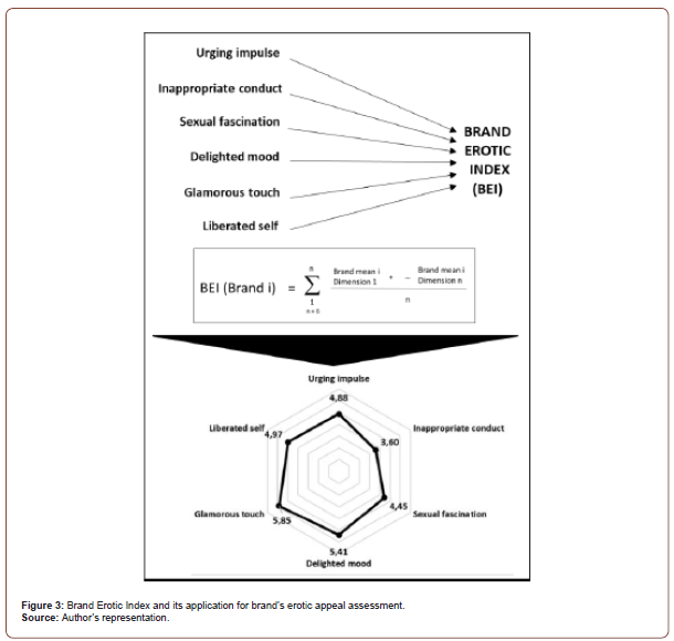
• Eroticism is a multidimensional construct. From the EFA, six factors were retained and were labelled in order of decreasing importance: Urging impule, Inappropriate conduct, Sexual fascination, Delighted mood, Glamorous touch and Liberated self (Figure 1). The geometric representation in the form of a hexagon refers to Sternberg’s Theory of Love [7], which was pictured by triangles. By far, the most prominent factor that explains eroticism is “Urging impulse”. This factor is exemplified by items coming exclusively from the desire and sexual desire dimensions. Therefore, desire and sexual desire drive the construct eroticism .

irispublishers-openaccess-textile-science-fashion

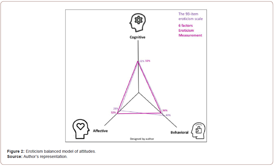
• Eroticism has a very balanced structure of attitudes relying on the Tripartite model of attitudes [8]: cognitive, affective and behavioral. Both for the 93-item scale as for the six-factor structure (Figure 2) each attitude type accounts for one third of the total.

irispublishers-openaccess-textile-science-fashion

• Based on an independent samples t-test, at p value < 0.01, women and men differ on their appreciation of eroticism’s two dimensions. For the dimension “Delighted mood”, women assess eroticism with even more pleasurable and positive aspects than men do. For “Glamorous touch”, women associate eroticism significantly more than men with beautiful and sensual aspects. Interestingly, for the two dimensions (“Urging impulse” and “Sexual fascination”) related to desire, sexual desire and sex, no significant differences between women and men were observed.

• Based on an independent samples t-test, at p value < 0.05, the older the people are, the more different their perceptions on eroticism’s three dimensions. For the “Inappropriate conduct” dimension: as expected older people (here between 40 and 69 years old) see, when linked to eroticism, a more reprehensible behavior than younger people (here between 20 and 39 years old). For the “Sexual fascination” dimension: older people associate eroticism more with sexual aspects than younger people would do. For the “Glamorous touch” dimension: younger people link eroticism with more flamboyance or stunning look than older people.

Recommendations for Fashion Brands to Benefit from the Power of Eroticism

Desire, eroticism’s strongest factor, underpins most consumption patterns and addresses a very broad spectrum of sectors. Intuitively one thinks of the luxury, fashion or beauty industries but this is not limited to them. From a managerial perspective, translating this theory on eroticism into today’s world of fashion brands is equivalent to answering the present paper’s second research question.

The erotic-based marketing strategy enables to shift from the classic macroeconomic approach of “Supply and Demand of Goods” to the “Supply and Demand of Desire”. The latter consists of the relationship between a brand and consumers both driven by desire. The brand, acting as supply, attracts consumers with its product or service offer. Conversely, the consumers, as demand, signal their consumption through their willingness to buy the brand.

This strategy is a two-way mutually reinforcing process between an erotic brand and the consumer. The brand has an erotic power over the consumer by seducing, attracting and touching him/her. At the same time, the consumer through his/her desire values erotic characteristics of the brand - in particular its brand’s personality [9] such as charming, successful, imaginative, cheerful, spirited, daring - and wants to be connected to the brand. Consequently, a cocktail of cognitive, affective and behavioral elements is mixed and contributes to a global and fast-growing branded fashion industry.

Similar to the well-known and respected concept of “Brand Love” [10], which has spread globally across sectors in the last decade, there might be an opportunity for marketeers to turn their brand into an erotic brand. According to the erotic-based marketing strategy, this can be achieved by following a five-building blocks process.

First, by implementing the Brand Erotic Index as a first step. This strategy’s starting point is the implementation of the newly developed Brand Erotic Index (BEI - see Figure 3). This index assesses the brand’s erotic appeal (on a scale from 1 to 7) along the construct’s six dimensions. From that process results a spider web that visually displays the brand’s erotic appeal ratings. In order to track the fashion brand’s erotic appeal and to maintain this relationship over time, this index can be repeated on a regular basis.

Second, by taking advantage of an increasing understanding of consumers’ decision making and customer journey. Based on the empirical results stated above regarding the balanced model of attitudes of the construct (approximately each of the three attitude accounts for one third of the total), eroticism has the ability to significantly lift the consumer-brand interaction that takes place in the consumers’ decision making process and along the customer journey.

With its balanced attitudes model relying on cognitive, affective and behavioral characteristics, eroticism helps to uncover the consumers’ five-step decision-making process [11]:

• The need (or desire) recognition step has the ability to assess the difference between the actual and desired state of affairs.
• The information search step consists of the knowledge, previous experience and the feedback of others on the offer.
• The evaluation of alternatives step helps to compare and evaluate the information necessary to make the right choice
• The purchase decision step includes the planning and execution of the buying act.
• The post-purchase evaluation step assesses the satisfaction of the offer and affects the next decision-making process.

Similarly, eroticism’s balanced attitudes model enhances the customer experience, which is conceptionalized in the seminal article from Lemon and Verhoef [12] as a “customer’s journey with a firm over time during the purchase cycle across multiple touchpoints”. This process is split into three phases: pre purchase, purchase, post purchase [12]. Along this customer journey, touchpoints are spread for the consumer to interact with the fashion brand. Those touchpoints prior, during and after the purchase can mostly relate to advertising, social media, online and offline experiences. The three attitudes at the core of eroticism are enablers of strong interaction between consumers and the fashion brand along the customer journey’s touchpoints.

Third, by adding value to the classical 4P’s Marketing Mix:

• Place: eroticism has the ability to foster imagination and to enhance affects and emotions. Implemented accordingly, the merchandising can re-enchant the customer experience in playing on a new atmosphere and in triggering customers’ imagination. Consequently, the in-store experience both through the adaptation of the fashion visual merchandising and a better trained sales staff should make customers’ visit a memorable experience.

• Product: product development especially in fashion should be adapted to some key characteristics of eroticism, namely the emphasis on senses, which act as a link to connect brand experiences to the inner core of human beings. Especially, the selection of the products’ raw materials is decisive as consumers place great emphasis on touch and smell when they intend to buy a product. The visual aspect is also critical both on the way the products are presented to the consumers and how they are wrapped once bought.
• Promotion: for an erotic brand, its communication should rely on eroticism’s key dimensions. As this present research’s findings have confirmed the multi-faceted aspect of eroticism should be reproducible in the brand’s communication and not limited to sexual aspects. The communication should subtly and implicitly be decoded by the customers. It should be teasing, desirable but never explicit as explicit is a promise of sex. • Price: thanks to the positive impact of eroticism on
customer’s experience and on their products’ perceived value, erotic brands can legitimally be considered as a value driver in their respective fashion categories. Hence erotic brand’s product offer can improve the average selling price of the brand’s assortment.

irispublishers-openaccess-textile-science-fashion

Fourth, by acting as a booster of the fashion brand’s intangibles. Eroticism is a decisive factor to raise and to nurture the brand’s intangibles. As it is much more difficult to copy intangibles than tangibles, this represents a critical and sustainable competitive advantage for erotic-loaded brand. Eroticism can be considered as a determining factor to boost brands’ intangibles. One can highlight its desirability, attractiveness, beauty, glamour, sex-appeal, transgressiveness, freedom, vanity and self-confidence. Thereby, eroticism impacts intangibles, which are prominent underlying drivers of both brand equity and brand identity, which ultimately affect the fashion brand’s positioning.

Finally, by delivering incremental revenues and profitability. Analyzing the financial soundness of embracing such a an eroticbased marketing strategy is recommended. The goal is to deliver incremental revenues and an increased profitability. In particular, a significant investment in selling and marketing expenses and capital expenditures is a pre-condition for this strategy to pay off. The tracking of key financial statement, especially the fashion brands’ P&L and cash flow statements, is mandatory. Turning a brand into an erotic brand is a process that might take time. The alignment of the brand’s financial resources with the strategy’s objectives (especially the investments at the POS, on communication and in people) is critical.

Limitations and Further Research

The present study has some limitations which should be amended by future research.

• The samples are rather small and homogeneous. Generality is achieved but generalizability is not complete. It would be helpful to incorporate to a new sample non-Western participant with additional socio-economic and cultural characteristics.
• Eroticism’s measurement relies only on EFA. It would be useful to find a new sample and to run a Confirmatory Factor Analysis. Therefore, possibly confirming the results from the EFA.
• The quantitative studies are based on self-report questionnaire. This can provide flaws in terms of validity. It would make sense to corroborate the findings from self-report questionnaire with additional in-depth interviews with experts.
• Eroticism seems to enjoy more applications physically at the POS and at product level. Future studies could more carefully examine further applications in an online environment and for services.

Conclusions

The primary aims of this article were to measure eroticism as a construct and to detect its ability to positively influence fashion brands. This empirically-validated research has discovered six dimensions that make eroticism a strong and fascinating construct to handle. Thanks to eroticism, fashion companies have a new and distinct avenue to pursue a differentiation strategy. As a starting point, companies can assess their brand’s erotic appeal by using the Brand Erotic Index. Brands can benefit from the added-value eroticism brings to the classical 4P’s Marketing mix. Furthermore, fashion brand’s intangibles are boosted by eroticism and this adds to the brand’s uniqueness. Companies have to align their financial resources with the erotic-based marketing strategy’s objectives. This strategy, built up on the newly designed 93-item hexagonal eroticism scale, can pave the way for fashion brands to pursue a new and distinct avenue thanks to the power of eroticism. It is up to fashion marketeers and general managers to consider turning their brand into an erotic brand.

To read more about this article....Open access Journal of Textile Science & Fashion Technology

Please follow the URL to access more information about this article

https://irispublishers.com/jtsft/fulltext/how-fashion-brands-can-benefit-from-the-power-of-eroticism.ID.000722.php

To know more about our Journals...Iris Publishers

To know about Open Access Publishers

Thursday, September 26, 2024

Iris Publishers-Open access Journal of Neurology & Neuroscience | Hereditary Motor and Sensory Neuropathy with Hypercapnic Respiratory Failure and Recurrent Rhabdomyolysis: A Case Report and Literature Review


Authored by Noor Sameh Darwich*,

Introduction

Diaphragmatic dysfunction due to phrenic nerve involvement in patients with hereditary motor and sensory neuropathy (HMSN) (formerly known as Charcot-Marie-Tooth [CMT]) is uncommon in the early stages of the disease [1]. However, respiratory compromise due to diaphragmatic dysfunction caused by phrenic nerve involvement can be silent and insidious with minimal symptoms such as orthopnea [2]. In advanced HMSN disease with diaphragmatic paralysis, patients can present with hypercapnic respiratory failure requiring ventilatory support [3]. The prevalence of obstructive and central sleep apnea is higher in patients with HMSN [4]. Mild elevation in serum levels of creatine kinase (CK) may be due to denervated muscle with increase permeability of muscle membranes to enzymes such as CK. Recurrent episodes of significant increases in serum CK level more than five times the upper limit of normal in patients with HMSN suggest rhabdomyolysis [5]. This condition could be triggered by prolonged immobilization from advanced physical disability, overuse of proximal hypertrophic extremity muscles, or a concomitant underlying genetic mutation associated with rhabdomyolysis [6].

Case Report

A 39-year-old caucasian woman with a history of HMSN presented with shortness of breath, right upper quadrant abdominal pain, and back pain associated with having tea-colored urine along with generalized muscle aches. She had been diagnosed with HMSN at age 8 without genetic testing. Her brother had HMSN as well. Her past medical history was notable for periodic episodes of weakness associated with rhabdomyolysis, obstructive sleep apnea (using a continuous positive airway pressure device at night), hypertension, and occasional dysphagia to solid food for several years. The patient’s respiratory complaints started five years prior when she presented with respiratory distress and elevated serum CK, transaminases, and dark urine consistent with rhabdomyolysis. She developed acute kidney injury managed with intravenous hydration and noninvasive positive pressure ventilation (bilevel positive airway pressure) was used during her hospitalization. The patient had similar previous presentations at least twice in the last few years before the current presentation that required hospitalization for respiratory failure and rhabdomyolysis. She had baseline twopillow orthopnea, which increased recently to four pillows. The patient attributed her current condition to overexerting herself in the previous few days at work as she works at a library and walks with bilateral ankle-foot orthotics without an assistive device. She was taking acetaminophen for pain as needed.

Her previous spirometry in upright position five years prior showed severe restrictive dysfunction with forced vital capacity (FVC) 1.45 L (38% predicted), forced expiratory volume in 1 second (FEV1), 1.22 L (38% predicted), and FEV1/FVC (84% predicted) consistent with a restrictive pattern. The patient was unable to perform spirometry in the supine position due to respiratory distress. Before the current presentation, she required moderate assistance for grooming and total assistance for lowerbody dressing and bed/chair/wheelchair transfer, and maximum assistance for rolling and supine-to-sitting positioning.

On admission to the hospital, her blood pressure was 142/96 mmHg, temperature 97.1 F (36.2 C), pulse 114 per minute, respirations 24 per minute, and oxygen saturation 100% on 2 L/min oxygen per nasal cannula. Her physical examination was notable for claw-hand appearance of all fingers in both hands with fingers held in the flexed position but could be passively extended almost to the neutral position. Her motor examination revealed normal tone in bilateral upper and lower extremities. Deep tendon reflexes were 1/4 in bilateral upper and lower extremities with no ankle clonus and absent Babinski reflex with intact light touch sensation while her sensory examination during a prior admission had shown impaired temperature and vibration sensation in bilateral upper and lower extremities. At current presentation, she could not sit up on her own without rolling over to one side and had difficulty getting into bed with intermittent numbness of her fingertips and intermittent burning of the top of her thighs lasting for 15–30 minutes that resolved spontaneously. Her chest examination was notable for bilateral diminished breath sounds at the bases.

Laboratory test results from the current and previous hospitalizations are displayed in Table 1. Hepatitis serology was negative with normal serum acetaminophen level and negative COVID-19 PCR nasal swab.

Ultrasound of the abdomen was unremarkable, and a chest X-ray revealed low lung volumes with elevated bilateral diaphragms. Computed tomography angiography of the chest revealed no signs of pulmonary embolism but was notable for bilateral lower lobes atelectasis. The patient was diagnosed with rhabdomyolysis and started on intravenous fluid therapy. On day 4 of hospitalization, the patient became increasingly lethargic, confused with questionable seizure-like activity in the extremities and acute respiratory distress. Arterial blood gases with comparisons to previous hospitalizations are displayed in Table 2.

Table 1:Laboratory test results from the current and previous hospitalizations.

irispublishers-openaccess-neurology-neuroscience


Table 2:Arterial blood gases from current and previous hospitalizations.

irispublishers-openaccess-neurology-neuroscience


The patient required intubation and mechanical ventilation and was transferred to the intensive care unit. An electroencephalogram revealed moderate nonspecific cerebral dysfunction of the left frontotemporal and right mesial frontal regions with severe nonspecific encephalopathy but no seizure or epileptiform discharges. Computed tomography of the head was unremarkable, and echocardiography revealed normal systolic function with ejection fraction 63%, grade 2 diastolic dysfunction with normal valve structure, and normal right atrial and ventricular function. She was weaned off the ventilator and was successfully extubated to noninvasive positive pressure ventilation (NIPPV) with a face mask. Her serum CK levels trended down during hospitalization. The patient had a history of occasional swallowing dysfunction with a solid diet and reported an event in which she nearly choked on solid food at a restaurant and had to undergo the Heimlich maneuver. Videofluoroscopy revealed laryngeal penetration with thin liquids without evidence of tracheal aspiration and moderate residue within the vallecula and piriform sinuses. The patient was started on a full liquid diet with aspiration precautions.

She was discharged home with oxygen at 4 L/min during the day and NIPPV with average volume-assured pressure support (AVAPS) mode during sleep and the day as needed. The patient refused inpatient rehabilitation or any more testing, including rhabdomyolysis genetic testing, phrenic nerve electrophysiologic studies, ultrasound of the diaphragm, and the sniff test. She was referred to a genetics clinic for counseling. We believe that our patient with HMSN had developed hypercapnic respiratory failure due to diaphragmatic dysfunction caused by underlying phrenic nerve involvement. Her recurrent rhabdomyolysis was most likely due to prolonged immobilization from advanced physical disability combined with overuse of her denervated distal muscles and proximal hypertrophic muscles in the lower extremities. We cannot rule out an underlying coexisting genetic metabolic disorder causing episodic rhabdomyolysis.

Discussion

HMSN is the most common inherited disease affecting the peripheral nerves. It is characterized by a heterogeneous inheritance pattern and characteristic pathological, electrophysiological testing, clinical, and presenting features [7]. HMSN has a global prevalence and affects all races and ethnic groups. The prevalence of HMSN is 10–40 per 100,000, depending on geographic region. Although there is no ethnic predisposition, some rare HMSN subtypes are restricted to certain racial groups. HMSN may be less common among African Americans in the US [8,9]. The prevalence of HMSN might be underestimated because some patients are asymptomatic, and some mild cases go undiagnosed or are misdiagnosed [10].

HMSN can be inherited with an autosomal dominant, autosomal recessive, or X-linked pattern. However, new spontaneous mutations (de novo) have been reported [11,12]. At least 80 gene mutations have been identified that encode proteins involved in myelin production, function, integrity, and maintenance. Other genes mediate functions in Schwann cells, axons, and other components of peripheral nerves. Some mutations affect genes responsible for mitochondrial proteins, while some are associated with unknown gene mutations [13]. Mutation types associated with HMSN can be whole-gene duplications, deletions, or point mutations. The majority of cases are related to mutations in four genes, PMP22, MPZ, GJB1, and MFN2. Duplications of the PMP22 gene account for 70–80% of cases [14]. HMSN is classified as types 1 through 7 with many subtypes within each type. Types one and two are the most common types. CMT1A accounts for 55% of all cases and 66.8% of CMT1 cases [15], while CMT2A accounts for most CMT2 [16]. The underlying pathology in peripheral nerves involves demyelination, axonal degeneration, or both [17].

Age of onset of symptoms varies by subtype, usually in the first decade for CMT1 and in the second or third decades for CMT2. Early-onset CMT2 can occur. A late-onset form of CMT2 presents between 35 and 85 years of age, while X-linked HMSN can be present early in infancy [18]. Disease severity is highly variable, and some individuals may show only minimal signs of weakness while others are disabled. The most common initial presentation of HMSN is distal weakness and muscle atrophy of the lower and upper extremities manifesting as difficulty walking, with frequent falls, poor finger control, and difficulty in hand manipulation and writing. There are also gait problems such as high-stepped gait, toe and flat foot walking, difficulty running and keeping up with peers [19], and frequently sprained ankles. Distal muscle atrophy causes stork-leg deformities with foot drop and deformities such as pes cavus (high arched feet) or pes planus (flat foot). These findings may be associated with impaired sensation presenting later, and they tend to be less prominent. They include a gradual decrease of proprioceptive, touch, vibratory sensation, and temperature sensation in the foot and hand. These symptoms predispose the patients to skin breakdown, non-healing foot ulcers, bony deformities of the foot due to loss of protective sensation. In all four limbs, pain sensation usually remains intact, accompanied by diminished or absent deep tendon reflexes [20]. Muscle cramps, numbness, and burning sensation with pain in extremities develop later [21]. Postural tremors and gait ataxia have been described in infantile-onset HMSN. Later in the course, foot deformities such as hammer toes, claw hand, hand weakness, and atrophy of the intrinsic hand and foot muscles can develop. Proximal muscles are less affected, and hypertrophy can develop as a compensatory mechanism in the early stage. However, proximal muscle weakness can develop late in the course of advanced disease. Intellectual disability with mental retardation has been reported in CMTX2 and CMTX4 subtypes with infantile onset.

HMSN usually affects long peripheral nerves. However, cranial nerve involvement such as optic neuropathy, sensory neuronal hearing loss [22], and dysphagia have been reported in CMT4B1, CMTX4, and CMTX5. There is sometimes slow conduction of electrical and magnetic stimuli in the facial, accessory, and hypoglossal nerves without clinical impairment of the cranial nerves [23]. Patients with CMTX1 can experience episodic acute or subacute onset of central nervous system manifestations characterized by transient neurological deficits associated with white matter abnormalities on brain magnetic resonance imaging (MRI). These deficits include transient ischemic attack, hemiparesis, quadriparesis, dysarthria, leukoencephalopathy, and ataxia, which resolve spontaneously without permanent deficit.

Nevertheless, brain MRI changes may persist for several years after the event [24]. Oculomotor nerve involvement presenting with diplopia is rare. Vocal cord paralysis, which can be unilateral or bilateral and unrelated to the degree of muscular weakness in patients with HMSN [25], is due to recurrent laryngeal nerve involvement; this has been reported in CMT2C, CMT4A, and CMT6 [26], and presents as dysphonia or stridor [27], increasing the risk of aspiration. These findings can be the presenting complaint of HMSN, often diagnosed with laryngeal electromyographic studies consistent with peripheral neuropathy affecting the vagus nerve and its laryngeal branch [28,29]. Autonomic nervous system involvement in HMSN can manifest as autonomic disturbances such as bladder dysfunction and orthostatic hypotension [30,31].

Although respiratory muscle and diaphragmatic weakness are uncommon in early disease of HMSN [32], the phrenic nerve may be involved. Phrenic nerve involvement manifests as diaphragm dysfunction or paralysis presenting with orthopnea and hypoventilation with hypercapnia that worsens during sleep, causing headache, fatigue, difficulty concentrating, and daytime somnolence. These findings may be relatively common in advanced CMT1 and CMT2 subtypes, including CMT2C, CMT1A, CMT4A, and CMT4B1 [33,34]. Observational studies attributed diaphragmatic dysfunction in a patient with HMSN due to coexisting diabetes mellitus. However, phrenic nerve neuropathy with diaphragmatic dysfunction was later described in a patient with HMSN without diabetes mellitus [35]. Respiratory compromise due to diaphragmatic paralysis can be silent and insidious and should be suspected in patients with HMSN presenting with minimal symptoms of orthopnea. The diagnosis should be considered before impending respiratory failure occur [36].

Phrenic nerve electrophysiological study in patients with CMT1 shows slow conduction velocity in most patients, and 24–30% have abnormal spirometry findings with reduced FVC. Even fewer patients (8%) reported having respiratory symptoms [37]. The leading underlying cause of respiratory failure in patients with HMSN is the denervation of the diaphragm and intercostal muscles [38]. However, thoracic cage abnormalities and neuropathic spinal arthropathy [39] with associated deformities such as scoliosis and kyphoscoliosis (reported in 42% of children with HMSN) can contribute to the restrictive ventilatory defect.

Demyelination abnormalities of the phrenic nerve were observed in patients with HMSN with the axonal form [40]. Patients with HMSN and diaphragmatic dysfunction and chronic hypoventilation are at high risk for developing central sleep apnea with desaturation and hypercapnia, mostly during rapid eye movement sleep. The latter occurs due to deficits in the functional utility of the diaphragm in respiration during rapid eye movement (REM) sleep, while the activity of the intercostal muscles is markedly inhibited during this sleep stage [41]. Due to pharyngeal nerve neuropathy causing upper airway collapse, obstructive sleep apnea was also described in patients with HMSN with or without diaphragmatic dysfunction. There is an increased prevalence of obstructive and central sleep apnea in patients with HMSN. The severity of sleep apnea and underlying peripheral neuropathy in patients with HMSN are highly correlated. Restless leg syndrome and periodic leg movement disorder are found in many patients with CMT2 [42]. Diagnosis of HMSN depends on history, including detailed family history. Physical examination findings include distal muscle weakness/wasting accompanied by diminished deep tendon reflexes in upper and lower extremities with possible palpable enlargement of the peripheral nerve, which may occur secondary to nerve hypertrophy. There is also possible skeletal deformity, usually occurring at later stages of the disease. These findings are supported by electrophysiological studies, including nerve motor and sensory conduction velocity study (NCVS) and needle electromyography (EMG), followed by genetic testing to confirm the diagnosis [43]. Electrophysiological signs of demyelination can usually be found in all peripheral nerves, including those clinically unaffected. In the demyelinating form of HMSN (CMT1), NCVS findings vary depending on the phenotype. There is usually significant slowing of conduction velocity in both the motor and sensory nerves, mostly the motor nerves with values < 38–40 m/s (normal range > 50 m/s). In the axonal-loss type (CMT2), the conduction velocity is normal or slightly reduced with markedly low compound muscle action potential amplitude (CMAP). Sensory nerve action potential (SNAP) amplitudes are usually reduced. NCVS may be reduced even in asymptomatic patients with HMSN. Classic findings on needle EMG indicate muscle denervation with the presence of fibrillation potentials. In general, there is no correlation between the severity of muscle weakness and motor impairment. The degree of slowing on nerve conduction studies can be detected even in asymptomatic individuals as early as one year of age. However, in some phenotypes like the CMTX, NCV slowing is proportionate to clinical severity [44].

Nerve biopsy was a critical diagnostic tool for diagnosing HMSN in the past. Currently, it is not routinely performed unless the clinical presentation is atypical when genetic testing does not reveal a molecular diagnosis and when NCVS and EMG are not diagnostic [45]. Classical findings of nerve biopsy in patients with HMSN show onion bulb appearance, which is a characteristic feature reflecting repetitive demyelination and remyelination in CMT1. In CMT2, sural nerve biopsy shows axonal degeneration and nerve fiber loss [46]. Muscle changes seen in HMSN are due to denervation and reinnervation with secondary myopathic changes, neurogenic atrophy, fatty infiltration, and edema but no inflammatory infiltrate. Muscle MRI reveals high signal intensity within skeletal muscles on T2-weighted images [47].

Diagnosis of diaphragmatic dysfunction due to phrenic nerve involvement in patients with HMSN is challenging [48]. Suggested studies include chest radiograph, spirometry, diaphragmatic ultrasound, chest fluoroscopy with the sniff test, and phrenic nerve electrophysiological studies such as cortical and posterior cervical magnetic stimulation [49]. Chest radiograph findings are neither specific nor sensitive but can show low lung volumes with the elevation of diaphragms. Vital capacity is a sensitive tool and reliable measure of respiratory function compared to supine and erect positions. FVC may fall >25% from upright to supine, suggesting a significant compromise of diaphragmatic function in patients with neuromuscular diseases [50].

Other findings on pulmonary function testing in patients with HMSN include reduced maximal inspiratory pressure (MIP). This parameter reflects diaphragmatic strength. In HMSN, maximal inspiratory pressure is usually lower than -60 cm H2O, and maximal expiratory pressure (MEP) can be mildly reduced. It should be noted that these tests are effort-dependent [51]. Fluoroscopy with sniff test involves quickly breathing in through the nose. It is a straightforward assessment of diaphragmatic motor function that confirms the absence of muscular contraction of the diaphragm during inspiration, where the diaphragm moves downwards as in healthy patients. There is a 1–2.5 cm excursion during normal quiet breathing, and in deep breathing, the excursion may be 3.6– 9.2 cm. In patients with diaphragmatic dysfunction, the affected hemidiaphragm does not move downwards during inspiration, or paradoxical motion can occur. Sniff fluoroscopy is helpful in the diagnosis of unilateral diaphragmatic paralysis. However, in bilateral diaphragmatic paralysis, the test can give the false appearance of caudad displacement of the diaphragm due to accessory muscle contracture in bilateral diaphragmatic paralysis [52]. Ultrasonography of the diaphragm with static measurement of the diaphragm thickness and changes in thickness during inspiration has been used to confirm diaphragmatic paralysis. However, the test is operator-dependent and has not been adopted for standard evaluation of diaphragm paralysis. Phrenic nerve enlargement sometimes is revealed on ultrasound in patients with HMSN [53].

Several electrophysiological studies are used to diagnose diaphragmatic paralysis in neuromuscular diseases with phrenic nerve neuropathy. Nevertheless, these diagnoses can be challenging, and expertise in interpretation is required [54]. Phrenic nerve stimulation testing accompanied by measurement of diaphragm CMAP using transcutaneous electrical stimulation or cervical magnetic stimulation of the phrenic nerve in the neck while recording diaphragm CMAP at the chest wall surface by two electrodes is usually an easy test with reliable results [55]. Another test is transdiaphragmatic pressure (Pdi), representing the difference between gastric pressure and pleural pressure. Pdi can be monitored using two trans-nasal thin-walled catheters. One is placed in the lower third of the esophagus above the diaphragm to assess changes in pleural pressure, and the second is placed in the stomach to measure abdominal or gastric pressure. Pdi can be measured during peak tidal volume inspiration and after phrenic nerve stimulation. At peak tidal volume inspiration, the value for Pdi is positive if the diaphragm is working normally and negative in diaphragmatic paralysis [56]. Diaphragmatic electromyography can also help differentiate between diaphragmatic paralysis due to underlying neuropathy and paralysis caused by myopathy. Nevertheless, it carries some risk from needle placement in the diaphragm. Dynamic MRI of the diaphragm is a promising noninvasive new technique for evaluating diaphragm excursion, motion, and volume [57].

Laboratory findings in HMSN may include elevated serum CK levels. As many as 50–75% of patients with motor neuron disorders such as amyotrophic lateral sclerosis (ALS) [58], spinalbulbar muscle atrophy (SBMA), and HMSN have elevated serum CK levels. These elevations are usually up to five times the upper limit of normal at rest. These values can be up to ten times the upper limit of the normal value after activity. These changes correspond to the degree of muscle denervation and compensatory reinnervation with muscle fiber hypertrophy and the degree of myopathic changes seen on muscle biopsy [59]. The significant elevation of CK in HMSN can be recurrent and episodic with flare-ups due to exacerbating factors such as period of prolonged immobility (which leads to rhabdomyolysis), emotional stress, alcohol use, pregnancy [60], and drugs such as vincristine, dapsone, metronidazole, and nitrofurantoin [61]. Vincristine, a chemotherapeutic agent used to treat various malignancies in both children and adults, can exacerbate and cause rapid deterioration of HMSN or reveal a previously undiagnosed case of HMSN [62]. In ALS, up to 40–75% of patients had elevated serum CK levels, while 80% of patients with SBMA has elevated serum CK levels. These elevations may be seen many years before the onset of clinical symptoms, while muscle biopsy shows changes of denervation atrophy without myopathic changes [63]. The precise underlying mechanisms of elevated CK levels in HMSN are not yet clear. However, one study suggested possible leakage of CK across cell membranes in denervated muscle tissue secondary to increased myocyte permeability or disturbed metabolism due to denervation and reinnervation processes. The latter lead to increased endogenous triphosphatase activity in the mitochondria of denervated muscle, leading to increased CK levels in patients with HMSN [64]. Finally, increased CK levels may result from the overuse of denervated muscle, increasing permeability, and compensated hypertrophic proximal muscle during ambulation in disabled patients with HMSN. The period of prolonged immobility may also contribute to the increased CK levels [65].

Correlations between elevated CK levels with the extent of muscle mass undergoing denervation and atrophy have been described. Prominent elevation of serum CK was more frequent among patients with the axonal phenotype of the MPZ mutation in HMSN [66]. In animal studies, muscles undergoing neurogenic atrophy were more permeable to CK than normal muscle. Between 3% and 30% of a subset of HMSN patients in a Japanese study were reported to have increased CK levels. Other muscle enzymes that may be elevated in HMSN are AST, ALT, lactate dehydrogenase, and aldolase [67]. Associated genetic mutations for rhabdomyolysis have been found in rare subtypes of HMSN. Recurrent episodes of rhabdomyolysis caused by LPlN1 gene mutations were described in a patient with CMT1A [68]. The C59T mutation in exon 1 of the MPZ gene has been associated with late-onset axonal type HMSN with elevated serum CK levels. Mitochondrial dysfunctions such as mitochondrial trifunctional protein (MTP) deficiency, a rare mutation autosomal recessive disorder of mitochondrial fatty acid in HADHB (hydroxyacyl-CoA dehydrogenase β-subunit) gene can cause recurrent rhabdomyolysis, and the presentation can mimic peripheral neuropathies such as HMSN [69,70,71]. Management of patients with HMSN includes genetic counseling and multispecialty supportive care to relieve symptoms, improve mobility, and improve flexibility and muscle strength to increase independence. There is currently no effective medical treatment to prevent or retard any of the pathophysiologies of HMSN, including abnormal myelin, myelin degeneration, and axonal degeneration [72,73].

Rehabilitation of HMSN patients relies on physical and occupational therapy to improve the symptoms and reduce or delay the risk of muscle contractures. These therapies involve moderate low-impact activities, including low-intensity exercises such as stretching, swimming, and weight training. These were found to be more beneficial than high-intensity exercise [74]. Orthoses to stabilize ankles, walking aids, fall precautions, ankle/leg braces, and podiatry consultations for appropriate footwear are needed occasionally. Various orthopedic procedures may be necessary to correct severe spinal or foot and joint deformities. These include osteotomy, arthrodesis, spinal surgery, and plantar fascia release, with pain management for musculoskeletal and neuropathic pain. Special care must be taken to avoid neurotoxic drugs such as neuromuscular blocking agents for patients undergoing anesthesia, and there must close monitoring postoperatively [75,76,77]. NIPPV is effective respiratory support when hypoventilation with hypercapnia and respiratory impairment occur in patients with HMSN. Long-term NIPPV is well tolerated in patients with neuromuscular disorders [78]. NIPPV can help manage muscle weakness with diaphragm dysfunction and sleep apnea [79]. NIPPV at night improved survival in patients with neuromuscular disease and respiratory failure. It also improved their quality of life. Continuous positive airway pressure therapy is not usually recommended in patients with neuromuscular disease, and bilevel positive airway pressure therapy is more effective than CPAP in these patients [80,81]. Diaphragm pacing has shown equivocal results in neuromuscular disease patients and has never been explicitly tested in HMSN patients.

Several potential experimental therapies for HMSN are being evaluated in clinical trials. These include the use of progesterone antagonists in animal models. These agents may slow disease progression [82]. High-dose ascorbic acid (vitamin C) [83] promotes myelination. However, it failed to show any significant benefit [84]. An ongoing trial of polytherapy (PXT3003) is studying the combination of baclofen, naltrexone, and sorbitol [85]. This combination was proposed to downregulate PMP22 overexpression. It may also improve myelination and axonal regeneration in patients with CMT1A, the most common form [86].

Stem cell and gene therapies are promising treatment options under investigation for patients with HMSN, including gene replacement, gene addition, suppression of mutant gene expression, or modulation of gene expression by using a highly modified virus as a vector to carry and insert a functional version of the gene to replace a defect gene into the cells such as Adenoassociated virus gene therapy technique. Implanting tonsil-derived mesenchymal stem cells (T-MSCs) after maturation into Schwann cells called T-MSC-SCs, then transplanting these cells into the muscles is another potential treatment for HMSN [87,88]. Although patients with HMSN should have a normal life expectancy, those with advanced disease complicated by respiratory failure have significantly shorter life expectancy than the normal population. Death in infancy in some subtypes of HMSN has been reported. Disabilities are common.

Conclusion

It is essential to have a high clinical suspicion of diaphragmatic dysfunction due to phrenic nerve involvement in patients with advanced HMSN disease. This complication should be recognized early in patients with orthopnea, daytime somnolence, and hypercapnia [89]. Closely monitoring of respiratory function status and sleep-disordered breathing is recommended in patients with HMSN [90]. Spirometry while supine and upright, phrenic nerve electrophysiological studies, the sniff test, and ultrasound of the diaphragm can be valuable diagnostic tools to aid in diagnosing diaphragmatic paralysis [91,92]. NIPPV has been used with success in patients with respiratory failure due to neuromuscular disorders in general and in HMSN disease in particular. It has been shown to reduce morbidity and mortality and improve quality of life [93,94]. While mild elevations in serum CK levels are expected in patients with HMSN, significant elevations should alert physicians to investigate underlying causes of rhabdomyolysis such as coexisting genetic metabolic disorders, medications or drugs, prolonged immobilization, overexertion, or infection [95].

To read more about this article....Open access Journal of Neurology & Neuroscience

Please follow the URL to access more information about this article

https://irispublishers.com/ann/fulltext/hereditary-motor-and-sensory-neuropathy-with-hypercapnic-respiratory-failure.ID.000733.php

To know more about our Journals...Iris Publishers

To know about Open Access Publishers
Iris Publishers-Open access Journal of Hydrology & Meteorology | Influence of Community Resilience to Flood Risk and Coping Strategies in Bayelsa State, Southern Nigeria

  Authored by  Nwankwoala HO *, Abstract This study is aimed at assessing the influence of community resilience to flood risk and coping str...