Authored by Mutlaq Naheitan Alsubaie*,
Abstract
Lipoma arborescens is a rare, mainly intra-articular lesion characterized by diffuse replacement of subsynovial tissue by mature fat cells. It can be primary or secondary. The secondary type of lipoma arborescens is the more common. It is associated with an underlying chronic joint inflammation and irritation and is usually affecting the elderly patients. In this paper, we report an unusual case of Lipoma arborescens secondary to gouty arthritis affecting the left knee of a young male associated with a giant popliteal cyst and degenerative changes. An open synovectomy and excision of the popliteal cyst was performed.
Introduction
Lipoma arborescens is a rare articular lesion consisting of subsynovial villous proliferation of mature fat cells [1]. Although its exact etiology remains unknown, two types of Lipomas arborescens have been described [2,3]. The primary type is rare, idiopathic and occurs in young patients [4,5]. The secondary type is the more common and is usually affecting the elderly patients. It is associated with an underlying chronic joint inflammation and irritation which include trauma, meniscal injuries, psoriatic arthritis, osteoarthritis, rheumatoid arthritis, diabetes mellitus and gout [6-8]. The knee is the most commonly affected joint [9]. Clinical presentation usually consists of chronic or recurrent swelling of affected joint accompanied by a limited range of motion or pain [10]. Magnetic resonance imaging (MRI) finding which shows synovial villous architecture with associated fatty signal and joint effusion is considered by some authors as pathognomonic for lipoma arborescens [9,11,12]. Surgical synovectomy is a recommended treatment [4]. In the current presentation we report a rare case of Lipoma arborescens of the knee secondary to gouty arthritis associated with a giant popliteal cyst.
Case
Clinical Presentation
38 years old male known case diabetes mellitus type two controlled, essential hypertension and gout on medications with multiple recurrent attacks. The patient presented to our clinic with the main complain of a painless swelling and stiffness in left knee for the last 6 years. The swelling had intermittent episodes of worsening accompanied his gout attacks. His symptoms affected his daily activity and his job as a swimming coach. He had no history of trauma and/or other associated systemic disorders. Physical examination revealed a painless, large-sized swelling in the posterior aspect of the left knee associated with fullness in the suprapatellar and parapatellar regions and knee effusion. There was no redness or tenderness. The range of motion of the left knee was 0-90 degrees. The restriction of the passive and active flexion was due to the posterior knee swelling. There was no other joint swelling or restricted range of motion. Distally, the neurovascular status was intact.
Imaging
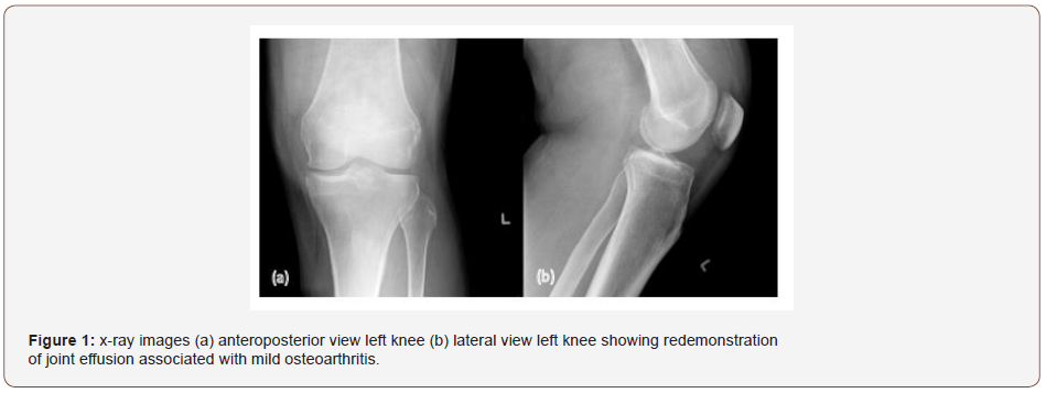
X-Ray Anteroposterior and Lateral Views Left Knee Reported
Redemonstration of joint effusion an area of dense soft tissue in the suprapatellar pouch and the popliteal fossa associated with mild osteoarthritic changes otherwise, no significant interval changes could be appreciated (Figure 1).
Magnetic Resonance Imaging of Left Knee Reported
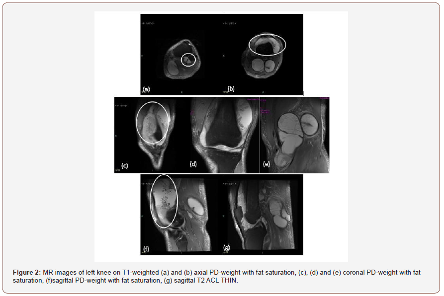
Moderate to severe fat joint effusion with significant synovial thickening and abnormal changes of the synovium particularly within the supra patella area as well as within the deep Hoffa’s fat pad.
Big septated relatively complicated Baker’s cyst measuring approximately 16 x 9 x 5 cm craniocaudally, transverse, and in anteroposterior dimensions respectively, causing significant mass effect on the surrounding soft tissues. Additionally, there is septated cystic change intimately related to the posterior aspect of the proximal tibiofibular joint. However, other structures of the joint are within normal (Figure 2).
Impression
1. Moderate to severe joint effusion with significant frondlike synovial thickening and inflammation appearance, likely representing lipoma arborescence with associated synovitis.
2. Big septated probably complicated Baker’s cyst, as described above.
3. Likely synovial cyst intimately related to proximal
Procedure
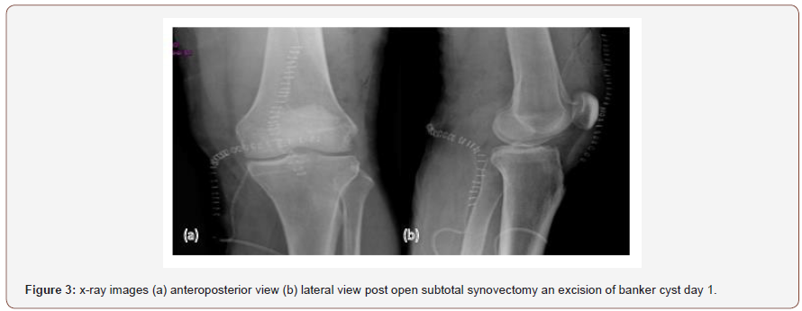
Patient underwent open subtotal synovectomy through a medial parapatellar approach and excision of the popliteal cyst through a posterior approach. The post-operative course was uneventful. The pain was controlled with medications and Physical therapy was started immediately (Figure 3).
Pathology Report
Cross Microscopic Reported
• Left Knee Baker Cyst: cyst lesion, measuring 13 * 7 cm with thick cystic wall. GROSS SHOWS variable intracystic whitish-yellowish growth with focal hemorrhagic changes.
• Left Knee Posterior Synovium: GROSS SHOWS non oriented variable size soft tissue fragments.
• Left Knee Synovial Tissue: Gross Show: projecting papillary-like structures.
Histopathology Reported
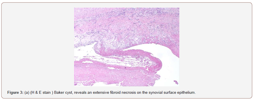
• Left Knee Baker Cyst: Histopathological Featues are Consistent with popliteal benign synovial cyst associated with marked superficial fibropurelent inflammation, Negative for Malignancy.
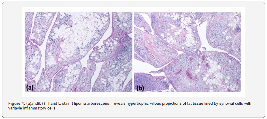
• Left Knee Synovial Tissue Biospy: Histopathology Features are Consist with lipoma arborescens associated with marked chronic synovitis, Negative for Malignancy.
• Left Knee Posterior Synovial Tissue Biospy: Histopathology Features are Consistent with lipoma arborescens associated with marked chronic synovitis, Negative for Malignancy (Figure 3a, 4).
Follow Up
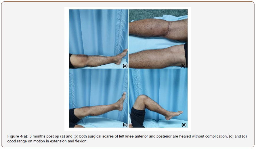
The post-operative follow-up at 7 months: the patient was doing fine, no pain or recurrent swelling with improvement of knee range of motion: 0/110 degree, patient has fully returned to his activities and duties without any active issue (Figure 4a).
Discussion
Lipoma arborescens also called villous lipomatous proliferation of synovial membrane, was first described by Hoffa in 1904, and more in detail in 1957 by Arzimanoglu [13,14].It is a rare and benign, mainly intra-articular lesion of unknown etiology characterized by synovial villous proliferation and sub-synovial connective tissue replacement by mature fatty tissue [1]. It most commonly involves the knee, preferentially the suprapatellar pouch, but other locations have also been described [3,9]. Although it is usually monoarticular, bilateral involvement and multiple joint involvement have also been reported [15,16]. It can be primary, which usually present in younger patients without obvious degenerative joint disease, or secondary to an underlying condition such as chronic synovitis, arthritis, trauma, meniscal injuries and inflammatory joint diseases, usually secondary causes are presented in the older age group [4- 8].
Patients with lipoma arborescens typically present with the history of slowly progressive swelling of the involved joint, which may be accompanied by effusion, decreased range of motion and pain. The intermittent exacerbations of symptoms may be related to mechanical trapping of the lipomatous villi inside the joint space [10,17,18].
The MRI is considered the gold standard for the diagnosis of lipoma arborescens. The MRI imaging findings include: a synovial mass with a frond like architecture, fat signal intensity on all pulse sequences, suppression of signal with fat-selective presaturation, associated joint effusion, potential chemical shift artifact and absence of magnetic susceptibility effects from hemosiderin [11,12]. In addition, in the secondary type of lipoma arborescens MR imaging may also demonstrate associated abnormalities such as degenerative changes, meniscus tear, synovial cyst and bone erosion [9].
The particularity of our case of secondary lipoma arborescens is the young age of the patient and the association with a giant popliteal cyst responsible of limitation of knee flexion. The surgical synovectomy is the recommended treatment of lipoma arborescens. Actually, arthroscopic synovectomy will take place of open synovectomy when applicable because of low rate of complications and rapid recovery. The early surgical treatment offers the best functional outcome [4,19].
Conclusion
Secondary lipoma arborescens is a reactive process of the joint, commonly involving unilateral knee due to underlying diseases. This pathology should be included in the differential diagnosis of recurrent effusion and chronic swelling of a joint. The early diagnosis and surgical synovectomy are recommended for the best functional outcome.
To read more about this article...Open access Journal of Orthopedics Research
Please follow the URL to access more information about this article


No comments:
Post a Comment