Authored by Brigitte Wendl*,
Abstract
Background: Orthodontic gap closure is an important alternative to prosthetic restorations on implants. However, the presence of bone of sufficient quality and quantity is critical to proper tooth movement in the maxilla, especially in patients with a markedly enlarged maxillary sinus. These two case reports demonstrate a new method to facilitate bodily gap closure through the maxillary sinus with the help of an interdisciplinary surgical approach (sinus lift and bone augmentation).
Material and methods: Two patients requiring orthodontic gap closure in regions with a markedly enlarged maxillary sinus and an area of atrophic bone that needed augmentation were treated with pre-orthodontic sinus lifts using the fully resorbable Guidor easy-graft Classic (Sunstar) augmentation material. Superelastic tension springs and miniscrews were chosen as orthodontic appliances. Target parameters were evaluated preoperatively, directly postoperatively and after the gap closure using CBCT datasets and models.
Results: The maxillary sinus was radiologically inconspicuous in all datasets. 3D evaluation of the tooth movement showed proper translational tooth movement.
Conclusion: Radiological and clinical evaluation of these two orthodontic cases showed successfully translational orthodontic tooth movement through an enlarged sinus supported by bone augmentation using a synthetic augmentation material. There were no significant signs of root resorption.
Keywords: Orthodontic cap closure; Sinus lift; Bone augmentation; Xenogeneic bone substitute; Translational orthodontic tooth movement
Introduction
Orthodontic gap closure is an important alternative to prosthetic restorations on implants. However, the presence of bone of sufficient quality and quantity is critical to proper tooth movement in the maxilla to achieve gap closure without root tipping or root resorption, especially in patients with a markedly enlarged maxillary sinus.
Kilic, et al. [1] showed that the distance between the sinus floor and the root tip was greatest for first premolars and shortest for the buccodistal roots of second molars on both the right and the left sides. Since molar roots are located close to the maxilla, the remaining bone height as well as the osseous elongation of the sinus will affect the available options for tooth movement. Sharan and Madjar [2] reported that the extraction of second molars led to greater expansion than did the extraction of first molars. They also reported that extractions of two or more adjacent posterior teeth caused greater sinus expansion.
Wehrbein, et al. [3, 4] proposed a quantitative description of the degree of tipping and kind of movement as a function of the configuration and extension of the basal maxillary sinus. It has been shown that with a more vertical extension in front of the tooth to be moved, a higher amount of tipping (about 10°) is obviously to be expected than for teeth moved through a more horizontally extended sinus base. A correlation was also found between the depth of the maxillary sinus recess and the amount of tipping: the deeper the maxillary sinus recess, the more pronounced the tipping. Re, et al. [5] showed in a case report that direct remodelling results in a dislocation of the lamina dura and, hence, complete remodelling of the sinus.
Tooth movement in the presence of anatomic limitations can be facilitated by surgical interventions such as corticotomies, which, like dentoalveolar distraction, have been proposed as effective and safe methods to abbreviate orthodontic treatments in adolescent and adult patients [6].
In addition, augmentative techniques can be used to advantage to move teeth more safely and to improve the physical transmission of force. Tooth movement through xenogeneic bone substitute materials has been shown to be successful in animal experiments demonstrating that tooth could be moved into an alveolar ridge area augmented with a biomaterial (Bio-Oss; Geistlich, Wolhusen, Switzerland) three months previously [7]. Furthermore, bone grafting material was also successfully used in moving mandibular front teeth in Class III patients receiving combined orthodontic and surgical treatment [8, 9].
Reichert, et al. [10] showed that orthodontic gap closure in extraction sockets filled with silica matrix-embedded, nanocrystalline hydroxyapatite bone substitute is possible without inflammation or root resorption. Kye-Bok Lee, et al. [11] demonstrated that augmented corticotomy effectively reestablished periodontal soft and hard tissue on the pressure sides after buccal tooth tipping, regardless of the type of grafting material used. The synthetic bone graft group, which had exhibited the greatest changes in angles during tipping, also showed most new bone formation and most intact soft tissue.
At present, no evidence-based protocol can be recommended for moving teeth through the maxillary sinus. According to the systematic review by Sun, et al. [12] the empirical application of constant and light to moderate force (by TAD, segment and multibrackets) to move teeth slowly through or into the maxillary sinus in adults appears to be the most secure way. Bodily movement was accomplished, but teeth appear to be easily tipped initially, potentially resulting in root resorption.
Given the absence of pertinent data in the literature. the question arises whether augmentative techniques might facilitate bodily tooth movement and the transmission of more physical force for gap closure through a markedly enlarged sinus.
Material and Methods
Two patients required orthodontic gap closure in regions with a markedly enlarged maxillary sinus and an area of atrophic bone that needed augmentation.
The bone height in the gap regions was reduced to less than 5mm, with the root tip presenting above the sinus floor in the panoramic radiographs.
Exclusion criteria were: severe general diseases, antiinflammatory medication, allergies to one or more of the materials used, tooth extraction in the gap region less than half a year previously, and insufficient oral hygiene.
Both patients were treated with pre-orthodontic sinus lifts using the Guidor easy-graft Classic augmentation material (Sunstar, Etoy, Switzerland). This material is fully resorbable and is replaced by autologous bones within months thanks to the phase-pure betatricalcium phosphate (β-TCP) it contains.
The direct sinus lift procedure employed was that described by Oscar Hilt Tatum. After uncovering the lateral wall of the jaw by reflecting a flap, the thin lateral wall of the jaw is weakened circularly with a spherical diamond drill in an area of approximately 1-2 cm². The resulting cover is “lifted” inwards together with the Schneiderian membrane adhering to the inner side, creating a fairly large cavity in which the synthetic bone substitute material is inserted.
Orthodontic appliance
Superelastic tension springs were chosen as orthodontic appliances as these are able to deliver forces of 1.5 Ncm in a standardized manner. Furthermore, lever arms and mini-screws were used for skeletal anchorage.
Bone heights, tooth positions, tooth movements and root resorption were evaluated preoperative, directly postoperatively and after the gap closure using CBCT datasets (Promax 3D Max, 10.0 × 5.9cm, 200 μSv, low dose, 96 kV, 6.3 mA; Planmeca, Helsinki, Finland) and models. Photographic documentation was carried out at the end of the treatment.
Translational tooth movement was verified clinically by way of stone casts and radiologically using two DICOM datasets. An A dataset (obtained postoperative but prior to the orthodontic tooth movement) and a B dataset (obtained at the end of orthodontic treatment) were superimposed using the coDiagnostiX (Dental Wings, Chemnitz, Germany) implant-planning software. The datasets were segmented digitally for a three-dimensional display of the hard-tissue structures, including the maxillary teeth and bones (nasal concha, nasal spine, borders of the upper maxillary sinus, parts of the zygomatic arch, parts of the sphenoid bone). The segmented A datasets was then imported into the B datasets. Their superimposition was performed on the basis of the segmented stable hard-tissue structures listed above and displayed as virtual 3D models. The coDiagnostiX software displayed virtual 3D models three-dimensionally in a separate window and within the axial, coronal and sagittal layers marking the borders of the previously segmented structures (bone and teeth).
Tooth positions and movements were measured in the axial and sagittal layers. To this end, the axial layer was adjusted to be parallel to the occlusal plane and positioned relative to the position of the alveolar crest. The sagittal layer was positioned to intersect the middle of the second premolar and first molar along the panoramic curve/dental arch. The distances between the positions of the premolar and molar adjacent to the gaps as well as tooth movement were measured at the level of the alveolar crest and of the root tips by superimposing the A and B datasets. Dimensions of augmentation (height, mesiodistal width, buccopalatal width) were measured separately in the A and B datasets (Figures 1, 2 for patients 1 and 2, respectively).


Results
Radiological findings
The A and B datasets showed age-appropriate bone structures in the maxilla for both patients. Molar roots had intruded into the maxillary sinus by at least the apical third of their length. No root resorption was seen in either the A or the B datasets. The maxillary sinus was radiologically inconspicuous in for both datasets. The postoperative A datasets showed a regularly positioned, welldemarcated sinus augmentation, while both B datasets showed a reduced volume of bone substitute material.
Patient 1
In the A dataset, the bone substitute exhibited a maximum height of 9.5mm, a maximum mesiodistal width of 8.3mm and a maximum buccolingual width of 11.0mm. At the end of the orthodontic treatment, the corresponding dimensions were 6.1mm, 4.7mm and 6.8mm, respectively.
Patient 2
In the A dataset, the bone substitute exhibited a maximum height of 10.8mm, a maximum mesiodistal width of 9.3mm and a maximum buccolingual width of 11.2mm. At the end of the orthodontic treatment, the corresponding dimensions were 6.6mm, 7.2mm and 10.2mm, respectively.
Patient 1
Diagnosis
Patient 1 was a 38-year-old woman with a skeletal Class II/2 dysgnathia showing an overjet of 1 mm and an overbite of 6 mm (Figures 3, 4). The profile appeared concave.


Cephalometric tracing yielded the following results:
SNA angle: 79°
SNB angle: 76.5°
Sum of angles: 387°
Angle of Inclination, upper anteriors: 83°
Angle of Inclination, lower anteriors: 84°
The patient exhibited bilateral half Class II malocclusion.
The panoramic radiograph and the CBCT sections showed a markedly enlarged maxillary sinus and atrophic bone at site 15 (Figures 5-7).


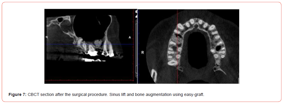
In view of the deep bite and Class II malocclusion, the right maxillary bridge was removed. The gap was closed by reciprocal gap closure (facilitated by augmentation by way of a sinus lift). In the left maxilla, a Class I occlusion was achieved by distalisation.
Treatment objectives and treatment alternatives
1. Periodontal evaluation and professional tooth cleaning prior to treatment
2. Skeletal discrepancy to be compensated dentally, as orthognathic surgery was not accepted by the patient
3. Transverse alignment of the maxilla
4. Full Class II molar relationship to be achieved on the right side by removing the existing maxillary bridge and performing reciprocal gap closure, and a Class I occlusion to be achieved by posterior distalisation
5. Correction of the sagittal and vertical overbites 6. Correcting the inclinations and torques of the anterior teeth
7. Uprighting of the mandibular right molars
8. Implant placement at site 35
9. Midline correction
10. Aesthetics: Broadening the smile and improving the smile curve
11. Periodontal: Slowing or halting progression of the existing recessions
12. Achievement of long-term stability
Treatment procedures
The maxilla and mandible were levelled and aligned using wires of ascending order (0.014-0.016 × 0.022 -0.017 × 0.025). Prior to gap closure, the sinus lift (Figures 8-12) and bine augmentation (Guidor easy-graft Classic) was done by an experienced maxillofacial surgeon. To facilitate translational tooth movement, custom 3D-printed lever arms were bonded to teeth 14 and 16. A tension coil (150g) on a 0.017 × 0.025 stainless steel wire was used for gap closure (Figures 13, 14).

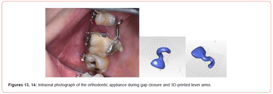
A correction to achieve a Class II relationship on the left side was performed using Class II elastics. To obtain good intercuspation, some vertical elastics were used towards the end of treatment. Orthodontic tooth movements and gap closure were carried out successfully, resulting in uprighted premolars and molars (Figures 15-18).
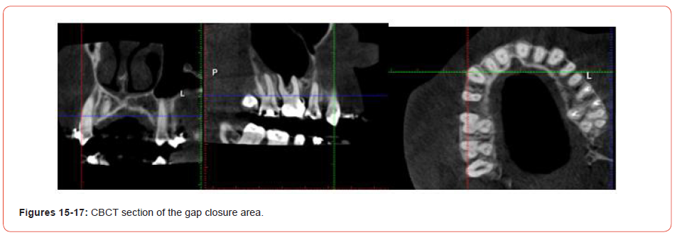
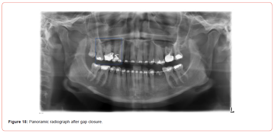
The mandibular teeth were aligned and uprighted, facilitating placement of an implant at site 35. The periodontal situation did not deteriorate. A Class I relationship in the canine region and a correct overbite and overjet were achieved on both sides. The facial profile improved thanks to the uprighting and proper torque of the anterior teeth (Figures 19-22).
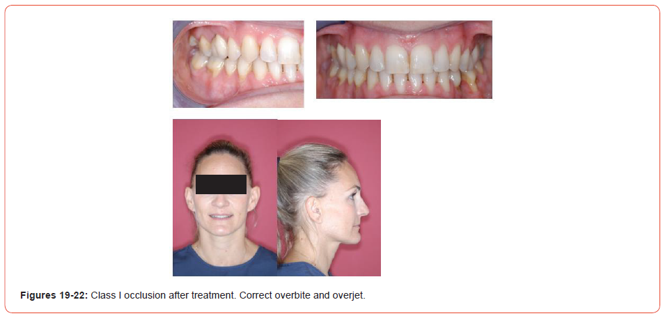
Fixed retainers were bonded to the maxillary and mandibular anteriors.
3D imaging with sagittal and axial superimpositions of tooth movement proved that bodily tooth movement had been successfully achieved (Figure 1).
Patient 2
Diagnosis
Patient 2 was a 46-year-old man with a skeletal Class II/1 dysgnathia (Figures 23-28).
Cephalometric tracing yielded the following results:
SNA angle: 87°
SNB angle: 84°
Sum of angles: 376°
Angle of Inclination, upper anteriors: 112°
Angle of Inclination, lower anteriors: 111°
(bialveolar protrusion)
The patient exhibited a full Class II malocclusion on the left side and a Class II occlusion of one-quarter premolar width on the right side.
Teeth 21, 16, 26 and 27 were missing. Tooth 45 was lingually blocked. Tooth 15 exhibited a crossbite. Some periodontal bone loss was visible in the radiographs (Figures 29-31).
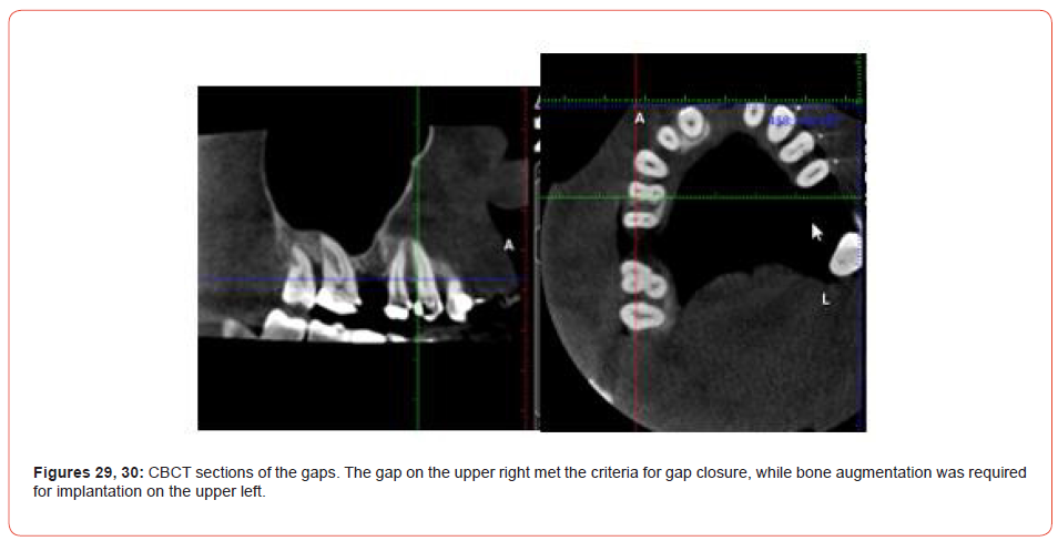
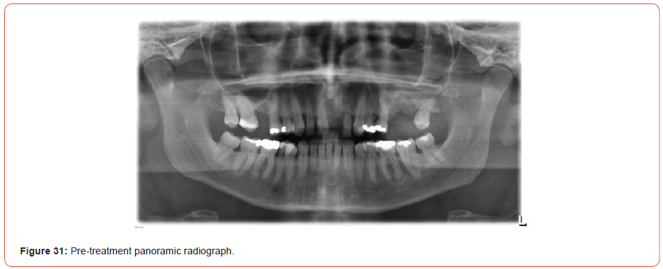
CBCT findings: The right maxillary gap side met the criteria for gap closure, while the left maxillary gab required bone augmentation and subsequent implant treatment.
Treatment objectives and treatment alternatives
1. Skeletal discrepancy to be compensated dentally
2. Gap closure on the maxillary right side through mesialisation of the maxillary second and third molars and distalisation of the premolars
3. Bone augmentation and implant placement on the maxillary left side
4. Creation of a Class I occlusion
5. Correction of the sagittal and vertical overbites
6. Correcting the inclinations and torques of the anterior teeth
7. Midline correction
8. Relieving the crowding and correcting the crossbite at tooth 15
9. Uprighting of teeth 46 and 47
10. Aesthetics: Broadening the smile and improving the smile curve
11. Periodontal: Slowing or halting progression of the existing lesions
12. Achievement of long-term stability
Treatment procedures
Following professional tooth cleaning (sub- and supragingivally), the teeth were aligned using a typical sequence of NiTi and stainless-steel wires. To achieve gap closure on the maxillary right side, custom lever arms were 3D-printed, and a mini-screw was placed on the palate to provide stable anchorage for mesialisation (Figures 32, 33). A sinus lift with bone augmentation (Guidor easygraft Classic) was performed (Figures 8-12, 34, 35).
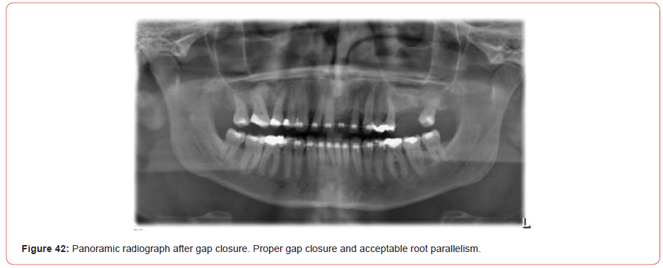
In the mandible, proximal stripping was performed to improve the inclination. Orthodontic tooth movements and gap closure was successfully performed. (Figures 36-38) illustrate the situation immediately after gap closure. The post-treatment radiographs (Figures 2, 39-42) show proper gap closure and acceptable root parallelism. The teeth were well aligned and uprighted. Orthodontic treatment was still in progress at the time of this writing.

Discussion
The floor of the maxillary sinus is similar to cortical bone in that it is a layer of compact bone lined with periosteal tissue. In adults, the size of the maxillary sinus is variable in its extension. Its floor extends between adjacent teeth or individual roots in about half of the population, creating elevations in the antral surface or protrusions of root tips into the sinus [13, 14].
Wehrbein and Diedrich [3] described a positive correlation between the length of root projection into the maxillary sinus as observed on panoramic radiographs and the amount of pneumatization that occurs after extraction. Sinus expansion following extraction can greatly decrease the amount of bone height available for implant placement. The relationship between the dental roots and the inferior sinus wall is known to influence orthodontic tooth movement, and the intrusion or bodily movement of teeth across the sinus floor that occurs with orthodontic treatment has been shown to cause moderate apical root resorption and a high degree of tipping [15].
The thickness of the inferior wall of the sinus and its relationship with the adjacent teeth is crucial for orthodontic tooth movement [4, 16]. This information provides a basis for the application of appropriate forces in orthodontic tooth movement and predicting the amount of tooth movement through the maxillary sinus. Augmentative techniques can facilitate tooth movement and a physical transmission of force leading to less tipping becomes possible, as illustrated by the cases presented here.
The treatment concept of bone augmentation consists of providing the bone with a stable osteoconductive structure that is gradually incorporated or replaced by the newly formed bone. Autologous bone grafts are considered the gold standard in bone regeneration surgery due to their osteoinductive potency [17, 18]. Synthetic materials consist mostly of β-TCP, a ceramic base that, depending on its surface texture, can have osteoconductive properties. Advantages of synthetic materials include their material characteristics and the reduced risk of allergies when bone augmentation is performed. The xenogeneic materials contain hydroxylapatite (HA) from animal bone or from algae and have a more complex three-dimensional structure offering improved osteoconductivity [19].
Bone augmentation materials should meet the following criteria for clinical use: scaffolding function to facilitate the ingrowth of bone cells from the surrounding graft (osteoconduction); support for growth factors or stem cells; surfaces for the migration, adhesion, differentiation and proliferation of cells; and stability to withstand the pressure of the covering soft tissue. Microporosities of at least 100μm to ensure the growth of blood vessels and pluripotent precursor cells and biocompatibility (not triggering any immune reaction) are important.
Among the most commonly used materials, HA and β-TCP have been successfully used for bone augmentation [20]. HA is considered almost a non-resorbable material and has the advantage that it serves as an osteoconductive scaffold for new bone formation. β-TCP, on the other hand, exhibits much greater biodegradability. To combine the positive aspects of the two calcium phosphates, biphasic materials with different ratios of HA and β-TCP have been developed [21-23].
The material used in the cases discussed here was Guidor easygraft Classic. Suitable for all dental applications requiring bone replacement, it is applied directly from the syringe into the defect, where it hardens and forms a porous but stable bone replacement. The fast-resorbable coating of polylactide ensures injectability, while the liquid Biolinker (N-methyl-2-pyrrolidone solution; Sunstar) promotes the mutual adhesion of the granules. The density of the coating is intended to prevent bacterial growth. The material as applied is characterised by high and interconnective porosity (and thus a haemostatic effect, promoting cell growth and bone formation) and forms a sticky mass. The size of the granules used in the study, of 450-1000μm is right for most medium-sized and large bone defects. The subsequent bone formation occurs simultaneously with the degradation of the filling material.
Roberts, et al. [24, 25] stated that modelling changes the size and shape of bones in response to mechanical loading, whereas remodelling is the turnover of bone that is related to bone maturation and mineral metabolism. Therefore, orthodontic tooth movement occurs as a result of a bone-modelling phenomenon, and the biologic balance of bone structures occurs by bone remodelling as a physiologic process.
Tooth movement through the maxillary sinus is another example of tooth movement through bone. In orthodontically induced tooth movement, the moving root is displaced in the cortical bone by frontal and undermining resorption. In this process, there is a balance between bone resorption and apposition in response to orthodontic forces. The modelling and remodelling mechanisms can readily adapt to changes in the mechanical loading of the alveolar bone.
Root resorption is associated with the removal of the necrotic material from the hyaline zone produced during the orthodontic tooth movement. The process is controlled by inflammation mediators. In this context, a distinction must be made between the treatment technique (type of appliance, duration of treatment, extent and type of tooth movement and size and type of force) and biological factors (innervation, abnormalities in bone density, inflammation, traumas and endodontic pre-treatments [26].
Tooth movement was successfully performed in both the cases presented here. In patient 2, For patient 2, dense structures could be seen in the radiographs, but no root resorption was observed.
The correlation between the topography of the maxillary sinus floor and related root position of posterior teeth using panoramic and cross-sectional computed tomography imaging was shown by Sharan and Madjar [27]. For most roots projecting into the sinus in panoramic radiographs, they report no vertical penetration into the sinus on CT images. Roots that did penetrate into the sinus on CT images exhibited much shorter penetration lengths than was apparent on panoramic radiographs.
Conclusion
Radiological and clinical evaluation of these two orthodontic cases showed successfully translational orthodontic tooth movement through an enlarged sinus supported by bone augmentation using a synthetic augmentation material. There were no significant signs of root resorption.
To read more about this article...Open access Journal of Dentistry & Oral Health
Please follow the URL to access more information about this article
https://irispublishers.com/ojdoh/fulltext/a-novel-technique-for-orthodontic-gap-closure-with-access-through-the-maxillary-sinus.ID.000629.php
To know more about our Journals...Iris Publishers
To know about Open Access Publishers


No comments:
Post a Comment