Wednesday, July 21, 2021

Iris Publishers- Open access Journal of Advances in Cancer Research & Clinical Imaging | A Simple Screening Test for Cancer


Authored by  AbulKalam M Shamsuddin*

Abstract

Comparative and correlative studies of the pathology and pathogenesis of colon cancer in animal models and human disease have resulted in conceptualization of ‘field effect” theory, and identification of a simple carbohydrate marker that is expressed early during carcinogenesis. This assimilated body of knowledge has resulted in development of a simple screening test for cancers of the lung, breast, colorectum, uterus, pancreas, and prostate. The marker galactose-N acetyl-galactosamine (Gal-GalNAc) is expressed in the cell surface and secreted glycoproteins of otherwise normal appearing tissues remote from cancer or precancerous lesions and is detected by enzymatic oxidation (10 minutes) followed by color reaction (1 minute). The high sensitivity, specificity, and cost-effectiveness of this point-of-care test makes it a great tool in our strategies for early detection, hence control of cancer. It would also reduce the number of unnecessary and expensive procedures, thereby decreasing the total national health-care cost to the societies and governments, globally.

Keywords: Screening test; Lung cancer; Breast cancer; Prostate cancer; Colon cancer; FOBT

Abbreviations: AOM: azoxymethane; FOBT: fecal occult blood test; Gal-GalNAc: galactose-N acetyl- galactosamine; GOS: galactose oxidase Schiff; MNNG: N-methyl-N’-nitro-N-nitroso-guanidine; NCI: National Cancer Institute; T-Ag or TF-Ag: Thomsen-Friedenreich antigen

Introduction

Regrettably, in 2020 approximately 10 million people were killed by cancer; now-a-days there is hardly a family that is not affected by cancer. Additionally, the healthcare cost, and loss of productivity owing to morbidity and poor quality of life, has been immensely draining to families, societies, and governments alike, everywhere. This scourge has been around for a very long time with no end in sight. As in SARS-Cov-2 now, in 1971 President Richard Nixon had declared ‘War on Cancer’ to combat this menace. Alas, 50 years later, conquering cancer remains an elusive goal despite progress in treatment, albeit modest. Again, akin to SARS-Cov-2, prevention is our best alternative. Cancer prevention includes (a) detection at the very early stage of the disease (secondary prevention) to reduce cancer mortality and increase the survival rate of patients; and (b) etiology prevention or primary prevention to reverse precancerous lesions or in situ carcinomas (a cancer that is still confined) to normal or stop them from progressing to invasive malignancies in populations at high risk – ‘nipping in the bud’. Thus, early detection is fundamental to prevention, and the key is to find the marker which is differentially expressed in highrisk tissues (cancer and precancer) but not in normal. Biomarker(s) meeting this criterion is/are logical choice for establishing accurate methods to detect cancers at infancy; it may also help in monitoring the efficacy of chemoprevention program by serving as intermediate endpoint marker [1].

Fundamental to the success of prevention programs of any disease is the detection of the problems. It includes identification of people with existing disease and those who are at risk of developing the disease. Identification of people with cancer is relatively simple for most cancers because of signs and symptoms from the disease; the individual, now a patient seeks medical attention. Extensive work-up including a battery of diagnostic tests are performed and appropriate therapy is administered; regrettably, it is usually too late for many, if not for most as it does not prevent the disease. Hence the adage: an ounce of prevention is better than a pound of cure. For an effective prevention program, we must actively seek individuals with cancer or high risk thereof from an apparently healthy noncompliant population. This is done by separating i.e., screening individuals into groups with high and low probability of cancer with the help of rapid, simple, accurate, and inexpensive tests (screening tests). Implicit in the definition of screening is a promise that there is a benefit for those who participate; they will be followed with further diagnostic tests and future management of the problem. But a screening test is never intended to give the full diagnosis, hence the distinction from diagnostic tests [2]. An individual who is screening test positive will need to undergo diagnostic procedures to confirm the presence of the disease. Ideal screening tests should have a high sensitivity (proportion of diseased subjects who are test- positive) and specificity (proportion of non-diseased subjects who are testnegative), be simple and non- invasive or minimally invasive, easy to administer – therefore enjoying a high acceptability amongst populations and of course cost-effective. It would not be successful if it is shunned for discomfort, cultural, religious, or other reasons or just too expensive [2]. Currently, except for colon cancer, there are no true good screening tests for other cancers at early stages. Imaging technologies such as mammogram, chest X-ray, low dose CT etc. cannot detect early cancers, for a tumor must attain a minimum size before it can be ‘visible’ by these. And that is too late!

The Galactose Oxidase-Schiff Test

The Galactose Oxidase-Schiff Test is one such screening test that fits all the criteria of an ideal screening test as it is rapid, simple, non-invasive, easy to administer, and inexpensive. Its high accuracy has been consistently and reproducibly demonstrated independently by numerous investigators in three continents. That is not surprising because of extensive scientific research on carcinogenesis and operation of i) field-effect of carcinogens and ii) a marker differentially and specifically expressed during carcinogenesis, shared by both cancer and precancer, but not by normal or regenerating tissue.

Field-effect

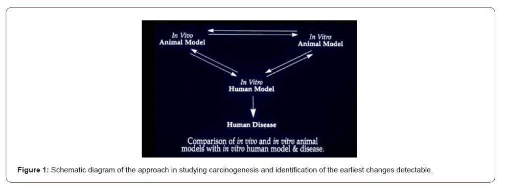
I studied early detection of cancer using colon cancer as a model since 1975 at the University of Maryland School of Medicine as my PhD dissertation. The advantages of colon as a model include a) relatively easy access to samples, b) well known precancerous changes (polyps) and conditions e.g., ulcerative colitis, Crohn’s disease etc.; c) well developed animal models with d) well characterized steps of carcinogenesis viz. initiation → promotion → progression; etc. Both in vivo and in vitro carcinogenesis experiments with rats and mice were performed to see how colon cancer forms in them, and to identify the earliest recognizable changes by microscopy and histochemistry [3,4]. Colon tissue from rats and human were cultured in the Petri dishes and exposed to the carcinogens N-methyl-N’-nitro-N-nitroso- guanidine (MNNG) for rat colon [3], and fecapentaene-12 a suspected human carcinogen, for human colon explants [5]. The findings were correlated with in vivo animal models, and ultimately to the human disease [6]: what does the human colon near and far from the cancer look like (Figure 1)?
irispublishers-openaccess-cancer-research-clinical-imaging

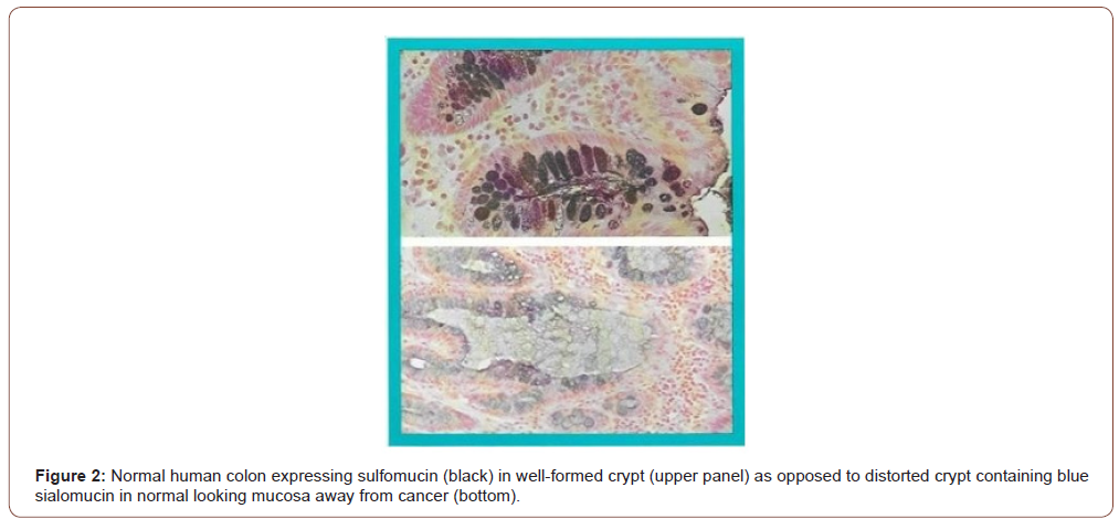
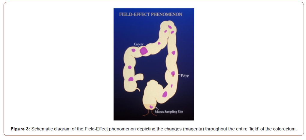
It has been observed that the earliest recognizable change in the carcinogen-exposed tissue is an alteration in the composition of secreted mucus that persists through precancerous and cancerous tissues. The microscopic and histochemical changes in the human colon away from cancer are identical to those seen in the colons of rats (mouse colon is distinctly different from human or rat colon [7]) treated with the carcinogen azoxymethane in vivo, or MNNG for rat explants and fecapentaene-12 for human explants in vitro in Petri dishes. These changes are therefore the earliest evidence (or markers) of cancer formation – even before a cancer has formed. Using high iron diamine alcian blue technique, the mucus in the colon away from the cancer that looked normal by naked eye appears as blue (Figure 2, lower frame) as opposed to black in truly normal human colon without cancer, in the upper frame (Figure 2). Also note that the colonic glands or crypts in a cancer bearing colon appear distorted – changes identical to the rat carcinogenesis models. It was reasoned that because of the generalized effect of the carcinogen throughout the entire field of the large intestine, it is most likely that the tissue away from an obvious cancer would be abnormal – Field-Effect. The carcinogens in our environment such as the air we breathe, the food we eat etc. expose the entire lung or large bowel. Therefore, it is logical that their effect would be observed throughout the exposed field. While the vast majority of the cells will undergo DNA repair, and other host defense mechanisms such as NK cell will prevent them to progress to cancer, a few cells will be promoted and even fewer progressed to precancer and cancer who may share the same early changes as the rest of the exposed tissue in the field. (Figures 2,3).
irispublishers-openaccess-cancer-research-clinical-imaging

irispublishers-openaccess-cancer-research-clinical-imaging

Field-Effect phenomenon indicates that the entire field bears the brunt of the carcinogenic assault and expresses variable changes throughout. In the schematic drawing of the large intestine (Figure 3), it is depicted that irrespective of where a cancer or a precancerous polyp may be, areas of the otherwise normal appearing mucosa will show expression of the marker perhaps in a patchy manner (it is not practical to sample the entire large intestine to see if every millimeter has the change; hence this assumption of “patchy”). Rectum being a part of the large intestinal “field” and a convenient sampling site is therefore likely to show the same changes. Since digital rectal examination is a part of routine physical examination (though not practiced diligently by many primary care physicians now-a-days), it is simple and noninvasive. It also allows the physician to examine the prostate in males, and uterus, cervix, and adnexa in females. Samples from lungs such as coughed up sputum, nipple aspirate from breast, endocervical mucus, or prostatic secretion are likewise simple and noninvasive.

The marker: gal-galnac

The observed mucin histochemical change has further been identified as due to a biochemical alteration in the cell surface and secreted glycoprotein – presence of the carbohydrate moiety D- galactose-ß-(1-3)-N-acetyl-D-galactosamine (Gal-GalNAc, also called Thomsen-Friedenreich antigen or T- Ag, or TF antigen, though it may be different). In normal cells, a terminal sialic acid blocks the T-Ag from being recognized by the lectin peanut agglutinin (PNA), or the enzyme galactose oxidase; in cancer and precancer owing to loss of the sialic acid it is now recognizable. D-galactose oxidase specifically oxidizes C-6 hydroxyl groups of D-galactopyranose and N-acetyl galactosamine residues of Gal-GalNAc, generating two vicinal aldehyde groups that react with basic fuchsin to give magenta/purple coloration. Thus, Gal- GalNAc can be visualized by a simple enzymatic reaction with galactose oxidase followed by Schiff’s reagent, resulting in the development of a simple test for early detection, initially of colorectal cancer [8] by using rectal mucus sample.

What is an Ideal Cancer Marker According to the National Cancer Institute (NCI)?

Kelloff et al of the Division of Cancer Control and Prevention (DCCP) at the National Cancer Institute [1] outlined six criteria for intermediate endpoint biomarkers of use in chemoprevention, and here is how Gal-GalNAc and STEDi live up to those expectations:

Is the intermediate biomarker differentially expressed in normal and high-risk tissue? YES!

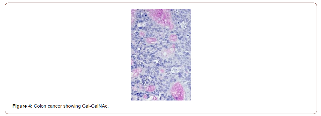
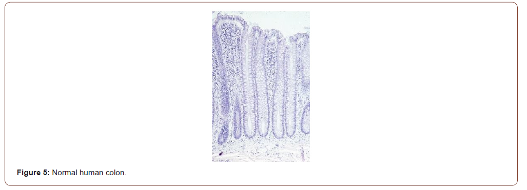
Figure 4 shows the expression of Gal-GalNAc (magenta) in a colon cancer but, not by the normal human colon (Figure 5). The normal tissues were obtained from healthy normal people without any cancer. Note the absence of magenta color in the mucus of normal colonic goblet cells. Also note that the colonic glands or crypts in truly normal humans appear uniform test tube shaped as compared to the those in cancer-bearing colon or in rats treated with the carcinogen azoxymethane (see Figure 4-6).
irispublishers-openaccess-cancer-research-clinical-imaging

irispublishers-openaccess-cancer-research-clinical-imaging

irispublishers-openaccess-cancer-research-clinical-imaging

At what stage of carcinogenesis does the marker appear?

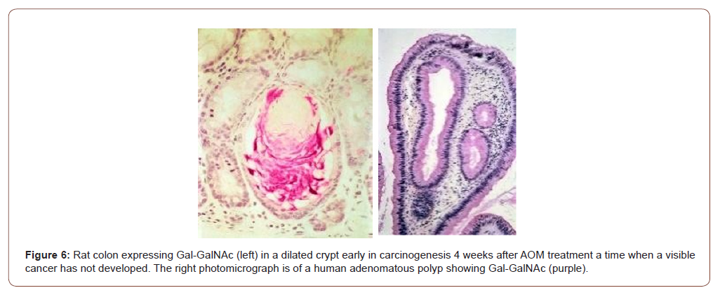
The earlier a reliable marker appears in the carcinogenic process, the greater is the chance for successful intervention. Answer: Gal-GalNAc is expressed very early during carcinogenesis. Not only the marker is expressed early during the carcinogenesis in rats in vivo (Figure 6 left panel) but also by human precancerous polyp (right panel appearing as purplish). The colors are slightly different owing to different batch of staining and tissue preparation. The mucus alteration is also observed in rat and human explants exposed to carcinogens in vitro [4,5]. Note the distorted appearance of the colonic glands as opposed to uniform test tube shaped ones in normal (Figures 5,6).

Does the marker and its assay provide acceptable sensitivity, specificity, and accuracy?

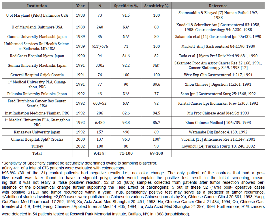
Answer: Both the marker [9,10] and the assay [11-14, Table I] enjoy 70-100% sensitivity and specificity. That it is not expressed by regenerating cells following wounding is an added evidence that Gal-GalNAc is carcinogenesis specific [10]. Please see the results on colon cancer in the following Table: results on other organ sites are provided after this: (Table 1)

Table 1: Performance Summary of GOS Test for Colorectal Cancer.
irispublishers-openaccess-cancer-research-clinical-imaging

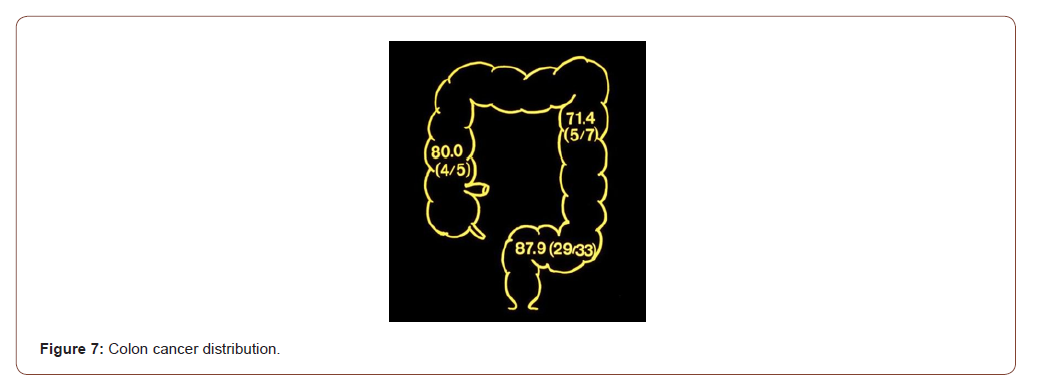
The proof of field-Effect: Schematic diagram of the human large intestine depicting the location of the cancers detected by the rectal mucus test. That the “field-effect” phenomenon is operational is proved by the detection of cancers in the various segments of the colon remote from the rectum where the mucus was sampled from. Note that 4 of 5 cancers (80%) of the ascending (or the right) colon, 5 of 7 (71.4%) of the descending (or the left) colon and 29 of 33 (87.9%) of the sigmoid colon were detected in the two studies by Sakamoto et al [11,12] (Figure 7). The widely used current fecal occult blood test (FOBT) for colon cancer screening has been notoriously inaccurate; “Occult blood testing is, at best an imperfect approach to the screening of colorectal cancer” concluded Dr. Ahlquist [15]. And that is not surprising since blood is not a marker of cancer or precancer. A newer test combining FOBT and DNA in stool appears to have better sensitivity and specificity albeit at a very high price.
irispublishers-openaccess-cancer-research-clinical-imaging

Gal-GalNAc is a common tumor marker: The usefulness of the tumor marker Gal-GalNAc in differentiating the benign from the malignant and pre-malignant lesions of the prostate was tested [16] yielding similarly high sensitivity and specificity (vide infra). Having determined that the principle is practicable in colon and prostate, studies were conducted on other cancers. The expression of Gal-GalNAc determined in a total of 133 tissue samples from 81 cases of the carcinomas of the breast, ovary, pancreas, stomach, and endometrium and 52 cases of respective normal controls [17]. None of the 52 cases of normal tissues (except 15 cases of stomach) showed expression of Gal-GalNAc. In contrast, 100% of adenocarcinomas from the breast (19 of 19), ovary (15 of 15), and pancreas (6 of 6), and 94.1% of stomach (16 of 17) cancers, and 91.7% (11 of 12) of uterine adenocarcinomas expressed Gal- GalNAc. The normal epithelia and their secretions in the vicinity of the carcinoma (within the “field”) in the breast, bronchus, endometrium, and pancreatic duct also expressed Gal-GalNAc in contrast to normal tissues obtained from non-cancerous individuals, which were totally non-reactive. Thus, the tumor marker Gal- GalNAc recognized by galactose oxidase-Schiff sequence was highly expressed not only by a variety of adenocarcinomas but also by the apparently normal-appearing epithelia and their secretions in the vicinity of carcinomas confirming the operation of a field effect phenomenon by carcinogenic agent(s) in these organs as well, setting the stage for identification of the marker in these secretions for mass screening for these cancers too [17].

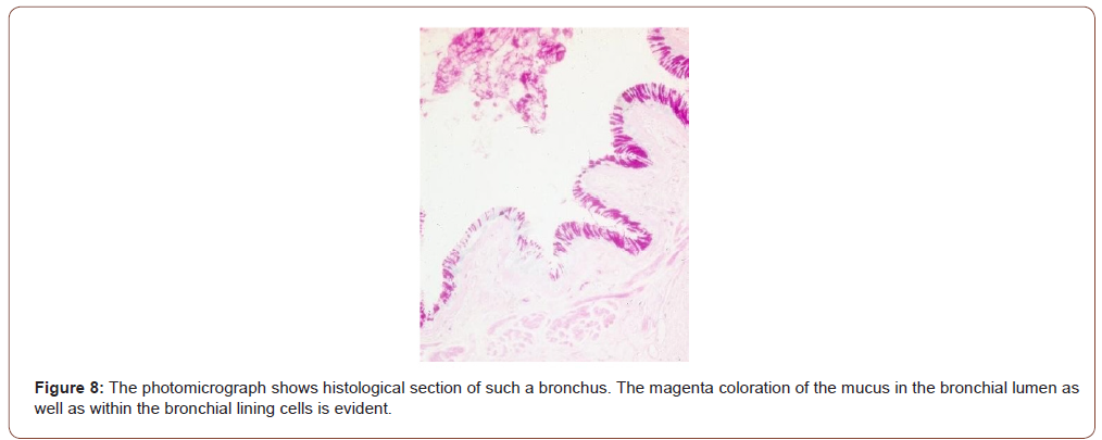
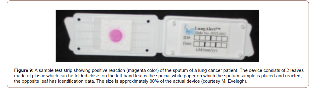
Studies on lung cancer: Twelve of 12 pulmonary adenocarcinomas expressed Gal-GalNAc. The bronchial tissue away from the cancer were available in 4 cases, all of whom also expressed the marker both in the epithelial lining cells as well as in the secreted mucus [17]. Coughed-up sputum therefore can be used to screen people for cancers of the lungs. Indeed, three clinical studies on lung cancer were performed on coughed up sputum [18-20]. Lai et al [18] reported the results of their study on sputum specimens from 116 healthy persons; and 216 cases of benign and malignant lung diseases were tested for the marker Gal-GalNAc. The result showed that 165 of the 182 patients (90.7% sensitivity) with lung cancer, confirmed by cytology and histology, had positive results, whereas 22 of 116 (19.0%) healthy controls were positive (81.0% specificity, Table 2). In 28 cases of patients whose sputum cytology showed various degrees of dysplasia-a precancerous condition that progresses to cancer, 21 were found Gal-GalNAc positive, of which 15 patients were identified to have lung cancer on further work-up! Thus, the concept of “false positive” as generally used does not apply to Gal-GalNAc and the GOS Test since they take the precancerous lesions and early cancers into consideration. In addition, three cases of early lung cancer in this study were also positive, supporting the fact that Gal-GalNAc is expressed at an earlier stage in the malignant process of the lung as well. The studies by Cox & Miller [19] and Miller et al [20] had smaller sample size and showed sensitivities of 64.7 – 88% and specificities of 77.8 – 93.6%. The test revealed 20 of 23 lung cancers among 76 patients. The other 53 patients were either healthy or had benign lung disease such as bronchitis. Even more germane to the issue of prevention is the fact that 13 of 15 cancers detected were early stage (Stage I and II). Of note is that a tumor must attain a minimum size before it can be detected by imaging techniques. And that is late! (Figures 8,9) (Table 2).
irispublishers-openaccess-cancer-research-clinical-imaging

irispublishers-openaccess-cancer-research-clinical-imaging

Table 2: Lung Cancer study.

irispublishers-openaccess-cancer-research-clinical-imaging

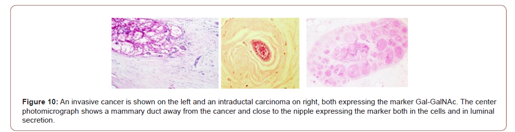
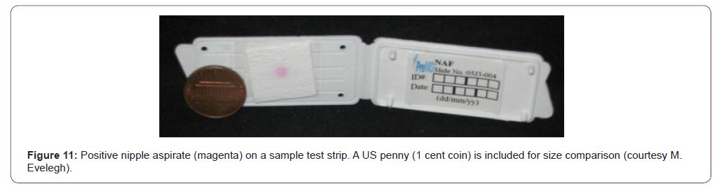
Breast cancer study: Gal-GalNAc is also expressed by the normal-looking breast tissue away from an obvious cancer by way of the field-effect phenomenon [17]. The marker was positive in 19 of 19 histological specimens of breast cancer giving it a 100% sensitivity. More importantly, and germane to our screening, the ducts away from the cancers and close to the nipple also express the marker both in the cells as well as in the secretions in the ducts. Thus, the nipple aspirate from a breast harboring a cancer should express the marker. Kumar et al [21] demonstrated that Gal-GalNAc (or TF antigen) is differentially expressed in nipple aspirate, albeit using a different technique – monoclonal anti- TF antibody. Nineteen of 25 cancer patients and none of the 25 healthy controls were positive yielding a 100% specificity and 76% sensitivity. The relatively low sensitivity of the test in nipple aspirate as opposed to tissue expression may be due to the use of different techniques for identifying the marker – galactose oxidase Schiff’s v monoclonal anti-TF antibody. Chagpar et al [22] also reported the utility of using nipple aspirate and Gal-GalNAc in screening for breast cancer. They investigated 23 women with biopsy confirmed, unilateral stage I or II breast cancer. They took samples (nipple aspirate by way of a suction cup attached to a syringe) from both breasts prior to surgery. Most, but not all the women were able to provide large enough fluid samples that could then be evaluated. Based on the resulting color of the test strips one could differentiate between a healthy and cancerous breast (Figures 10,11).
irispublishers-openaccess-cancer-research-clinical-imaging

irispublishers-openaccess-cancer-research-clinical-imaging

irispublishers-openaccess-cancer-research-clinical-imaging

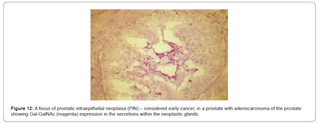
Prostate cancer study: The current screening test-PSA is inaccurate primarily because it is non-specific; its level goes up in benign conditions as well. The marker Gal-GalNAc is differentially and specifically expressed in prostate has also been observed in histological sections taken from 65 cases of adenocarcinoma [16]. While none of the 35 benign prostates and 11 foci of adenosis expressed Gal-GalNAc (100% specificity), 62 (95.4% sensitivity) of 65 adenocarcinomas expressed the marker. That the expression of the marker is not non-specific (meeting the NCI’s criteria #3) has been demonstrated by the absence in 25 samples of benign prostatic hyperplasia (BPH) and 11 of adenosis, neither of which are precancerous, nor were the normal prostates (10 samples) giving the marker a specificity of 100%. Notably, foci of prostatic intraepithelial neoplasia when present in some of the cancer specimens were also positive (Figure 12), demonstrating the useful of this marker in identifying early cancers [16]. As for colon, lungs, and breast, testing prostatic massage secretion or seminal fluid with GOS test should yield similar results (Figure 12).

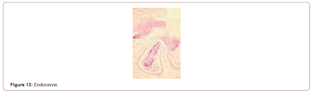
Uterine cancer: Currently there are no screening tests for uterine cancers except for maybe the Pap smear wherein a uterine cancer could be detected by chance; but it is usually advanced when the cancer cells shed from endometrium are detected in Pap smear. Expression of Gal-GalNAc with high sensitivity (91.7%) was observed in 12 cases of uterine endometrial adenocarcinoma [17]. In all three cases where endocervical tissues was available, the endocervical mucus was positive for Gal- GalNAc. The photomicrograph (Figure 13) shows histological section of endocervix of a patient with endometrial adenocarcinoma; magenta coloration of the mucus in the endocervical glands and in endocervix is evident. Again, this supports the Field-Effect of carcinogenesis. More importantly, this makes it a simple, convenient, and non-invasive method of screening for endometrial adenocarcinoma while a gynecologist routinely collects samples for Pap smear (Figure13).
irispublishers-openaccess-cancer-research-clinical-imaging

irispublishers-openaccess-cancer-research-clinical-imaging

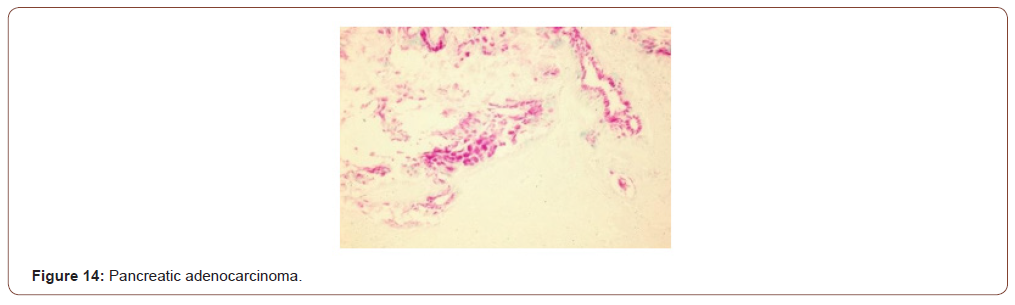
Pancreatic cancer: Pancreatic cancer is one of the deadliest of all cancers, primarily because of its silent inception; and owing to its location, signs and symptoms do not show till very late. All 6 of 6 cases of adenocarcinoma of pancreas were positive for Gal- GalNAc, and the normal ducts away from the cancers also showed positive reaction [17]. Thus, GOS test could be performed on fluid samples including pancreatic juice obtained during ERCP (endoscopic retrograde cholangiopancreatography) thereby providing additional value to the screening of the cancer. Figure 14 shows an adenocarcinoma of the pancreas with magenta color in the cancerous ducts and glands. (Figure14).

How easily can the marker be measured?

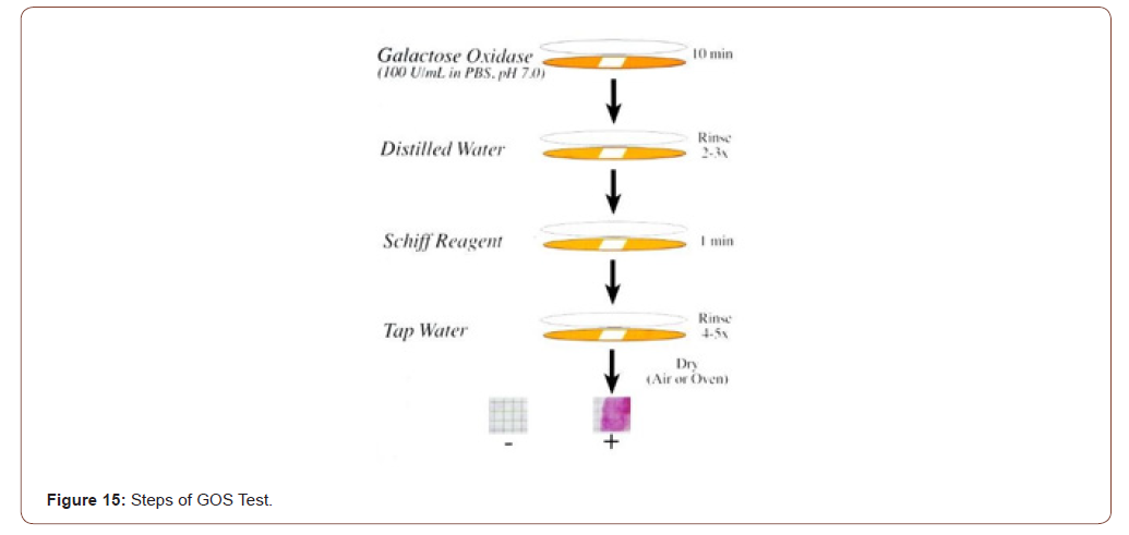
Answer: GOS test for detection of the marker is non-invasive done on mucus sample obtained during routine digital rectal examination, coughed-up sputum, nipple aspirate, prostatic massage secretion, endocervical mucus etc., and the entire assay period is ~15 min. This is the only point-of-care test for breast, lung, colon, prostate, and uterus; the results are available before the individual is ready to leave the doctor’s office. The test sample is placed on a special paper and reacted with galactose oxidase for 10 minutes, rinsed with distilled water, reacted with Schiff’s basic fuchsin for 1 minute, washed with tap water and dried. A pinkmagenta- purple color is positive, no color is negative (Figure 15). Positive indicates the presence of the marker Gal-GalNAc which is correlated with the presence of precancer, precancer or a high-risk thereof (Figure15).
irispublishers-openaccess-cancer-research-clinical-imaging

irispublishers-openaccess-cancer-research-clinical-imaging

Note: Though desirable, quantitative evaluation of the color reaction is neither practical nor meaningful as studies have shown no correlation between the intensity of color and the underlying disease.

Can the marker be modulated by chemo preventive agents?

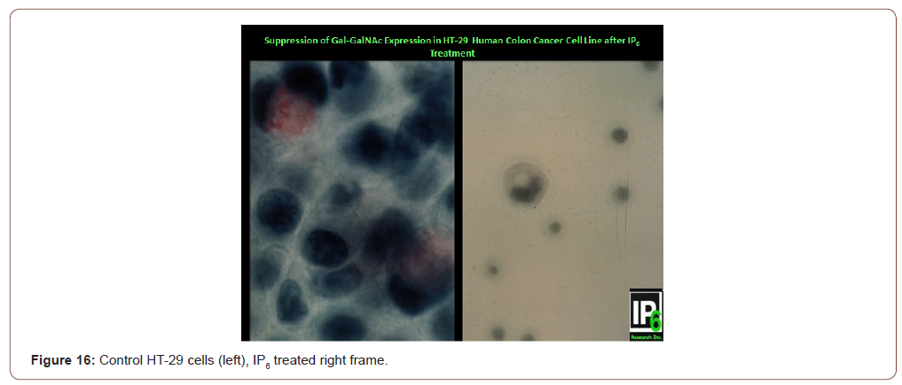
Answer: YES! Sakamoto et al. [23] and Yang & Shamsuddin [24] have demonstrated that; indeed Gal-GalNAc expression can be suppressed by the chemo preventive agent IP6 [25,26]. Figure 16 shows that HT-29 human colon cancer cells express the marker Gal- GalNAc (magenta color in mucus of cells in left panel). Following IP6 treatment, HT-29 cells terminally differentiate and produce mucin, yet not Gal-GalNAc, akin to normal goblet cells (right panel showing a differentiated HT-29 cell that has a mucus vacuole yet not expressing the marker [23,24]. Clinical studies are needed to validate this, but that would take a very long time. In this regard of particular importance is the study on colon cancer by Vucenik et al [13] where 32 of 53 (60%) samples collected from patients after tumor resection showed persistence of the biochemical change; 5 out of these 32 (16%) post-operative cases with positive GOS Test had tumor recurrence within a year. Thus, persistently positive test may serve as a predictor of tumor recurrence (Figure16).

Does modulation of the intermediate biomarker correlate with a decrease in cancer rate?

This would require a long time-years if not decades, and additional resources.

In conclusion, GOS test is a point-of-care screening test that is very simple, rapid, non-invasive, and inexpensive yet accurate for identifying asymptomatic people who may be at high risk of cancer or precancer of the lung, breast, colon, pancreas, prostate, and uterus. It is based on robust scientific background; researched and validated over three decades in >20 independent clinical studies in North America, Europe, and Asia. It exploits a disaccharide marker expressed early during carcinogenesis that persists in precancer and cancer and, satisfies all the practical rigid criteria set forth by NCI for an ideal marker. Given the fact that cancers of the lungs, breast, colon, prostate, and uterus comprise the most cancers, it is in the interest of public health that they are identified at a very early stage, or even before they are formed. The sooner it is used, the more lives could be saved from these cancers.

To read more about this article...Open access Journal of Advances in Cancer Research & Clinical Imaging

Please follow the URL to access more information about this article


To know more about our Journals...Iris Publishers

To know about Open Access Publishers

Tuesday, July 20, 2021

Iris Publishers- Open access Journal of Pediatrics & Neonatal Care | Prevalence of Different Types of Leukemia and Associated Factors among Children with Leukemia in Children’s Cancer Units at Al-Kuwait Hospital, Sana’a City: A Cross-Sectional Study


Authored by Hassan A Al- Shamahy*


Introduction

Leukemia is the most common type of cancer in childhood, accounting for 25 percent of all cancers that occur before the age of 20. There are two main types of childhood leukemia - acute lymphocytic leukemia (ALL), which accounts for about three quarters of leukemia, and acute myeloid leukemia (AML) accounting for most of the rest of the leukemia cases. ALL is a disease that affects about three quarters of leukemia. The hematopoietic tissue in the bone marrow is characterized by the overproduction of immature lymphocytes (a type of white blood cell). ALL occurs at all ages, from birth to adulthood, but the incidence peaks between 2 and 6 years of age. In the United States, there is a majority of white and male children and young adults with ALL. Improvements in treatment have led to remarkable gains in survival, estimated at 79 percent at 5 years. Acute myeloid leukemia is a cancer of the myeloid white blood cell line that occurs at all ages from childhood. The outcome for AML is poorer than for ALL, with a 5-year survival rate of 41 percent [1,2]. The precise cause of leukemia is not up till now obvious. Nevertheless, a lot of factors, mainly genetics, genetic mutations, epigenetic lesions, ionizing radiation, other chemical and occupational contacts, curative drugs, smoking and some viral agents, have been concerned in the development of leukemia [3-8]. Commonly 2 types of classification systems are used for leukemia: (the Franco-American and British classification system (FAB), which relies on morphology and cytochemical staining to identify specific types of leukemia, and the World Health Organization (WHO) that reviews classification information, cytomorphology, cell chemistry, immune profiling, cytogenetics and clinical features to identify and classify clinically significant disease entities [9,10]. Lymphoma malignancies correspond to a heterogeneous group of illnesses separated into four classes established on tumor cell maturity and disease distribution such as acute lymphocytic leukemia (ALL), chronic lymphocytic leukemia (CLL), lymphoma malignancies, plasma cell tumors and hairy cell leukemia [11].

Globally Leukemia involved 200,676 males and 151,289 females (with ASR 5.6 and 3.6, respectively). There were 46,449 males and 35,880 females diagnosed in 2012 with leukemia in Europe. In Australia, Asia and the USA about 233,451 residents were diagnosed with leukemia by 2017. In contrast, in Africa by 2012, it has a rate of incidence equal to 23,928 cases (ASR 3.0 per 100,000) [12-14]. Leukemia was the most common type of cancer among children (including 29% of 3,707 cancer cases in children, and acute leukemia counting 89% (91% of which were ALL and 9% AML) of all cases of leukemia in children [15].

In developing countries, the influence of leukemia is massive attributable to premature death of children, loss of parents, failure of productivity due to disability, and high medical costs affecting the social, economic and health well-being of the population [16- 18]. While leukemia is treated very well in the developed world, there is little evidence of the current state of the disease in Yemen in general and in the study area in particular. On the other hand, in Yemen as is the case in most Arab countries, there are few specialized epidemiological records devoted to this area, and for this reason it is important to encourage, update, build and continue to present studies on childhood leukemia with the goal of achieving greater impact on public health, with early diagnosis and appropriate treatment aimed at enhancing survival and minimizing potential consequences. According to the limited Yemeni Cancer Studies, the most common types of cancer among Yemeni children and adults were leukemia (33.1%), lymphoma (31.5%), central nervous system tumors (7.2%), and bone tumors (5.2%) [19- 21]. This cross-sectional study aims to determine the prevalence of different types of leukemia and the associated factors among children with leukemia in the pediatric cancer units of Al-Kuwait Hospital, Sana’a City.

Patients and Method

A cross-sectional study was conducted on children with leukemia who were treated selectively in the pediatric leukemia units of Al-Kuwait University Hospital in Sana’a. Group diagnostics and histopathological diagnoses were formed in line with the French, American and British classifications of leukemia in children in the pediatric leukemia units, over a period of 5 years from January 1, 2014 to December 31, 2018. Factors associated with become infected with leukemia that were studied included ages, sex, and outcomes. The association of death and recovery with different age groups and leukemia types was also studied through rates and calculation of OR, CI, X2 and p values through probability tables (2x2 tables).

Statistical Analysis

Data were recorded using appropriate descriptive statistics (including frequency, mean, and standard deviation). The odds ratio (OR) was used to determine the strength of the association between two events, such as leukemia, age, gender, and residence. The association between death, age, and type of leukemia. In addition to calculating the relationship between cure, age of patients, and types of leukemia. The two events in the current study were independent if and only if OR was equal to 1. For sample constraints of odds ratio in small numbers (less than 5), Fisher’s exact test was used as an alternative estimator for the association between events in the current study.

Ethical Approval

Ethical approval was obtained from the Medical Research & Ethics Committee of the Faculty of Medicine and Health Sciences, Sana’a University. All data, including patient identification were kept confidential.

Results

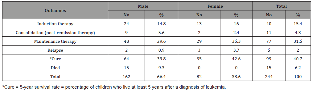
Table 1 shows the age and gender distribution of children with childhood leukemia in Sana’a, Yemen. The mean ± SD age of all cases was 6.44 ± 3.7 years. Most of the cases were in the age group 1-5 years (50%), followed by the age group 6-10 years (32.1%), while only 17.9% of the cases were in the age group 11-15 years (disease decreases with increasing age). As for gender, most of the cases were males (66.7%), while the percentage of females was 33.3% (male to female ratio = 2-1). Table 2 shows leukemia outcomes among children suffering from childhood leukemia in Sana’a, Yemen. The cure rate was 40.7% while the death rate was 15 cases (6.2%), all of them male (male mortality rate = 9.3%). The relapse rate was 2%. The rest of the cases were in maintenance therapy (31.5%), induction therapy (15.4%), and consolidation (post-remission therapy) for 4.3% of cases. Table 3 shows the age-group association of death among children with childhood leukemia in Sana’a, Yemen. The highest mortality occurred in the 6-10 year age group (8/78; 10.2%), with an associated OR = 2.6, CI = 1-7.4, X2 = 3.4, p = 0.06. However, the low mortality rate was 3.3% in the 1-5 year group, without association (OR = 0.34, p = 0.06). Also, in the 11--15 yearold group, the death rate was 6.8% roughly similar to the overall death rate (6.1%).

Table 1:Age and gender distribution of children with childhood leukemia in Sana’a, Yemen.

irispublishers-openaccess-pediatrics-neonatal-care


Table 2:Leukemia outcomes among children suffering from childhood leukemia in Sana’a, Yemen.

irispublishers-openaccess-pediatrics-neonatal-care


Table 3:The association of death with age groups among children suffering from childhood leukemia in Sana’a, Yemen.

irispublishers-openaccess-pediatrics-neonatal-care


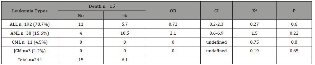
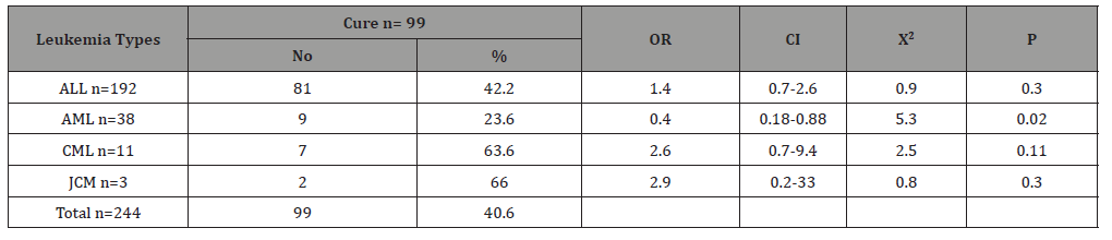
Table 4 shows the type of leukemia association of death among children with childhood leukemia in Sana’a, Yemen. Acute lymphoblastic leukemia was the most common, accounting for 78.7% of the total, while the other types were less common, with acute myelogenous leukemia count of 15.6%, chronic myelogenous leukemia at 4.5% and Juvenile myelomonocytic leukemia only at 1.2. %. The highest mortality occurred in the AML (4/38; 10.5%), with an associated OR = 2.1, CI = 0.6 – 6.9, X2 = 1.5, p = 0.22. However, the mortality rate was 5.7% in the ALL patients, without association (OR = 0.72, p = 0.6).There was no death occurred in CML and JCM cases (0%). Table 5 shows the age-group association of cure among children with childhood leukemia in Sana’a, Yemen. The cure rates were roughly similar in the different age groups. It is ranged from 38.5% in 1-5 year group to 43.2% in 11-15 year group. Table 6 shows the type of leukemia association of cure among children with childhood leukemia in Sana’a, Yemen. The highest cure rate occurred in the JCM (2/3; 66%), with an associated OR = 2.9, CI = 0.2 – 33, X2 = 0.8, p = 0.3. The second high cure rate occurred in the CML (7/11; 63.6%), with an associated OR = 2.6, CI = 0.7 – 9.4, X2 = 2.5, p = 0.11. However, the cure rate was 23.6% in the AML patients, without association (OR = 0.4, p = 0.02). The cure rate was 42.2% in the ALL patients, with association (OR = 1.4, p = 0.3).

Table 4:The prevalence and association of death with type of leukemia among children suffering from childhood leukemia in Sana’a, Yemen.

irispublishers-openaccess-pediatrics-neonatal-care


Table 5:The association of cure with age groups among children suffering from childhood leukemia in Sana’a, Yemen.

irispublishers-openaccess-pediatrics-neonatal-care


Table 6:The association of cure with type of leukemia among children suffering from childhood leukemia in Sana’a, Yemen.

irispublishers-openaccess-pediatrics-neonatal-care


Discussion

Information of the prevalence of leukemia in a population may envisage pathogenic hypotheses for disease control and aid in the effective management of leukemia and other malignant hematomas. In developing countries, and especially in Yemen, there is little information about the burden and patterns of haematological malignancies, especially leukemia. In the current study, with regard to gender, most of the cases were male (66.7%) while the percentage of females was 33.3% (male to female ratio = 2:1). This finding differs from that reported in Africa where the ratio of males to females is roughly equal, although females dominate slightly (1: 1.06) [22], but similar to that reported from the United States where the American Cancer Society estimates for leukemia in 2021, about 5,690 new cases, 3,000 in males and 2,690 in females [23]. The current results of different leukemia prevalence rates among gender are in line with the facts that the prevalence of leukemia must be varies according to gender due to the biological factors [11,15,24,25].

Leukemia may appear at all ages, from newborns to the elderly, but the distinctive forms have different age distributions [26]. In the current study, the mean age of ± SD for all cases was 6.44 ± 3.7 years and most of the cases were in the age group 1-5 years (50%), followed by the age group 6-10 years (32.1%), while only 17.9% of the cases were in the age group 11-15 years (Table 1). This is similar to what has been reported elsewhere for pediatric leukemia where the mean age ± SD of pediatric leukemia cases was 6.0 years with a peak incidence at 6-10 years [2,12,14]. This differs from the leukemia hypothesis with age in which older children may develop leukemia more frequently than younger children due to advancing age, as many environmental exposures to carcinogens, irradiation, and malignant mutations due to clonal expansion occur more often [27,28]. However, most of the younger children in the current study could be explained by the fact that prenatal and early life exposure is thought to be important determinants of childhood leukemia. Several mechanisms have been identified through which external and internal factors can influence the risk of developing leukemia in children. Exposure to a carcinogen or toxic substance early in a female’s life may cause permanent damage. Since no new oocytes are formed after birth and their maturation begins during pregnancy, the exposure that occurs during this critical time can be of great importance. During pregnancy, exposure to agents such as ionizing radiation may act directly while others may act indirectly by transporting the placenta. On the other hand, offspring may be exposed after birth to environmental exposure, either directly or indirectly [29]. Since most of the children are from rural areas, they may have been exposed to various environmental exposures during their stay with their parents who are farmers.

Environmental factors, even though not well articulated, influence the chance of developing leukemia. In this study, the highest proportion of 68.9% out of the total of 244 patients diagnosed with leukemia was observed among rural residents (p < 0.05). In Yemen, rural residents’ lifestyle is based on agricultural activities such as farming and plantations agriculture; especially Gat, fruits and vegetables plantation are the major practice around the study area, thus this may lead to the repeated use of chemicals such as pesticides, herbicides, and fertilizers for agricultural activities which will result in genetic mutations conferring leukemia [30].

Leukemia types were determined using the FAB classification method [10,11], Wright-stained morphological examination, and cytochemical staining with Sudan black B staining to differentiate the cell lineage. In this study, acute lymphocytic leukemia was the most common, accounting for 78.7% of the total, while the other types were less common, with acute myelogenous leukemia count of 15.6%, chronic myeloid leukemia 4.5%, and juvenile myeloid leukemia at only 1.2%. This result was consistent with results from Ethiopia, Nepal, and Pakistan [22,31], while it was contradictory with a study from Albania [32]. In the current study, the highest mortality occurred in the 6-10 year age group (8/78; 10.2%), with an associated OR = 2.6, CI = 1-7.4, X2 = 3.4, p = 0.06. However, the low mortality rate was 3.3% in the 1-5 year group, without correlation (OR = 0.34, p = 0.06). These results are consistent with the American Cancer Society’s fact that most cases of leukemia occur in young children, but most deaths occur in older children. Very young children may perform better than older children due to differences in the nature of leukemia in children, differences in treatment (often young children’s bodies can handle aggressive treatment better than older children), or a combination of these [23]. The highest mortality occurred in AML (4/38; 10.5%), cure rate = 23.6%. However, the mortality rate was 5.7% in ALL patients, with cure rate = 42.2%. These results are in line with findings by the Leukemia and Lymphoma Society of the USA where the 5-year survival rate for children and adolescents under the age of 15 years diagnosed with ALL was 91.8% in the United States between 2007 and 2013. While the survival rate for children under 15 years of age with AML was only 66.4% for the same period [33].

Conclusion

ALL is the most common type of leukemia in Sana’a city; and males and young children are affected the most by leukemia. In the current study there was an association between leukemia and younger age group, with males. There was an association between high mortality and the 6-10 year age group, with AML. Also, there was no association between ages and cure rate, but a high cure rate occurred with JCM and CML. More comprehensive investigations of relevant factors and predictors are needed with more modern diagnostic methods and investigate correlation factors with the treatment protocols used.


To read more about this article...Open access Journal of Pediatrics & Neonatal Care

Please follow the URL to access more information about this article

https://irispublishers.com/gjpnc/fulltext/prevalence-of-different-types-of-leukemia-and-associated-factors-among-children.ID.000569.php

To know more about our Journals...Iris Publishers

To know about Open Access Publishers

Monday, July 19, 2021

Iris Publishers- Open access Journal of Otolaryngology and Rhinology | Endoscopic Transnasal Resection of Adenoid Cystic Carcinoma of the Nasal Septum: A Case Report and Literature Review


Authored by Tessei Kuruma*,

Introduction

Adenoid cystic carcinoma (ACC) is the most common tumor of salivary gland tissues in the head and neck area and the second most common tumor in the nose and sinuses [1,2]. Sinonasal ACC accounts for 10–25% of all head and neck ACCs [3]. However, this tumor rarely develops in the nasal septum. In fact, very few cases of ACC in the nasal septum have been reported to date [4-6]. Sinonasal tumors are often asymptomatic and are frequently associated with inflammatory disease, leading to delayed diagnosis. The clinical features of ACC differ from those of squamous cell carcinoma, and the features must be recognized for diagnosis and treatment. We report a case of ACC that occurred above the anterior septum.

Case Presentation

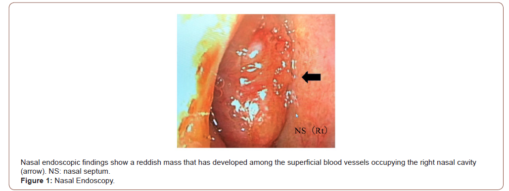
A 61-year-old woman presented to a local otolaryngologist with a 1-year history of right epistaxis. A mass was identified in the right nasal cavity and she was referred to our department for further examination. Nasal endoscopic findings on the first visit revealed a reddish mass that had developed among superficial blood vessels occupying the right nasal cavity (Figure 1). The tumor readily bled simply by inserting gauze into the nasal cavity under local anesthesia for biopsy. Judging from the surface properties of the tumor, it was considered to show abundant blood flow.

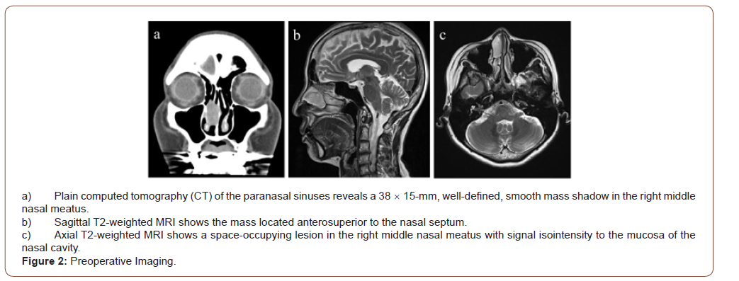
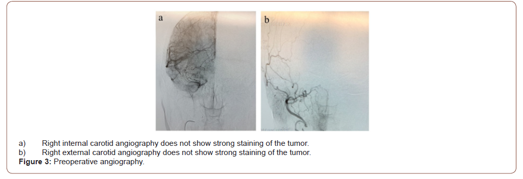
Sinus Computed Tomography (CT) showed a 38  15mm, welldefined, smooth mass shadow in the right middle nasal meatus. The mass excluded the nasal septum but showed no infiltration (Figure 2a). In sinus magnetic resonance imaging (MRI), T2-weighted image, a space-occupying lesion was found in the middle of the right nasal meatus, appearing signal isointense to the mucosa of the nasal cavity. The tumor was in wide contact with the inferior turbinate, but the boundary was clear (Figure 2b,c). The patient had a history of asthma and was unable to undergo diagnostic imaging using contrast media. However, because the mass was considered to have a rich blood supply, we consulted with a radiologist about the utility of angiography. We decided to perform angiography while paying close attention to potential allergies to contrast media. Angiography was performed from the internal carotid artery and external carotid artery, but no deep tumor staining was observed (Figure 3a,b). From this result, vascular tumors were ruled out. Biopsy of the tumor was performed again at an outpatient clinic. At the time of tumor biopsy, the tumor was squeezed outward with a detacher, and it was found that the tumor originated from the nasal septum. The biopsy was performed under a rigid microscope with an assistant sucking blood from the biopsy site. As a result, bipolar ablation and bleeding could be stopped in a short time.

irispublishers-openaccess-textile-science-fashion

Whole-body positron emission tomography (PET)/CT showed no integrated images of cervical and distant metastases other than the nasal cavity. Right nasal ACC (T1N0M0) was diagnosed. The surgery was performed endoscopically under general anesthesia. Since the tumor was located in front of and above the nasal septum, the nasal wing was lifted with a hook to improve the field of view of the endoscope.

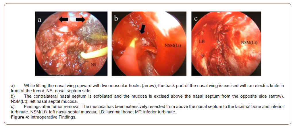
The mucosa of the nasal septum above the tumor was confirmed endoscopically, and an arc was incised upward from the nasal septum above the tumor, then the lacrimal bone and anterior surface of the inferior concha were resected with an electric knife (Figure 4a). In addition, the nasal septal mucosa and nasal septal cartilage were incised while maintaining a sufficient safety margin in front of the tumor, and the contralateral nasal septal perichondrium was peeled off. When the inferior and posterior nasal septum mucosa had been sufficiently exfoliated, the nasal septum and lamina perpendicularis of the ethmoid bone were exfoliated, and excision was performed from the opposite side toward the tumor-bearing surface with a safety margin (Figure 4b). The tumor was removed from the olfactory fissure site together with part of the mucosa of the lacrimal bone and middle and inferior conchae (Figure 4c). Rapid intraoperative pathology confirmed the absence of residual tumor, especially in the upper mucosa.

irispublishers-openaccess-textile-science-fashion

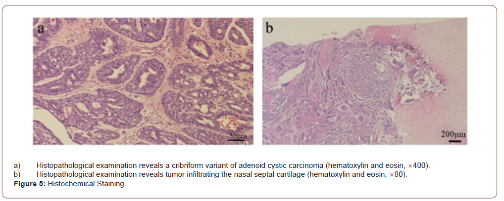
Postoperative pathological examination revealed cribriformtype ACC. The tumor was 30  14mm in size and showed a biphasic structure of myoepithelial cells and glandular epithelial cells, forming a glandular cavity and growing in a cribriform pattern (Figure 5a). The tumor was found to be infiltrating the nasal septal cartilage but showed no clear perineural or vascular infiltration. The margins and depths of the resected tumor were negative. Based on the results of pathological examination and consultation at a conference with a radiologist, postoperative radiotherapy was considered unnecessary (Figure 5b).

irispublishers-openaccess-textile-science-fashion

Local findings in the nose were observed every month for 1 year after surgery, and were evaluated by CT of the neck, chest and abdomen every 6 months to thoroughly check for local recurrences and distant metastases. From 1 year postoperatively, medical examinations were performed once every 2 months. As of 3 years since the surgery, the patient has shown no local recurrences or metastases of the tumor.

Discussion

ACC is of epithelial origin and shows a high frequency of local recurrence and distant hematogenous metastatic spread to the lungs, bone, and liver, although lymphatic spread is rare [7]. ACC is the second most common tumor in the nasal cavity and paranasal sinuses after squamous cell carcinoma [1,8]. ACC in the nasal cavity and paranasal sinuses origin often shows a worse prognosis than those in other areas of the head and neck. However, nasal cavity tumors have been found to have better outcomes than sites in the paranasal sinus.

ACC is most commonly an asymptomatic, slowly expanding mass, often found in the head and neck [6]. However, nasal ACC may manifest as nasal congestion, epistaxis, secretions, and facial swelling [9,10,4]. Pain and paresthesia may also be reported, potentially reflecting a tendency of the tumor toward perineural infiltration [6]. ACC originating from the sinuses is generally asymptomatic and can often be found in a fairly advanced state. However, as in the present case, nasal septal ACC may be more likely to be detected early because symptoms may appear early in the disease course [10].

Local findings in the nasal cavity alone do not allow easy determination of the base and extent of tumor extension, and diagnostic imaging with CT and MRI is very helpful in assessing tumor characteristics and enlargement. In particular, when selecting a surgical method (endoscopic surgery, skull base surgery or removal by external incision), the presence of tumor infiltration along the cranial nerves extending to the skull base is an important consideration. Low-grade ACC in the paranasal sinuses may mimic the characteristics of simple polyps that fill the nasal cavity and show slight bone remodeling on both CT and MRI, while high-grade ACC can manifest as a large, irregular mass with bone destruction on both CT and MRI [11]. Contrast-enhanced imaging is necessary to confirm tumor infiltration into the skull along the cranial nerves and to clarify whether the tumor has abundant blood flow. In the present case, contrast-enhanced MRI could not be performed due to bronchial asthma. However, angiography completely ruled out vascular tumors, and preoperative biopsy was able to be performed safely.

Tissue biopsy is required for definitive diagnosis. However, malignant tumors such as this tumor often bleed. When a tumor develops in the nasal cavity, sinusitis often occurs as a secondary effect, so bleeding from the nasal mucosa is also increased. Since the nasal cavity is narrow, firm contraction of the inside of the nasal cavity with adrenaline gauze is necessary, along with confirmation of the site for the base of the tumor. Furthermore, properties of the tumor surface need to be confirmed under endoscopy and a solid sample must be obtained from the tumor body. In outpatient biopsy, profuse bleeding may prevent collection of sufficient samples. Biopsy can be completed within a short time by cauterizing and stopping bleeding while having an assistant perform blood suction.

Perzin, et al. [12] examined 62 cases of salivary gland ACC. Histologically, they classified three growth patterns: solid, cribriform, and tubular. The cribriform pattern is the most common histological subtype [12]. All nasal septal ACCs reported so far, including this case, have been this type [5,6,13]. Perzin et al. [12] also examined tissue type and prognosis. Five-year recurrence rates for solid, cribriform, and tubular-pattern tumors have been reported as 100%, 89%, and 59%, with average survivals of 5, 8, and 9 years, respectively [1]. Patients with solid ACC display the worst prognosis [12].

Conventionally, among the histological types of adenoid cystic carcinoma, the tubular type has been considered to represent the most differentiated type, and the solid type has been considered to represent the least differentiated type [12,14]. Yamamoto, et al. [15]. suggested that ACC changes from well-differentiated to poorly differentiated over time, resulting in increased tumor growth potential and the possibility of recurrence and metastasis [15]. Szanto, et al. [16] subdivided ACC into three groups: Grade 1, solid pattern 0%; Grade 2, solid pattern >0% but <30%, and Grade 3, solid pattern ≥30%, depending on the mix of ACC tissue types. They reported 5-year survival rates of 92%, 65%, and 14%, respectively, and 10-year survival rates of 76%, 26%, and 5%, respectively. A detailed pathological assessment of the proportion of solid pattern thus has important prognostic value in ACC [16].

Surgical excision is the modality of choice for the treatment of nasal septal malignancies. The surgical approach is modified for the individual tumor according to the size and location of the tumor. Recently, some authors have deemed surgical resection of ACC of the sinonasal tract via an endoscopic approach as an effective, minimally invasive method that in well-selected patients could achieve satisfactory application, and together with adjuvant therapies providing good results, complete removal of the tumor and a high survival rate [17].

Surgery for malignant tumor of the nasal septum requires consideration of the most suitable method for removing the tumor with a sufficient safety margin. Endoscopic surgery is superior to external incision and intracranial surgery in terms of esthetic outcomes and side effects. However, limits exist to the indications for endoscopic surgery. According to Stammberger, et al. [18] the limitations of the endoscopic technique result from the anatomical spread of the tumor, when there is extensive infiltration of the orbit, dura/brain, and other vital structures [18]. The key anatomical structures include the internal carotid artery, optic nerve, cavernous sinuses, and ethmoid roof [19].

Most researchers reserve the endonasal method for experienced surgeons and consider the possibility of its application to depend on TNM staging [18]. According to Hanna, et al. [20] endoscopically treated tumors are usually of lower stage, i.e., T1- T2 [20]. They emphasized the necessity of cooperation between the endorhinolaryngologist and neurosurgeon [18]. Our hospital has some specialists in skull base surgery and ocular plastic surgery, so we consult with them regarding tumors near the orbit or skull base and decide on the surgical method without sticking to endoscopic surgery. The combined use of an endoscope is also useful for external incision and intracranial surgery. Substantial developments in endoscopy have been made in recent years, and improvements in image quality have made it possible to ensure that endoscopy provides a much safer margin than tumor removal under the naked eye.

In endoscopic surgery, if the field of view deteriorates with bleeding, etc., or if the expectation is that the surgical field cannot be expanded well due to the narrow working space in the nasal cavity, an auxiliary external incision will be required. For anterior nasal septal tumors, lateral rhinotomy is the preferred surgical approach, whereas sublabial incision with Denkers’s approach is considered more appropriate for posterior nasal tumors [10].

The primary lesion in this case was early-stage T1. However, because the tumor was well anterior and superior to the septum, the point was whether the upper part of the nasal septum above the tumor and the back surface of the ala of the nose could be clearly seen. The operation was performed by one assistant lifting the nasal wings with a hook and another assistant suctioning blood or cauterizing with an electric scalpel. By performing endoscopic surgery using these 4-5 hands, the area around the tumor was clearly visible. The extent of tumor progression and the surgical procedure could be shared by all surgeons at the preoperative conference.

ACC is considered to be radiosensitive, but not radiocurable, so the rationale for administering postoperative radiation therapy is to treat any residual microscopic disease, which is present in most patients [21]. Postoperative radiation is now generally recommended. However, the benefit of adjuvant radiotherapy has never been clearly demonstrated in the absence of a randomized prospective trial [22-24].

A meta-analysis by Amit, et al. [25] revealed margin status and tumor site as significant predictors of outcome in ACC patients with tumor in the sinuses and skull base, whereas perineural infiltration was not associated with prognosis. As an important result, Amit, et al. [25] identified no additional benefits from adjuvant therapy [25].

As surgery of the primary site was the only demonstrated clinical intervention noted to confer survival benefit on patients diagnosed with ACC in the head and neck region, evidence supports surgical therapy remaining as the mainstay of therapy, at least for now. Therefore, in the treatment of ACC of the nasal septum, removal with a sufficient safety margin is of primary importance. Postoperative radiotherapy is required if the surgical margin of the tumor appears microscopically positive.

Conclusion

We encountered a 61-year-old woman with ACC of the nasal septum. Endoscopic removal surgery was performed. Since the tumor was located above the anterior septum, 4- or 5-hand endoscopic surgery could be performed and the tumor was removed with a sufficient safety margin, even in the narrow surgical field. Following thorough examination with the radiology department, postoperative radiotherapy was not performed. When performing nasal septal surgery for ACC, the extent of lesion extension should be grasped by local and imaging findings, and a method for tumor removal with a sufficient safety margin should be devised. ACC shows a high incidence of local recurrence or distant metastasis, so careful, long-term follow-up is warranted.

To read more about this article....Open access Journal of Otolaryngology and Rhinology

Please follow the URL to access more information about this article

https://irispublishers.com/ojor/fulltext/endoscopic-transnasal-resection-of-adenoid-cystic-carcinoma-of-the-nasal-septum.ID.000600.php

To know more about our Journals...Iris Publishers

To know about Open Access Publishers

Iris Publishers-Open access Journal of Hydrology & Meteorology | Influence of Community Resilience to Flood Risk and Coping Strategies in Bayelsa State, Southern Nigeria

  Authored by  Nwankwoala HO *, Abstract This study is aimed at assessing the influence of community resilience to flood risk and coping str...