Tuesday, January 30, 2024

Iris Publishers-Open access Journal of Neurology & Neuroscience | Kuru: The Neurodegenerative Disorder; Roles of PrPC and PrPSc in infection, Effects in Blocking Neural Synapses and its Comprehensive Observation


Authored by Ahnaf Ilman*,

Introduction

Professor Luisetto M et al. introduced that KURU, the first human transmissible spongiform encephalopathies, can be penetrated in humans. And can impact the brain and immune system by the primary process of the oral intake of affected materials by cannibalising. Also revealed that no boundaries of the genome had not been determined and added that the Kuru Disease efficient process works explicitly against the Brain and immune system as well as creates a fatal neurodegenerative disorder [1].

Methods & Materials

• Observation of the risk factors and impacts on the human body caused by KURU represented through the vigour of research articles.

• What are the must to alleviate the criticisms and analysis of pathogenesis?

• References have been referred based on the information collected from reliable and high impact journals, videos of related lectures by prominent professors of the world.

Results & Discussion

Background: Dr Collinge J. recently introduced that possibly, the disease and incubation somehow sign any future epidemic. Besides, oral intake is the principal factor of infection. It is regarded as a prion disease. The appearance of Kuru indicates the assumption of a disease which occurs rarely. Cannibalism can be the wherewithal to get rid of the prion disease infection [2]. Background: Dr Collinge J., et al. represented that the neurological and genetic analysis on some Kuru infected enlightened one person having Kuru infection in the tonsil biopsy. In the end, this relevance has created a firm belief that it is positively related to oral as a medium of disease [3].

Background: Dr Collinge J., et al. revealed that the incurable and neurodegenerative disorder created by Kuru disease attacks in the brain system. It is a disease caused by prions. Three echelons of Kuru infection - (i) Ambulant Stage, (ii) Sedentary Stage, and the most criticism, and the last stage is considered as (iii) Terminal Stage. The ambulant echelon represents the disorder in operating body organs, and synchronisation in speech can be found while speaking. Then, in the step of Sedentary, jerking in body, bursting out with laughter, and mental disabilities have been analysed. Besides, the infected become unable and cannot do any work without the support of anyone. Finally, in the last stage- Terminal Stage, infected become more incompetent, and other symptoms and orders turn more synchronised. Kuru disease was first identified in the tribal society of Papua New Guinea during the 1950s. As a disease which is rare and transmits positively from one to another, it transferred to several people. And sex variation analysis mentioned that the chance of the women and the children is very high because of cannibalism and getting a heightened touch to the corpses which can be attacked with Kuru [4].

Dr Liberski, PP. introduced that Kuru, the first transmitted prion disease had been manifested from Chimpanzees to humans. It has been considered acting as a neurodegenerative disease. Neurodegenerative disease is regarded as an illness in the human brain that mainly attacks neurons or creates neuron criticism. Neurons combination builds the nervous system- brain and spinal cord. Cannibalism is taken as a medium of infection [5].

Dr Kompoliti, K. mentioned that this very neurodegenerative disease infects the human body by an unconventional medium that acts against DeoxyRiboNucleic Acid and RiboNucleic Acid. And it has been created from the usual precursor protein and Cross Beta Pleated contamination. Since it is a brain system infecting disease, its presence in the very tiny neurons can be observed by only the microscopic system. One of the most common and fearful signs and symptoms alongside the risk factor of this disease is a disorder in regular movements. But soon after, it turns into a high fatal neurological disorder. It means criticism and synchronisation in the brain and the immune system. Consequently, brain and movement-related problems like coma and inabilities in activities are analysed. Women and children are having a high chance to get an infection and can have highly infected brain tissue through the following cannibalism in the regions of Papua New Guinea. Besides, they highly get contact with the Kuru infected- people and even animals. In some areas, ritual-like endocannibalism is a must to be followed by them as their culture, so like their culture, they consume the dead body of Kuru infected or non-infected. And this can be probably considered as the principal wherewithal transmission although a hypothesis found that people who don’t follow cannibalism, can get infected by Kuru Disease [6].

Ferguson-Smith, MA. and his study recently introduced that at the beginning of the disease transmission in 1957, 5%-10% infected patients were found in some regions. His study revealed that negative-fastness in movement, shivering while working had been effectively recognised. Alongside problems had been found while they talk- sometimes get muted and sometimes speak unsteadily. In the critical stage, patients sometimes go into a coma. Death took place within 1year of this disease. For consuming, preparing the fleshes, brains or other tissues of estimated cannibalising material; women do these processes. That’s why getting in touch with fluid of the materials, and they have a high chance of infection. And several studies revealed that the percentage of women is more than that of men. Besides, men do not involve themselves in this process. That’s a very relevant evidence of this study [7].

Structural Protein Effects

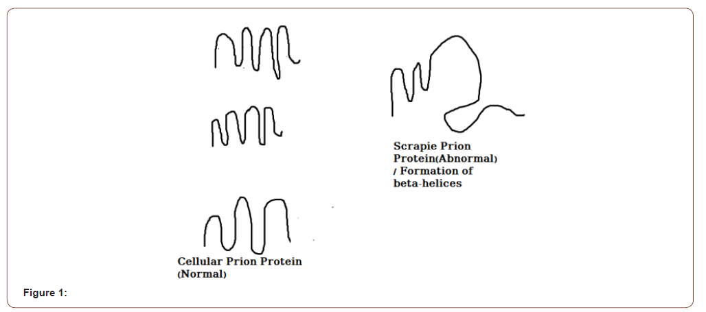
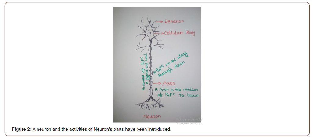
In the 20th century, researchers discovered that any disease could be caused by protein and the protein that causes disease-most often neurodegenerative illness, disorder and other diseases is called Prion Protein; in short PrP. PrP has two effective forms- PrPC that is called Cellular Prion Protein, and the other one is- PrPSc, which is called Scrapie Prion Protein [8]. In the chromosome number 20 of the human body, PRNP bears a protein called PrP [9]. PrPC is the normal protein and also found in heal the human body; it is formed as Alpha-helices form [10]. But because of the amino-acid formation and polypeptides, Alpha-helices of PrPC transform into PrPSc and beta sheets from the transforming receptacle named Molten Globule [11]. PrPSc is an abnormal and unstable phase that plays a role in mutation of PrP. PrPSc impacts on the standard and cellular prion protein-PrPC and it turns into PrPSc and creates a beta-sheet that is abnormal and misfolded/improperly folded [12]. The misfolded or PrPSc effects on the Central Nervous System and final cause is the failure of activities in the brain [13]. The centrum of the mind is Neuron. Neuron is consistent with axon, dendron, cellular body. Combination of Axon and Dendron features Neural Synapses. Due to the misfolded PrPSc, Neural Synapses get blocked and heal cells start to be deceased and damaged [14]. Microbials soon begin to kill the damaged cells, and the spongiform phase gets created.

Finally, death occurs by average 1year onset of the disease infection [15] (Figure 1).

In the above-added Figure: 01, two types of Prion Protein- PrPC and PrP Sc have been introduced by several signs and graphical work. In the figure, the regular phase of Cellular Prion Protein and abnormal phase of Scrapie Prion Protein have been accurately figured out with the accurate analysis and study on Alpha-helices and Beta-sheets. As beta-sheets belonged to an improper phase of protein, it contains a complicated gene-coding cycle that acts for mutation. And be ta-sheets containing Scrapie Prion Protein inspire other Alpha Helices normal prion protein. And if the appearance of one PrPSc is found in the human body, it transforms into many and many heal PrPC, and the brain and immune system get positively affected (Figure 2). A neuron is consistent with a cellular body, axon and dendron. PrPC or Cellular Prion Protein generally remains in the human body that does not play any dangerous role in the case of disease production. It has a connectivity to the human brain and its neurons. Cellular Prion Protein moves to the Neurons and the human brain through the Axon and axonal transport. So, axon is considered as the media [16]. Actually, PrPC plays a role in the comprehensive management and activities of the brain, body and immune system. If there remains any misfolded Prion Protein anyhow, that can cause life hampering disease.
irispublishers-openaccess-neurology-neuroscience


irispublishers-openaccess-neurology-neuroscience


Transmission

Kuru is a prion disease that is transmitted by practising cannibalism and consuming affected human brain [17] Or no other natural criticism and complications can be acknowledged as the wherewithal of the transmission of the protein-based disease. Principally, improperly appeared protein is the responsible because, through experiment, it has been revealed that the misfolded protein-based PrPSc blocks the neural synapses. Finally, it impacts the brain and immune system and causes death. Thus, kuru disease occurs and effects [18]. Besides, PrPSc contains scrapie and scrapie is positively related to the Transmissible Spongiform Encephalopathies [19]. In the 19 mid’s, it was revealed that Kuru is the first human prion disease transmitted to the Chimpanzees. For this reason, it was proved that the principal cause of transmission of Kuru is allowing and practising Cannibalism [20]. Later, cannibalistic people were found having the prion disease. And from then, Kuru started to transmit to humans. Finally, in 2004, when the government of Papua New Guinea and other infected regions headed to avoid the practice, the cases of Kuru were declined and discarded [21]. Kuru is under the section of Infectious TSE-Transmissible Spongiform Encephalopathies that effectively affects the brain and immune system. TSE is formed from Infectious, Sporadic and Familial features. Besides, Kuru cannot be defined in Sporadic and Familial Sections. As Kuru can be transmitted, it has been remarked under the Infectious TSE or Transmissible Spongiform Encephalopathies. After studies introduced that the patients who were infected in and died of Kuru Disease, they anyhow got contact with the brain and materials got contacted with PRPN. The pathophysiology of Kuru disease is quite unimaginable since it extracts growth hormones and central nervous system [22].

Death and Affected Analyses with Epidemiology

The cases of Kuru features fatality and left untreated for those patients who were infected because there was neither any proper treatment nor any kind of reliable and recommended medication. In the 20th Century, precisely in 1957-1959, frequency of cases of neurodegenerative disease-Kuru was about 2700, and mostly 200 infected died because of the lack of treatment [22]. A study by Alpers, MP., et al. revealed that during 1987-95, 66people died of being attacked by the neurodegenerative disorder creating Kuru Disease; in which 49 people were in female sex, and the rest 17 were male. Every year, about 3-12 people died from it. Including that in 1987, a one-year-old person and 1991, a 30 years person died from the suspected epidemic creating disease. In one study, the incubation period of the disease and its infection was introduced 30 years and here on this study, ranging to 1995, showed 35 years. The study by Alpers, MP., et al. also introduced that in 1987, overall 12 people died from the Kuru in which eight people were females and 4 were males. People ranging from 50-59 & 30-39 died most- 10(5people+ 5people=>50-59ages + 30-39ages) in number then in 1987. And 1death was found below 20, and ranging 20-29, 1 person died. In 1988, 8 people died; 7 were females, and 1 was male. In that year also, people aging 30-39 and 50-59 were mostly infected- 6people (3+3=>30-39+50-59). In 60-69 and 40-49, 2 deaths were found one by one in every range. In 1989, 11 patients died while female and male in number were respectively 6, 5. But in 1989, ages from 40 to 49 suffered most considering the death- 4 in number. For the people of 30-39 and 50-59 ages, death cases were respectively 3,3. And also in the year, among people aged 60-69, 1 death was caused. In 1990, death cases started to decline, occurring nine deaths; in which seven were female and 2 were male. People ageing from 40 to 49 faced five deaths. 50-59 aged people faced three deaths, and only one death was found in 30-39 aged people. But a flabbergasting feature is that in 1991, there were three deaths and three of them were female. In between 40-49 and 30-39 age, respectively 2 & 1 deaths were revealed. But death cases increased by 1992, causing seven new deaths. Here females and males were consecutively 5 and 2. In 50-59 ages, four deaths and 40-49 ages, three deaths were found. But death cases declined by 1993. Then, overall, four deaths were introduced and all of them from female gender. In age variation, it has been found that from 50 to 59 age, three people’s death were found and one additional death was found from the 40- 49 range. Five new deaths were newly started in 1994 in which four were female, and 1 was male. From the age range of 60-69, 1 new death; in 50-59 ages, three new deaths and 40-49 ages, one death were revealed. Seven new deaths were found in 1995. 1, 4, 2 deaths were found respectively in the age ranging of 40-49, 50-59, 60-69 [23].

Symptoms

Like other diseases, Kuru has several complicated and problematic symptoms which indicates the possibility of Kuru. Pain in the body and headache(especially in joint arms) is one of the symptoms in most cases [24] Serious management complications by improper movement in the human body, problems in activities performing, jerking in Human Body are also the effects and symptoms [25]. Random and unreasonable Laughing and brain working system complications [26] and Coma(in serious and final feature- not always happens) are the impacts on heal human body [27].

Prevention

As Kuru is a prion and rare disease caused from the infected brain tissue and spinal cord fluid [28], it is tough to get the proper medication as while the PrPC transforms into PrPSc(misfolded protein), PrPSc forms the Beta Sheets is the leading causing reason of complications due to its genomic abnormality. It is also a discovery that a disease can be occurred by any protein. So, currently, no medication has been accurately defined [29]. A study by Louisiana State University revealed that Kuru cases were clearly declined by unfollowing Cannibalism. So, avoiding cannibalism can be the wherewithal to prevent it [30].

Conclusion

Kuru has been identified as the first human TSE. It indeed penetrates the human body by oral intake-through practising cannibalism. As for citizens of Papua New Guinea practised cannibalism, the disease has been transmitted by the conduct; proved by experiment. Besides, it is also revealed that Chimpanzees are remarked as the media of transmission to humans. There were three stages- Ambulant, Sedentary and Terminal Stage in the discussion of Pathophysiology. Cellular Prion Protein remains in an ordinary human body that does not affect the human body by any disease. But on the contrary Scrapie (beta-sheet containing and abnormal) Prion Protein is the main reason for Kuru disease. And day by day, it modes Cellular Prion Protein into Scrapie Prion Protein. That finally plays a role in blocking the Neural Synapses, and then the brain gets affected. Later on, the condition of the patient attacked with Kuru gets complicated, and in the terminal stage, people worsen into Coma and then to Death. As currently, no vaccination or medication has been defined, there is no alternative of Prevention in our conducts while prevention is just to prohibit cannibalism. If cannibalism can be banned, the entire world can get rid of a suspected future epidemic.

To read more about this article....Open access Journal of Neurology & Neuroscience

Please follow the URL to access more information about this article

https://irispublishers.com/ann/fulltext/kuru-the-neurodegenerative-disorder-roles-of-prpc-and-prpsc-in-infection.ID.000717.php

To know more about our Journals...Iris Publishers

To know about Open Access Publishers

Friday, January 26, 2024

Iris Publishers-Open access Journal of Archaeology & Anthropology| Italian Studies and Graffiti, A Complex Relationship


Authored by Nicola Guerra*,

Abstract

Despite its massive presence in our cities, the accusations of vandalism have caused scholars of Italian language and culture to shy away from the analysis of graffiti. Another factor might also have been a certain traditional elite perspective, to the detriment of ordinariness and of the everyday. In today’s society of widespread control of both physical and virtual spaces through capillary video surveillance and online monitoring which sees the usable spaces for dissent reduced graffiti art still represents an opportunity for free communication. Documenting and studying graffiti means participating in a work of democratization of linguistics, lightening it from the redundant burden of the classics, by the rigid reference to standard Italian compared to popular Italian (intended as a variety of speakers from the lower social classes characterized by poor social prestige), and by a certain purism that translates into fear of those evolutionary dynamics of the language that find representation in graffiti, not by chance creator and diffuser of numerous neologisms and linguistic contact.

Keywords: Graffiti; Italian studies; Street art; Italian linguistics.

Graffiti; Italian studies; Street art; Italian linguistics.

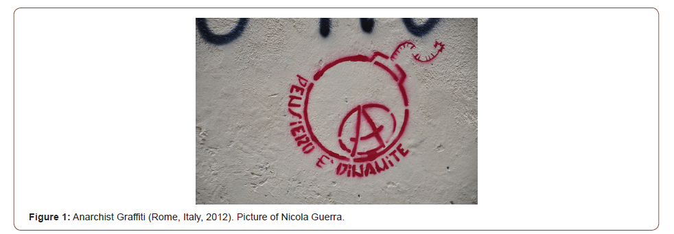
Both attributable to the birth of humanity and contemporary protagonist of the urban space, graffiti art does not find consensual definitions on either an artistic or a linguistic level. Despite its massive presence in our cities, the accusations of vandalism have caused scholars of Italian language and culture to shy away from the analysis of graffiti (Guerra [1], Guerra [2],Telmon [3], Radtke [4]. Moreover, this reticence to study them might also have been exacerbated by the fact that graffiti is used by subcultures that represent a counterpower to the organization of society. Another factor might also have been a certain traditional elite perspective, to the detriment of ordinariness and of the everyday Guerra [5] ,Zagrebelsky [6]; Williams [7]. Yet, a careful study of graffiti would help to follow the development of everyday language, to understand the dynamics of ‘young’ lingo, to grasp the sociolects of subcultures and metropolitan tribes, and to grasp the diametrical melting pot of the city, consisting of a kaleidoscope of communication channels in which orality and writing coexist and hybridize Guerra [1,5]; [2,8]. Figure 1. Anarchist Graffiti (Rome, Italy, 2012). Picture of Nicola Guerra.

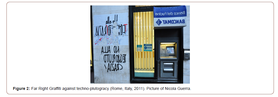
An effective way of defining graffiti is to consider as such every intervention in which the semantic element, i.e. the set of graphemes, words and their articulation in texts, has the upper hand over the semiotic element represented by symbols, figures and abstractions, predominant instead in muralism Guerra [1,2,5] This definition takes into consideration how graffiti is a functional opportunity to blend linguistic and urban space Guerra [1]. As for the accusations of vandalism, we must not overlook the fact that contemporary graffiti have a counter-hegemonic essence, and although this is exercised by breaking the rules of urban decorum and disrespecting private property, it allows us to escape the stringent regulations required to access mass media communication classics such as newspapers and television. Therefore, graffiti can be seen as an exposed form of writing that communicates to the masses but not to the apparatus, because exercised in ordinariness and spontaneity, leaving the channels of high cultural and political control. It thus becomes a factor of communicative democratization. To put it in the manner of graffiti: «clean walls, silent people». Figure 2. Far Right Graffiti against techno-plutogracy (Rome, Italy, 2011). Picture of Nicola Guerra.

irispublishers-openaccess-archaeology-anthropology

The walls speak

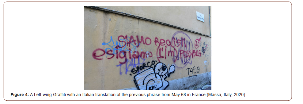
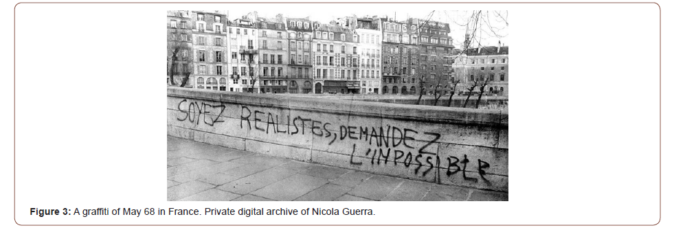
It is no coincidence that graffiti, together with paste-ups and poster-art, is the protagonist of the social and political revolt of May 68 in France Tesson & Barbery [9] Kugelberg and Vermès [10]; Rohan [11] and becomes an important channel of communication of the Protests of 1968 in Italy Guerra [8,12,13]. The post-war political scene in Italy, characterized by high technicalities and parliamentary equilibriums, was set apart by the use of a language generally known as ‘politichese’ (‘political-speak’), characterized by the tendency to mask rather than clarify contents, in which much-thought lexical choices gave a high register and a strong bureaucratic feel to political language Guerra [12,13,14]. If usually the overcoming of ‘political-speak’ is fixed at the year 1994, to the advantage of the ‘gentese’ (‘people-speak’) as a consequence of the political vacuum determined by «Mani Pulite», a more careful analysis shows how the reaction to it is antecedent to the events of the time and is undertaken by groups and political movements antagonistic to the system, starting from 1968 and for all the period known as Years of Lead Guerra [12,13]. Figure 3. A graffiti of May 68 in France. Private digital archive of Nicola Guerra.Figure 4. A Left Wing Graffiti with an Italian translation of the previous phrase from May 68 in France (Massa, Italy, 2020). Picture of Nicola Guerra. Those were years in which, also through graffiti, the use of a political language that moves from inhibition to participation and from cryptic to clear is asserted and consolidated, with the adoption of a linguistic register that maintains a certain intellectuality combined with usability and intelligibility, blending together instances of contestation, political propaganda, and civic sense within a high linguistic spontaneity Mori [15,16]. «We want it all», «Let’s take the city» - graffiti becomes an existential claim and affirmation of a different vision of the urban space, mirror of a new social order. If, too hastily, graffiti are considered works of a lower artistic level compared to murals, in which the semiotic element predominates and therefore the visual solicitation of the user, it is necessary to highlight how the artistic nature of graffiti is both linguistic and in the ability to condense, in the short time of realization and in the little space available, complex concepts that represent an ideology, a creed, a lifestyle Guerra [17,18]. The art of graffiti is mainly the art of language joined to graphic art in the choice of font and calligraphy. Both in the graffiti of 1968, more characterized by generational unity, and in those of the Years of Lead, pervaded by the dynamics of opposite extremisms fueled by social tension, the yearning for participation, clarity, social transformation and inclusion of those who were excluded from the strict logic of the party, parliamentary balancing acts, and a television dominated by control and censorship is loud and clear.

irispublishers-openaccess-archaeology-anthropology

Documenting and studying graffiti means participating in a work of democratization of linguistics

In today’s society of widespread control of both physical and virtual spaces through capillary video surveillance and online monitoring which sees the usable spaces for dissent reduced Zuboff [19],Della Porta , Reiter [20] graffiti art still represents an opportunity for free communication. In the freely used and built urban space, graffiti offers an opportunity for expression to those who are excluded, in whole or in part, from institutional communication, as in the case of numerous heterogeneous subcultures: ultras, casuals, skinhead, hip-hop, rap, Hispanic and Latino gangs, political radicals, and ecologists. Documenting and studying graffiti therefore also means participating in a work of democratization of linguistics, lightening it from the redundant burden of the classics, by the rigid reference to standard Italian compared to popular Italian (intended as a variety of speakers from the lower social classes characterized by poor social prestige), and by a certain purism that translates into fear of those evolutionary dynamics of the language that find representation in graffiti, not by chance creator and diffuser of numerous neologisms and linguistic contact Guerra [1,2,5,8].

To read more about this article...Open access Journal of Archaeology & Anthropology

Please follow the URL to access more information about this article

https://irispublishers.com/oajaa/fulltext/italian-studies-and-graffiti-a-complex-relationship.ID.000551.php

To know more about our Journals...Iris Publishers

To know about Open Access Publishers

Iris Publishers-Open access Journal of Ophthalmology & Vision Research | Demographics and Pattern of Referral of Participants at a One-Week Long Free Glaucoma Screening Event


Authored by Elizabeth A Awoyesuku*,

Abstract

Aim: The aim was to elucidate the demographic characteristics of the participants who presented at a week-long free eye screening programme to mark the World Glaucoma Week 2019 at the Ophthalmology department, University of Port Harcourt Teaching Hospital and their sources of referral.

Methodology: Members of the public were invited for free eye screening at the department of Ophthalmology, University of Port Harcourt Teaching Hospital using several channels of information dissemination including electronic media (Radio), Posters /Banners and Social media. Each had a comprehensive ocular examination done. Those identified with glaucoma were referred for follow-up in the glaucoma clinic. Data obtained was analyzed using SPSS version 21. The age groups gender, other demographic distribution of the subjects amongst other were presented using frequency tables and charts.

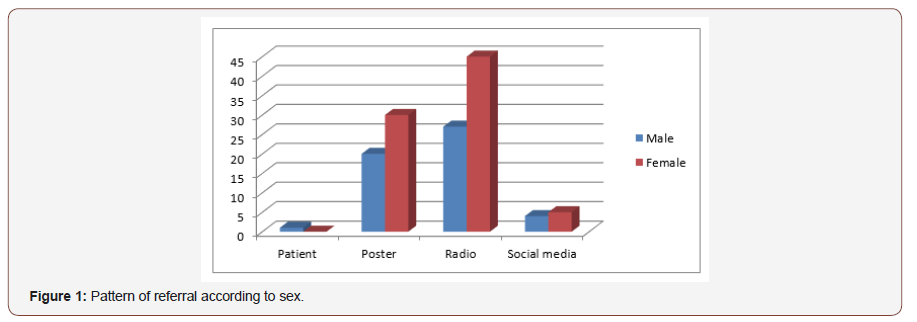
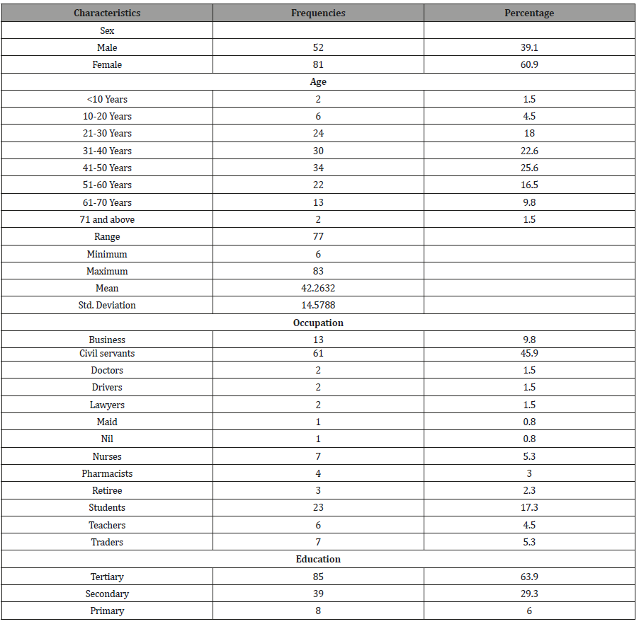
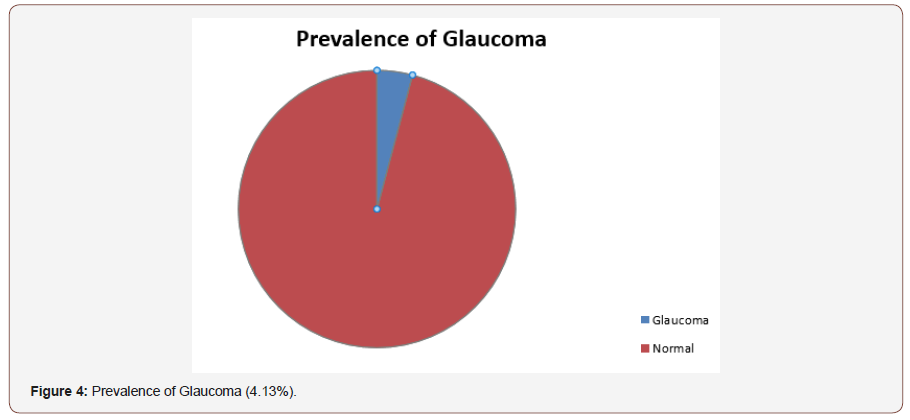
Results: A total of 133 participants (266 eyes) responded to the invitation for free eye screening. 39.1% were male and 60.9% female with a mean age of 42.26 years ±14.58. 48.2% were in the age group of 31-50years. 45.9% of the participants were civil servants with 63.9% of them having a tertiary form of education. 54.5% of participants presented for the screening after listening to radio announcement. The Prevalence of glaucoma in this study was 4.13%.

Conclusion: Women accessed free eye screening more than men in our study and the mass media (Radio announcement) resulted in the most means of referral. The prevalence of glaucoma from our study was 4.13%.

Keywords: Demographics; Glaucoma screening; Pattern of referral

Introduction

Primary Open Angle Glaucoma (POAG) is a chronic condition characterized by loss of retinal ganglion cells. Increased intraocular pressure, positive family history, older age and African descent place an individual at an increased risk for glaucoma [1].

The prevalence of POAG ranges from 1.1% to 3.0% in Western populations, and from 4.2% to 8.8% in populations of African descent [2]. Subgroups of population at risk for developing glaucoma have insufficient knowledge and need to be identified and targeted [3]. In developing countries detecting glaucoma access to care is further limited by poor access to health facilities [4].

In an effort to reverse the trend the World Glaucoma Association and World Glaucoma Patient Association through a joint initiative put together the ‘World Glaucoma Week’ which encourages annual week-long intensive advocacy and mass enlightenment programs to increase awareness of the disease as well as screening to improve case finding [5].

This annual event has been celebrated yearly by the department of Ophthalmology, University of Port Harcourt and this year 2019 a study was undertaken to elucidate the demographics of the population as well as the best means of information dissemination which resulted in access of care with the aim of planning future glaucoma screening activities and improving case finding.

Materials and Methods

Members of the public were invited for free eye screening at the department of Ophthalmology, University of Port Harcourt Teaching Hospital using several channels of information dissemination. A total of 133 persons participated and each had a comprehensive ocular examination done including a visual acuity examination (unaided, Pinhole), Slit lamp examination of anterior segment, Dilated Slit lamp biomicroscopy with +78D lens, Non-contact tonometry, Pachymetry and perimetry. Those identified with glaucoma were referred for follow-up in the glaucoma clinic. Data obtained was analyzed using SPSS version 21. Mean and standard deviations were determined for age. The age groups gender, other demographic distribution of the subjects amongst other were presented using frequency tables and charts Statistical significance was put at p ≤ 0.05. A diagnosis of glaucoma was made following dilated slit lamp biomicroscope with VCDR >0.7, demonstrable perimetric changes and elevated intraocular pressures above 21mmHg

Results and Discussion

As the population increases in developing countries so also will the number of persons with glaucoma increase leading to worsening socioeconomic burden of the disease [6]. In Nigeria, the National Blindness and Visual Impairment Survey had a prevalence of glaucoma related blindness in above 40years was 5.02-6.9% [7]. The World Glaucoma Week is a joint initiative between the World Glaucoma Association and the World Glaucoma Patient Association. It has been celebrated over 9 years all over the world including Nigeria with a total of 626 events were recorded during the week March 10th-16th 2019 [5]. Most of the events are geared towards improving public awareness of the disease. The World Glaucoma week has also been used to gather data on causes of visual impairment as was seen in the World Glaucoma screening in 2016, 449 persons were screened during the week [8].

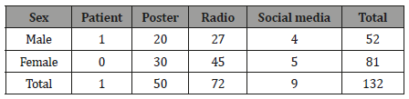
The World Glaucoma Week was celebrated in our department by having free glaucoma screening. Using many channels of information dissemination members of the public were invited for a week-long free glaucoma screening. The demographic characteristics of the population are found in the table below (Table 1&2) (Figure 1).

irispublishers-openaccess-ophthalmology-vision-research

Table 1: Demographic characteristics of study population.

irispublishers-openaccess-ophthalmology-vision-research

Table 2: Demographic characteristics of study population.

irispublishers-openaccess-ophthalmology-vision-research

Gender

In a report by Janicijevic K, et al. [9] a review of 10 years of annual glaucoma screening activities showed a preponderance of females accessing the free screening which corroborates our study findings where women were more in number. A study in the South East of Nigeria by Kizor Akairaiwe [10] however found more men availing themselves for screening than women. There is gender inequality in accessing healthcare as most women do not have the finances required to do this, it’s no wonder they may resort to free eye care activities such as outreaches.

Age

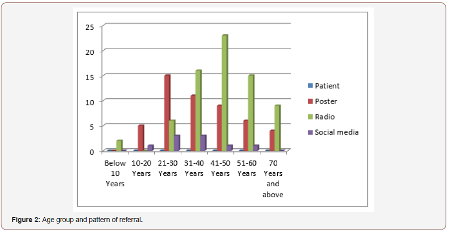
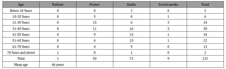
(Table 3) Several studies show most of the participants of free eye care programmers tend to be in the 5th to 6th decade of life [10-13]. Glaucoma typically is asymptomatic until late stages and is more likely to affect those of black race with positive family history and advancing age [14]. this was emphasized during various forms of information dissemination and is therefore not surprising that our study had a lot of people in the 5th and 6th decades of life presenting. This may also be postulated to be same in other studies aside from the fact that visual disorders increase with increasing age and therefore the elderly are more likely to access care since they would be symptomatic. Age group 21-30years had a significant use of print media (Figure 2).

irispublishers-openaccess-ophthalmology-vision-research

Table 3: Pattern of referral of patients presenting by Age.

irispublishers-openaccess-ophthalmology-vision-research

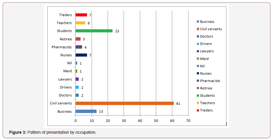
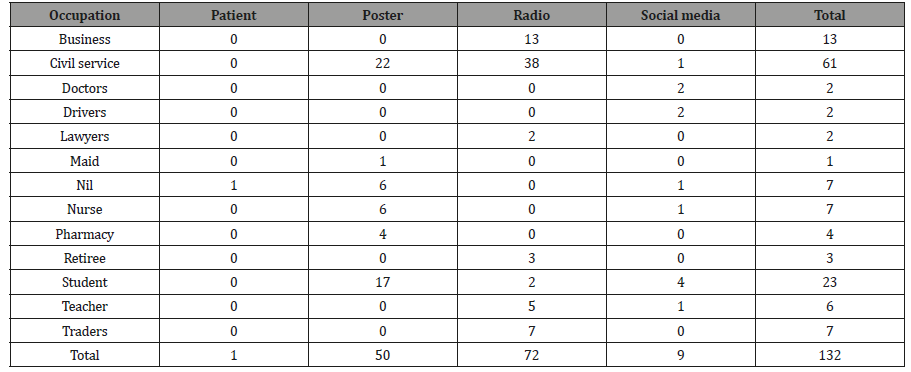
Majority of the Figure 3participants in our study were civil servants. This maybe because Port Harcourt (South-South Nigeria) being an oil producing community has a lot of office workers. This is unlike that in Enugu [10] (South- East Nigeria) where majority were businessmen or Odukpani, Cross Rivers State and Ethiopia where farmers topped the number Majority of the population assessed screening because of radio announcement followed closely by information on posters [15,16] (Figure 3) (Table 4).

irispublishers-openaccess-ophthalmology-vision-research

Table 4: Pattern of referral of patients presenting by Occupation.

irispublishers-openaccess-ophthalmology-vision-research

Sources of referral /means of information dissemination

Electronic Media (Radio) was the highest means of information dissemination in our study Most of the referrals were by electronic media, Posters (print media) featured in age group 21-30years while social media (Digital media) didn’t play a significant role across all age groups. This was same as that of Akaraiwe, et al. [10]. In the study by Adekoya, et al. [12] most patients were self-referred. Electronic media did not however feature significantly in the study at Ethiopia by Tenkir, et al. [15]. This can be explained by the fact that most rural areas in Nigeria have access to the radio unlike in Ethiopia where radio services were sparse at the time of the study.

Educational level

Most of the participants in our study had tertiary level of education same as the study in the South East Nigeria [10] while in Ethiopia [15] mainly secondary school educational graduates accessed the outreach. This is in sharp contrast to Amoomo A [17]. in Namibia where 64.8% of the respondents were unemployed and 44.5% had not completed primary school education.

Prevalence of glaucoma

(Figure 4) The population-based survey of the prevalence and types of glaucoma in Nigeria: results from the Nigerian National Blindness and Visual impairment survey have put the overall prevalence of Glaucoma in above 40years at 5.02%. Risk factors for increased prevalence were illiteracy, increasing age, males and Igbo ethnic group [18]. The prevalence of glaucoma of glaucoma in our study (South-South Nigeria) was 4.13%, 14.5% in the South-East Nigeria [10], and 7.3% in South-Western Nigeria [19].

Case finding for glaucoma is a continuous process though it may not be economically viable, in spite of this; screening is a great challenge in low economies such as Nigeria. [20] The World Glaucoma Week is a good opportunity to improve public awareness of Glaucoma as well as improve case finding in Nigeria.

irispublishers-openaccess-ophthalmology-vision-research

Conclusion

Women accessed free eye screening more than men in our study and the mass media (Radio announcement) resulted in the most means of referral. The prevalence of glaucoma from our study was 4.13%.

To know about Open Access Publishers
Tuesday, January 23, 2024

Iris Publishers-Open access Journal of Pharmacy & Pharmacology Research | Expanding the Kidney Donor Pool through Use of Hepatitis C-Infected Donors: is it Time to Dive in?


Authored by Dinesh Kannabhiran*,

Introduction

The survival benefit of kidney transplantation for patients with end-stage renal disease (ESRD) is well established. However, the demand for kidney donor organs greatly exceeds the current supply. The use of hepatitis C infected donors could increase the number of kidneys available for transplantation. The use of highly effective second-generation direct acting antivirals (DAAs) has been recently studied for the prevention of chronic hepatitis C virus (HCV) infection in kidney transplant recipients who are HCV negative and receive HCV infected kidney allografts. Small, open-label trials have demonstrated the feasibility of using DAAs as either pre- and post-exposure prophylaxis or as treatment after detection of HCV transmission. Short term outcomes illustrate 100% prevention of chronic HCV infection with renal function and allograft survival that are comparable to recipients of non-HCV infected kidney donors. Long-term allograft and patient outcomes are required to determine whether the use of HCV infected organs should be considered for all patients with ESRD waiting for kidney transplant. The survival benefit of kidney transplantation for patients with end-stage renal disease (ESRD) is well established [1,2]. However, the demand for kidney donor organs greatly exceeds the current supply which encourages organ procurement organizations and transplant centers to look for innovative strategies to increase the donor pool. Recently, the opioid crisis has increased the number of overdose deaths exponentially [3]. Additionally, the number of hepatitis C virus (HCV) seropositive donors increased from 452 organs per year to 1506 per year between the years of 2000 and 2016 but only 57% of the HCV seropositive kidneys were transplanted in 2016 [4]. The use of hepatitis C infected donors could increase the number of kidneys available for transplantation but this strategy has historically been avoided because of risk of HCV transmission as well as inadequate treatment response and risk of rejection with interferon-based regimens. The advent of highly effective secondgeneration direct acting antivirals (DAAs) has increased viral cure of patients with chronic HCV infection to more than 96% [5-7]. Ongoing research is investigating whether DAAs can be prescribed post-transplant to HCV negative recipients receiving HCV infected donors to increase the donor pool.

The Transplanting Hepatitis C Kidneys Into Negative KidnEy Recipients (THINKER) trial, was a prospective, open-label, nonrandomized trial that included a total of 20 kidney transplant HCV negative recipients who received hepatitis C infected kidneys, determined by HCV nucleic acid testing (NAT) [8,9]. Recipients were included if they were expected to have prolonged times on the wait list while being maintained on dialysis. Recipients could not have any contraindications to liver transplantation or evidence of liver disease detected by FibroScan imaging or serologic testing. Only HCV genotype 1 infected donor kidneys were included. Patients were assessed for HCV transmission on post-op day 3 and were started on elbasvir-grazoprevir (ELB-GRZ) upon detection of HCV infection. Participants with genotype 1a HCV were tested for baseline nonstructural protein 5A (NS5A) resistance mutations. Patients with genotype 1a/1b were treated with ELB-GRZ for a total of 12 weeks while patients with genotype 1a with NS5A resistance mutation were started on ELB-GRZ and ribavirin for 16 weeks total. Outcomes included HCV cure determined by sustained viral response at 12 weeks (SVR12) and adverse events attributable to HCV infection or DAA therapy with 1year follow-up. All patients received rabbit antithymocyte globulin induction therapy and triple immunosuppression maintenance with tacrolimus, mycophenolate mofetil, and prednisone. Twenty patients were included in the trial, of which 19 became HCV PCR positive by post-operative day 3 (range 2-4). The last patient became HCV PCR positive on day 5 after transplant. Seventeen donor allografts were infected with HCV genotype 1a and only 3 donor allografts had NS5A mutations. All patients had undetectable HCV PCR within 4 weeks of therapy. Renal function at 12 months was similar between patients who received a hepatitis C infected kidney donor and matched recipients who did not accept hepatitis C infected kidney donor. No episodes of allograft rejection occurred during treatment. Four patients developed low-level de novo donor specific antibodies (DSAs) that were detected during routine surveillance. Five patients experienced increased but transient elevations in aminotransferase levels which returned to baseline without intervention. One patient with ESRD due to IgA nephropathy was diagnosed with focal segmental glomerulosclerosis post-transplant with no evidence of recurrent IgA nephropathy. Of interest, 50% of donor organs were excluded in THINKER because of HCV infection that was non-genotype 1 [10]. For this reason, a second trial started, called Exploring Renal Transplants Using Hepatitis C Infected Donors for HCV Negative Recipients (EXPANDER), to utilize these discarded organs [11]. Overall, the methodology of EXPANDER was very similar to THINKER, however, EXPANDER included kidney donor organs infected with genotype 1–4 and DAA therapy was administered as pre- and post-exposure prophylaxis. One dose of ELB-GRZ was administered pre-operatively as pre-exposure prophylaxis. After transplant, patients with genotype 1a/1b and 4 were treated with ELB-GRZ for 12 weeks total. Donors with HCV genotype 1a were tested for NS5A mutation and recipients who received a HCV genotype 1a with NS5A mutation kidney allograft were prescribed ELB-GRZ and ribavirin for 16 weeks total. Patients who received genotype 2 or 3 organs were treated with ELB-GRZ and sofosbuvir (SOF) for 12 weeks total. If the donor allograft genotype was unable to be determined due to insufficient viral load, the recipient was treated with ELB-GRZ for 12 weeks.

Ten hepatitis C negative patients were transplant with hepatitis C infected kidneys. Each patient received one dose of ELB-GRZ prior to transplant. Post-transplant, 7 patients received 12 weeks total of ELB-GRZ for genotype 1,4, or unknown genotypes. No patients received an organ infected with genotype 1a with N5SA mutation. The remaining 3 patients had non-genotype 1 or 4 HCV and were treated with ELB-GRZ and SOF. SVR12 for all patients was 100%. No adverse events occurred related to HCV infection or DAA therapy. Like THINKER, one patient experienced increased aminotransferase levels which increased to 5 times greater than the upper limit of normal (ULN) and then returned to normal without clinical symptoms or intervention. While renal function of HCV infected recipients was not compared to non-HCV infected, matched controls, the median creatinine level at week 12 was 1.05 mg/dL (IQR: 0.9 – 2.0 mg/dL) and GFR was 63.5 mL/min (IQR: 47.8 – 69.9 mL/min). THINKER and EXPANDER both illustrated the feasibility of using highly effective DAA therapy as post-exposure prophylaxis in HCV negative recipients who receive HCV NAT positive donors. The participants of both trials were carefully selected under IRB and the DAA was supplied by pharmaceutical companies. A recent, single center experience by Molnar et al reported outcomes for the use of hepatitis C infected donors for hepatitis C negative recipients when used as standard of care treatment [12]. The standard of treatment was offered to all waitlisted recipients regardless of time of the waiting list or dialysis. Allograft donors were considered if they were HCV NAT-positive, donor age of 45 years or younger, and had a donor biopsy with less than 10% glomerular sclerosis. Recipient HCV PCR was not tested until 4–8 weeks after transplant. Once HCV RNA was positive, HCV genotype was tested and patients were started on glecaprevir-pibrentasvir (GLE/PIB), sofosbuvirvelpatasvir (SOF/VEL), or sofosbuvir/ledipasvir (SOF/LED) for at least 12 weeks as determined by hepatology. The hospital planned to pay for treatment if the third-party payor denied any patient HCV treatment. Standard immunosuppression was rabbit antithymocyte globulin induction with triple immunosuppression of tacrolimus, mycophenolate mofetil, and prednisone maintenance. Between March 1, 2018 and December 31, 2018, the program transplanted 53 hepatitis C negative kidney transplant recipients with HCV NAT positive donors. All patients had undetectable HCV RNA by 12 weeks of treatment. Renal function was similar to THINKER trial at 6 months post-transplant with eGFR 66 mL/min (IQR: 54–79). Forty-seven patients (89%) were treated with GLE/PIB, 5 patients (9%) were treated with SOF/VEL, and 1 patient (2%) was treated with SOF/LED. All insurance plans approved treatment within a median of 5 days of insurance application (IQR: 2–8). Eleven patients originally were denied by the third-party payor but were approved after appeal. Ten patients had AST > 3 times upper limit of normal and 8 patients had ALT > 3 times the ULN. All returned to baseline without intervention. One patient developed fibrosing cholestatic hepatitis. This patient was started immediately on DAA therapy and experienced compete resolution. Thirty-two (60%) of patients experienced low level CMV infection without tissue invasive disease. Eighteen patients (34%) developed BK viremia and 2 patient developed BK nephropathy on biopsy. Sixteen patients (30%) developed de novo DSA. Three patients had acute cellular rejection (ACR) and 1 patient had an episode of combined antibody-mediated rejection and ACR.

The short-term outcomes for kidney transplantation of HCV negative recipients using HCV infected kidney donors are excellent. The renal function and allograft survival in clinical trials are comparable to recipients of non-HCV infected kidney donors. Furthermore, the use of HCV infected kidney donors for transplantation decreases the patient’s wait time on dialysis [8- 12]. Emerging evidence is addressing some of the initial concerns about the use of HCV infected donors including cost-effectiveness of this strategy [13,14]. The study performed Molnar et al further illustrates that third-party insurance companies are paying for the therapy with approval of DAA therapy occurring in less than a week in most cases [12].

While initial studies increase the collective understanding of managing this patient population, some questions remain unanswered [8-16]. First, what is the optimal timing of DAA therapy? EXPANDER was the first study to attempt pre- and postexposure DAA prophylaxis strategy [11]. While the number of patients in this study was small, less patients in EXPANDER had elevated transaminase levels compared to either THINKER or the Molnar et al studies leading to the conclusion that avoidance of HCV infection through pre- and post-exposure prophylaxis may help to decrease transaminitis associated with HCV transmission [8-9,11-12]. Moreover, one patient in the Molnar et al. study developed fibrosing cholestatic hepatitis. The authors mention that DAA therapy was started early for this patient and the fibrosing cholestatic hepatitis resolved completely but no additional information was documented. Originally, the treatment protocol for this study started DAA therapy between 4–8 weeks posttransplant but evolved over time to start DAA therapy < 3 weeks after transplant. Further studies will help to elucidate if pre- and post-exposure DAA therapy is associated with less transaminitis compared to waiting to treat after confirmed HCV transmission. Another interesting finding of the Molnar et al study that is not mentioned in either THINKER or EXPANDER is the development of low level CMV viremia in 60% of patients and BK viremia in 34% of patients [8-9,11-12]. Two patients with BK viremia developed biopsy proven BK associated nephropathy (BKAN) but no patients had allograft loss during follow-up. It is unclear whether HCV viremia contributed to the concurrent CMV and BK viremias or whether this population of patients were at higher risk of these infections at baseline.

The detection of DSAs was noted in both THINKER and the Molnar et al study [8-9,11-12]. While no rejection episodes occurred during DAA treatment in the THINKER trial, 4 episodes of rejection were documented in the Molnar et al study; 3 episodes of ACR and 1 episode of mixed ACR and antibody mediated rejection. The grades and timing of these rejections were not discussed. It is unclear whether the rejections occurred during or after DAA therapy but it should be noted that the overall incidence of ACR for this cohort is similar to national outcomes [17]. Patients received rabbit antithymocyte globulin for induction therapy with a mean total dose of 4.9 mg/kg (±0.9 mg/kg). Target tacrolimus goal levels post-transplant and goal mycophenolate mofetil doses were not noted. Future studies should continue to monitor the development of DSAs and rejection in this patient population.

Overall, the utilization of HCV infected kidneys for HCV negative recipients is a promising strategy to increase access to transplantation and should continue to be studied. Current evidence suggests that HCV negative recipients who receive pre- and postexposure DAA prophylaxis or DAA therapy upon detection of HCV transmission can be transplanted with HCV infected kidneys with no documented chronic HCV transmission to date and excellent shortterm renal outcomes. Future studies should investigate the optimal timing of DAA therapy to decrease adverse events described in the literature including the development of DSAs, CMV and BK viremia, and elevated transaminase levels. Long-term allograft and patient outcomes should also be collected to determine whether the use of HCV infected organs should be considered for all patients with ESRD waiting for kidney transplant.

To know about Open Access Publishers
Iris Publishers-Open access Journal of Anaesthesia & Surgery | Brief review on the Trauma Score


Authored by Behzad Saberi*,

Abstract

Trauma Score is used clinically to estimate the severity of injury in the patients with trauma. The score is a numerical grading system and consists of cardiopulmonary function measurements and Glasgow Coma Scale with reduced total value. Respiratory rate, Respiratory expansion, Systolic blood pressure and Capillary refill are the parameters which would be measured in the Trauma score. Each of these parameters is given a number and by adding up the numbers, the severity of the injury would be estimated. The measured score would be between one and sixteen. This is a brief review on the Trauma score which having knowledge about that would be of help for the clinicians to estimate the severity of injury in the patients with trauma.

Keywords: Trauma Score

Body

Respiratory rate, Respiratory expansion, Systolic blood pressure and Capillary refill are the parameters which would be measured in the Trauma score. If the Respiratory rates would be between 10 to 24 per minute the score would be 4. Score 3 is given when the respiratory rates would be between 25 to 35 per minute. Respiratory rates which are measured equal to 36 per minute or more would be given score 2. Respiratory rates between 1 to 9 per minute would be given score 1 and if there would not be any respiratory rate, the score would be 0. If the Respiratory expansion would be normal the score would be 1 and if it would be retractive or there would not be any respiratory expansion the score would be 0. If the Systolic blood pressure would be 90 mmHg or higher the score would be 4. Score 3 is given if the Systolic blood pressure would be between 70 to 80 mmHg. Systolic blood pressures which are between 50 to 69 mmHg would be given score 2 and ones which are between 0 to 49 mmHg would be given score 1. If there would not be any Systolic blood pressure the score would be 0. Normal Capillary refill would be given score 2 while the delayed one would be given score 1. If there would not be any Capillary refill the score would be 0. If the patients have Trauma scores equal or more than 13, their survival rates would be more than 90 percent and if they have the scores equal or less than 6, their survival rates would be less than 10 percent.

Conclusion

This brief review tries to summarize the Trauma score which can be practical and easy to use for the clinicians. Having knowledge about the Trauma score would help the clinicians to approach the patients with trauma with more precision.

To read more about this article...Open access Journal of Anaesthesia & Surgery

Please follow the URL to access more information about this article

https://irispublishers.com/asoaj/fulltext/brief-review-on-the-trauma-score.ID.000556.php

To know more about our Journals...Iris Publishers

To know about Open Access Publishers
Iris Publishers-Open access Journal of Hydrology & Meteorology | Influence of Community Resilience to Flood Risk and Coping Strategies in Bayelsa State, Southern Nigeria

  Authored by  Nwankwoala HO *, Abstract This study is aimed at assessing the influence of community resilience to flood risk and coping str...