Thursday, September 5, 2024

Iris Publishers- Open access Journal of Gastroenterology & Hepatology | Complex Sources of Hyperbilirubinemia


Authored by Adel Ekladious*,

Introduction

The authors present a case of a patient with severe hyperbilirubinemia due to acute hepatitis B infection which resolved without intervention with subsequent normalization of synthetic and excretory liver function. Despite this, the patient remained jaundiced due to unconjugated hyperbilirubinemia in the context of a recent diagnosis and flare of hereditary spherocytosis. Due to the complex clinical presentation, the patient underwent a liver biopsy with the additional genetic diagnosis of Gilbert’s Syndrome being established. This illustrates an unusual case of both Hepatitis B and hereditary spherocytosis being a potential cause for a flare of Gilbert Syndrome.

Case Report

A 59-year-old Australian man returned from a 3-week holiday in Taiwan. On his return to Australia, he became progressively unwell with nausea, loss of appetite, and dark urine. During his holiday, he reported having unprotected sexual intercourse but denied heavy alcohol use, other illicit substance use, or other high-risk behaviours. He was otherwise well man with no other significant medical history and not on any regular medications. Social history revealed a non-smoker and social alcohol intake of two glasses of wine each evening. In regard to family history, the patient was adopted, and family history could not be traced.

He was up to date with vaccinations including Covid-19. On review at the General Practitioner (GP), he had normal vital signs but was markedly jaundiced with the yellow sclera. His abdomen was soft and non-distended with mild tenderness in the epigastrium and right upper quadrant. There was no organomegaly. The remainder of his examination was unremarkable.

His initial investigations included

Full Blood Count (FBC), Urea, Electrolytes, Liver function tests, ESR, CRP, serology and Polymerase chain reaction (PCR) for Hepatitis A, B, C, D, and E, serology for Chlamydia, Herpes simplex virus (HSV), Gonorrhoea, Human immunodeficiency virus (HIV), Syphilis, Trichomoniasis, Human papillomavirus (HPV), Epstein Barr Virus (EBV), Cytomegalovirus (CMV), Coxsackievirus and Toxoplasmosis, Thin and Thick blood film for Malaria, and Ultrasound for the abdomen in addition to Urine and Stool examination. Of the investigations mentioned above, abnormalities were detected in the following results-Liver function was grossly abnormal with Alanine aminotransferase (ALT) of 3000 IU/L (20- 45IU/L), Aspartate aminotransferase (AST) of 2000 IU/L (20-40 IU/L, Gamma glutamyltransferase 170 IU/L (10-30 IU/L), Bilirubin 400 umol/L(2-20umol/L) with 50 % conjugated bilirubin, Alkaline phosphatase 150 IU/L (2-17 IU/L), Albumin 30 g/L (40-60g/L). INR 1.1, PTT 40 seconds (30-40), D Dimer 0.3(<0.50), APTT 60 seconds (60-80).

Serology for hepatitis A showed positive IgG and negative IgM (either past infection or immunization), and serology for hepatitis C, D, and E were all negative for recent infection. However, hepatitis B serology was consistent with acute infection- Hepatitis B surface antigen (HBsAg) was positive, Hepatitis B e-antigen (HBeAg) was positive, Hepatitis B surface antibody (HBsAb) was negative, Anti- Hepatitis B core IgM (anti-HBc IgM) was positive, Anti-Hepatitis B core IgG (anti-HBc IgG) was negative, serum hepatitis B DNA was 2000 IU/ml. HIV serology and p24 antigen were negative, ultrasound for the liver was unremarkable, and COVID rapid test and PCR were negative.

Based on the above investigations, the patient was diagnosed with acute hepatitis B infection and advised to stay at home with close monitoring of liver function tests. The patient started to improve after two weeks with improvement in clinical and biochemical parameters. After 6 weeks, the patient seroconverted and tested positive for HBsAb and Hepatitis B e-Antibody (HBeAb), ALT, and AST started to normalize. Despite this, the patient remained jaundiced. Repeat bilirubin after two months was 450 umoles /L(20-40umoles/L), and repeated blood testings showed normal liver function, however, the Hemoglobulin dropped to 12g/dl (14- 16g/dl) but all other blood testings including repeat viral hepatitis screen were either negative or inactive. The patient went on to have an autoimmune liver panel with the following investigations tested either negative or normal-Antinuclear antibody (ANA), Anti-dsDNA, Anti- Smith (Sm) antibody, Anti-soluble liver antigen, Anti-liver-kidney microsomal (LKM) antibody, anti-mitochondrial antibodies, Serum and urine copper, Serum ceruloplasmin, alpha one antitrypsin (AAT) protein, iron studies and genetic tests for hemochromatosis, carbohydrate-deficient transferrin, Thyroid function and Synacthen test.

Subsequently, the patient was referred to a hepatologist who proceeded with a liver biopsy. The histopathology was examined by two pathologists in two different hospitals, both pathologists agreed there were no structural abnormalities, apart from the increased agranular endoplasmic reticulum, and mild deposition of glycogen, in addition to increased lipofuscin pigment in the centrilobular region, these features had been reported before in Gilbert syndrome. The patient then went on to have genetic testing for Paroxysmal Nocturnal Haemoglobinuria (PNH) with flow cytometry, and for Gilbert and Crigler-Najjar syndrome. UG1A1 *28 (uridine diphosphateglucuronosyltransferase) mutation confirmed the diagnosis of Gilbert syndrome.

Blood film results received from a haematologist regarding the same patient confirmed a few spherocytes, a mild increase in reticulocytes, and a direct antiglobulin test was negative which ruled out autoimmune hemolytic anaemia. Additionally, the patient tested positive for eosin-5′-maleimide (EMA) binding by flow cytometry, and a repeat ultrasound of the abdomen did not show any gallstones or splenomegaly. Testing for cryoglobulin was negative, Glucose 6 phosphate dehydrogenase (G6PD) level was within normal range and Parvovirus IgM was negative. The patient was diagnosed with Gilbert syndrome and mild asymptomatic hereditary spherocytosis and was reassured and discharged back to his GP with advice that should he have any abdominal pain it could be due to pigmented gall stone.

Discussion

Jaundice is defined as the yellowish pigmentation of the skin, sclera, and mucosa due to hyperbilirubinemia. It is clinically detectable when the circulating bilirubin is greater than 35umol/L. Hyperbilirubinemia can be further classified as Conjugated (Direct) and Unconjugated (Indirect) hyperbilirubinemia. Unconjugated hyperbilirubinemia is due to increased production, impaired hepatic uptake, and decreased hepatic conjugation of bilirubin.

The common causes of Unconjugated Hyperbilirubinemia in adults are

A. Hemolytic Anemia: Immune-mediated (ABO incompatibilities, Auto-immune hemolytic anemia); non- Immune mediated (Sepsis, Microangiopathic hemolytic anemia, Liver disease); Extra-vascular causes (G6PD deficiency, Pyruvate Kinase Deficiency, Hereditary spherocytosis, PNH, Hemoglobinopathies, hypersplenism)

B. Congenital: Gilbert Syndrome and Criger Najjar syndrome

C. Drugs induced: Chloramphenicol, Gentamicin, pregnaneidol

Conjugated hyperbilirubinemia is caused by hepatic or cholestatic diseases. The common causes of conjugated hyperbilirubinemia can be further classified into

1) Hepatocellular: Hepatitis, cirrhosis

2) Cholestatic causes- Primary biliary cirrhosis, Congenital (Dubin-Johnson syndrome. Rotor Syndrome); Drugs, Hepatitis, obstructive CBD pathologies.

Fractionation of total bilirubin and test for urinary bilirubin help to determine the type of hyperbilirubinemia. If conjugated bilirubin is less than 20% of the total- it is predominantly unconjugated and if it is more than 50% it is predominantly conjugated hyperbilirubinemia. Moreover, the presence of urinary bilirubin is consistent with conjugated hyperbilirubinemia [1- 3]. Gilbert syndrome is the most common inherited cause of unconjugated hyperbilirubinemia. It is caused due to mutation in the promoter region of the UGT1A1 gene, which results in reduced UDP-glucuronosyltransferase (UGT) production [4,- 8]. The most common genotype of Gilbert syndrome is the homozygous polymorphism A(TA)7TAA in the promoter of the gene for UDP-glucuronosyltransferase 1A1 (UGT1A1), which is a TA insertion into the promoter designated UGT1A1*28. Patient usually has asymptomatic mild unconjugated hyperbilirubinemia usually triggered by fasting, haemolytic reactions, febrile illnesses, menstruation, physical exertion, infection, and dehydration [14]. Gilbert syndrome can coexist with hereditary spherocytosis and present with moderate unconjugated hyperbilirubinemia [15]. It has been found in patients who are hepatitis B carriers but not active hepatitis B. However, there are no case reports for acute Hepatitis B infection as a trigger for Gilbert syndrome so far.

In conclusion, Gilbert syndrome is a benign autosomal recessive liver disease characterized by unconjugated nonhaemolytic hyperbilirubinemia due to defective glucuronidation of bilirubin secondary to a reduction in the activity of UGT with a good prognosis. Its management is conservative with observation and does not require any pharmacotherapy. The present case has shown the association of acute Hepatitis B infection as a trigger for Gilbert syndrome and highlights the importance of considering it as one of the differentials diagnoses for precipitating undiagnosed Gilbert syndrome. However, diagnosis of Gilbert syndrome requires careful clinical assessment and ruling out other common causes of unconjugated hyperbilirubinemia including haemolytic anaemia before proceeding with the genetic analysis of the UGT1A1 gene [9-13].

To read more about this article...Open access Journal of Gastroenterology & Hepatology

Please follow the URL to access more information about this article


To know more about our Journals...Iris Publishers

Iris Publishers-Open access Journal of Otolaryngology and Rhinology | Ankyloglossia is to be Treated Properly and in Time


 Authored by Fahim Ahmed Shah*,

Introduction

The medical term for tongue-tie is Ankyloglossia, The piece of skin joining the tongue to the base of the mouth is called the frenulum. This is known as the lingual frenulum in medical terminology Frenulum is a general term for a small fold of integument (skin) or mucous membrane that limits the movements of an organ or part. The lingual frenulum is a tag of either skin or mucosal tissue which is attached to the inferior surface of the tongue to the floor of mouth. Ankyloglossia (tongue-tie) is the general clinical term for the short frenulum which limits the range of movement of the tongue; there is still no accurate classification for this condition [1]. Although Tongue tie is of varied degree in severity but for all practical purposes clinically it can be divided into four types, this depends on how close the frenulum is attached to the tip of the tongue

Type I Attachment of the frenulum is to the tip of the tongue, just in front of the alveolar ridge

Type II Attachment is of two or three or four mm behind the tongue tip and on the alveolar ridge.

Type III Attachment is to the mid-tongue and the middle of the floor of the mouth.

Type IV Attachment is to the base of the tongue and is thick.

The lingual frenulum length (short) and position of insertion (anterior) can lead to speech disorders and may affect postnatal feeding [2].

Tongue-tie is a birth defect that affects 3-10% of newborn babies. Incidence varies widely with associated syndromes like Opitz syndrome, orofacial digital syndrome, Beckwith-Wiedemann syndrome etc. with no racial predilection it is more common in boys than girls with male preponderance ratios varying from 1.5 to 2.6 [3].

In tongue-tie, this lingual frenulum is unusually short, tight and thick, restricting the movements of tongue. This prevents the baby from feeding properly and also causes speech problems. Clinically tongue tie is the name given to the condition arising when the frenulum is unusually thick, tight and short. The lingual frenulum length (short) and position of insertion (anterior) can lead to speech disorders and may affect postnatal feeding [4]. Genetic factors are suspected, as tongue-tie is frequently familial [5].

The Gravity of the Problem

Tongue tie is a rare but definite congenital deformity.- Etiologycally during early development, the tongue is fused to the floor of the mouth. In the embryological development the tongue gets free, with the frenulum left as the only remnant of the initial attachment. Tongue-tie is the result of this short fibrous lingual frenulum and is often blamed for slow speech development. Child who have real limitation of movement as a result of tongue tie have a history of difficult milk feeding as well because it adds to the baby’s difficulties in taking the breast with poor protractility and severe limitation of the tongue movement resulting in inability to suck. If the tongue-tied infant cannot maintain the tongue over the lower gum during sucking, the “phasic bite reflex” (chewing) is triggered [6]. This chewing motion is sufficient to transfer milk from the bottle, but is clearly problematic at breast, because breastfeeding requires well-defined peristalsis from the front to the back of the tongue as well as tongue-palate synchronization. Some tongue-tied infants cannot even manage a bottle

Ankyloglossia is a common congenital anomaly that is usually detected soon after birth. It is characterized by partial fusion or in rare cases, total fusion of the tongue to the floor of the mouth due to an abnormality of the lingual frenulum which connects a moveable part to a fixed part and stabilizes the part from undue movement it stabilizes the base of the tongue without interfering with the tongue tip movement. In ankyloglossia, this lingual frenum has an anterior attachment and may be unusually short causing virtual adhesion of the tongue tip to the floor of the mouth therefore it is a physical defect. Tongue is the main organ for speech. If this tissue short, its mobility is affected resulting in difficulty in speaking. This cord like tissue that extends from the mid-portion of the floor of the mouth to the midline of the inferior surface of the tongue blade. This tissue is considered important for speech as it is assumed to give mobility to the tongue. Tongue-tie or ankyloglossia refers to a condition where this tissue is short, thereby restricting tongue free movement Ankyloglossia (tongue-tie) limits the range of motion of the tongue, impairing the ability to fulfill its functions. But if a child can protrude his tongue beyond the lower lip and elevate his tongue to touch the upper lip with his mouth half open or the tip of the tongue is able to protrude outside the mouth without clefting then probably the child will have no difficulty in speaking Interestingly, it is the function not the shape that determines speech ability.

Current Trends in the Early Treatment of the Problem

Earlier tongue tie has been described as a myth of hoary antiquity but the condition is not entirely mythical although surrounded by an aura of superstition and old wives’ tales”. In the past practicing physicians were taught that treatment of tonguetie, (ankyloglossia) is an outdated concept - a relic of times past but current trends is the early treatment of the problem to avoid complications such as feeding problems, refusal of breast feeding, speech difficulties such as inability of proper pronunciation of many alphabets, vowels, delay in speech or speech- usually sounding interdental because of the restricted movement of the tip. Tonguetie has emerged as a recognized cause of breastfeeding difficulties and a very easily corrected one. Approximately 25% of newborns with ankyloglossia have feeding problems, as the child grows older, there may be difficulty in moving a bolus in the oral cavity and clearing food from the sulci and molars. This leads to chronic halitosis and dental decay or dentition- causes a pulling effect on the gingiva away from the teeth and even cosmetically it may look abnormal and tongue has a forked or serpent look.

Clinical Assessment

The clinician should first have inspection of the tongue and then evaluation of its functions inspection should include tongue’s appearance when it is lifted as the infant cries or tries to extend the tongue. While lifting, the frenum should be palpated and its elasticity determined attachment of the frenum to the tongue should normally be approx. 1cm posterior to the tip. Tongue tie can vary widely depending on the length of attachment of the frenulum in some babies it extends to the tip of the tongue. The thickness and elasticity of the frenulum and its effect on tongue movements should be assessed because the dictum is to be able to speak properly with good pronunciation one must have a free tongue movement, the tip of the tongue should particularly be free enough to lift and touch the roof of mouth in pronouncing many alphabets especially vowels. Therefore any speech difficulties especially of any pronunciation in preschool age child should be evaluated.

The mother should be asked in particular about child’s ability to breastfeed. Thorough evaluation should determine the adequacy of latch during feeding Assessment of range of motion of the tongue should include the degree of extension of the tongue beyond the lower dental ridge and lip, [7] elevation to palate with mouth wide open, and transverse movement from one corner of the lips to the other without twisting the tongue. Elevation seems to be the most important tongue movement for breastfeeding and should be weighted most heavily in the assessment [8].

FRENULOTOMY• Frenotomy, frenectomy, and frenuloplasty are the main surgical treatment options to release or remove an ankyloglossia.

Frenotomy is a simple procedure in which tongue-tie release only involves cutting the short, tight piece of skin that connects the underside of the tongue to the floor of the mouth but it should be carried out only by those who have been trained in the procedure since the orifices of submandibular and lingual salivary glands open under the tongue over the floor of the mouth, therefore the dividing of the freunim should be closer to the base of the tongue rather than the floor of the mouth. At times frenuloplasty is required which is transverse cutting of frenulum and its vertical repair. Care should be taken to avoid cutting the deep lingual vein just lateral to the midline otherwise significant venous bleeding could occur also injury to the more inferiorly placed submandibular ducts in the floor of the mouth should be taken care of Complications of frenotomy include infection, excessive bleeding and injury to the salivary ducts.

Conclusion

To conclude Tongue-tie is a significant clinical entity, which when symptomatic, should be treated timely and properly to minimize the later complications of difficulty in speech and difficulty in breast feeding. This simple procedure usually resolves feeding problems straight away and brings good quality of speech.

To read more about this article....Open access Journal of Otolaryngology and Rhinology

Please follow the URL to access more information about this article

To know more about our Journals...Iris Publishers

To know about Open Access Publishers

Wednesday, September 4, 2024

Iris Publishers-Open access Journal of Dentistry & Oral Health | Comparison of Dentin Debris Extrusion After the Use of WaveOne Gold and Procodile Q NiTi File


Authored by David E Jaramillo*,

Introduction

The goal of root canal treatment is to prevent and treat apical periodontitis, by eliminating inflamed pulp tissue as well as microorganism and necrotic pulp tissue by chemomechanical debridement and providing a good quality root canal filling. According to Sjögren, et al. [1] the success rate of root canal treatment goes as high as 96% in teeth without periapical lesions. Salehrabi, Rotstein [2] metanalyses, found a 97% retention rate of root canal treated teeth while Hannahan, Eleazer [3] showed no statistical difference in the success rate between single implants and teeth with non-surgical root canal treatments. The root canal instrumentation is perform using different type of hand files or mechanical driven rotary or reciprocation instruments. However, during root canal instrumentation dentin debris, pulp tissue, and bacteria are likely to get extruded through the apical foramen. This extrusion might be responsible for the presence of severe postoperative pain and/or swelling [4].

The design of the file is important to prevent the extrusion of the debris during the cleaning and shaping phase. The amount of extruded debris was first measured using a model invented by Myers and Montgomery [5]; showing that there is a close relationship between the instrument, the technique used and the quantity of debris extruded through the apex. Balanced force technique has demonstrated least extrusion when compared to linear filing motion [6].

Procodile Q (Komet, USA) is a standard nickel titanium (NiTi) file with a positive rake angles that maximizes efficiency and flexibility, and additionally is heat treated pre-grinding; since post production treatments dulls the cutting capacity of files. The system combines a variably tapered core with controlled memory for unrivaled flexibility for a safer and more efficient root canal preparation and performance, with a unique design to resist cyclic and torsional fatigue and to prevent debris extrusion.

The system offers three different sequences for narrow, medium, and wide canals with taper .06, .05 and .04 respectively. The narrow sequence consists of three files with taper .06 and tip size 20, 25 and 30. The sequence for medium canals is comprised of two files taper .05 with a tip size of 35 and 40, and lastly the wide canal sequence includes two files with a taper .04 and a tip size of 45 and 50. To determine the canal size the manufacturer suggests to slowly introduce a file iso #15 to working length and if the file meets resistance, then the canal is considered narrow in size, on the other hand if the file meets little or no resistance the canal is “medium” or “large”.

The new trend is to reduce the chair time while improving the efficiency of the root canal treatments, thus manufacturers are fabricating and proposing single file systems to meet the needs. Different types of alloys make instrument characteristics different than conventional nickel titanium files. A new M-wire NiTi in endodontics instruments provides instruments with more flexibility and fatigue resistance. WaveOne Gold (Denstply Tulsa, OK) has been one of the most utilized M-wire single file system used by practitioners.

WaveOne Gold is a reciprocating system that runs at 10 cycles per second with a 360° rotation every three reciprocating motions. Four files are available to fit different canal sizes, the Small file is a tip size #20 with .07 taper, the Primary file offers a tip size #25 and a .07 taper, the Medium file has a tip size #35 and .06 taper, and the Large file is equivalent to a #45 tip size with .05 taper.

The Step Back Technique, is a technique that allows to produce a continuous taper from the orifice entrance to the apex of the canal. The procedure was introduced in 1964 by Clem [7], and is made in two stages. Stage one grants the preparation of an apical constriction at the end of the canal, meanwhile on stage two, by increasing the file size and gradually stepping back we produce a sustained taper which will facilitate the obturation process.

Since there have been other studies evaluating the degree of debris extrusion of different file systems, we wanted to measure the amount of dentinal debris extruded through the apical foramen by the Procodile Q system since is a system that is new to the market and there are no studies about this particular file.

Materials and Methods

The protocol was approved by the Ethics Committee for the Protection of Human Subjects of the University of Texas Health Science Center School of Dentistry at Houston (protocol no. HSCDB- 22-0174).

Freshly extracted mandibular incisors were collected from urgent care and periodontal clinic and stored in a 5% thymol solution until use. Teeth were subjected to multiple angled radiographs to confirm the presence of a single canal, no evidence of calcifications or previously treated. Root curvature was measured following the Schneider [8] method and teeth with a curvature of <5˚ were selected for the study. A total of 60 freshly extracted human mandibular incisors with mature apices, straight root canal and single root canal, were selected for the study.

Elected teeth were standardized using a diamond disc to a length of 16mm going from the incisal edge to the apices of the root end.

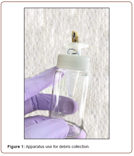
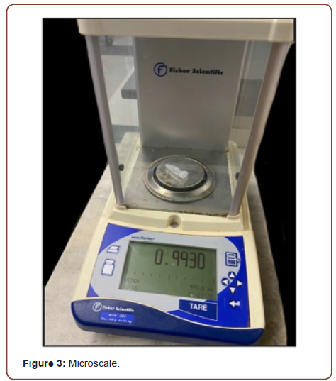
The debris were collected and measured using a modified Myers and Montgomery model [5], that consisted of an Eppendorf micro test tube and a second Scintillation vial. Two perforations were drill to Eppendorf micro test tubes caps, a small orifice that served as a released for the build-up pressure, and a bigger one that held the teeth in position for instrumentation. Then the micro test tubes were weighted two times in an electronic scale to obtain a mean initial weight and the results were recorded. A Scintillation vial was used to hold the Eppendorf microtube in place.

Each tooth granted coronal access using a round #2 carbide bur, then apical patency was established by inserting a # 10 K-file from the access opening to the apical terminus, thus confirming permeability of the canal and working length. The total number of samples (n=60) was divided into four subgroups of 15 teeth each (n=15) and instrumented with a different shaping technique. Every tooth was mounted in the modified Montgomery model and a dental dam was used to hide the apex of the root from the operator. Instrumentation was done following manufacturer instructions and apical patency was preserved using a #8 K-File past of the working length.

Step back group

The first phase of the canal preparation was done using hand files from #15 through #40 at the predetermined working length (16mm). In between files; one mL of distilled water was used as irrigation solution and a #08 K-file was employed to keep patency at the apex. A hand file #40 was standardized as the master apical file for each sample. In the second phase of the shaping process, the continuous tapper was created by incrementing the size file and stepping back progressively.

WaveOne gold group

The first file utilized for the WaveOne Gold group was the Small file (##20/.07) using a pecking motion with light apical pressure (following manufacturer recommendations) until reaching working length. Once working length was reached, apical patency was corroborated with a #8 K-File passing working length measurements and one mL of distilled water was used as an irrigation agent. This procedure was repeated with the Primary and Medium files.

Procodile Q group

After utilizing a #15/.02 hand file as a path glide, the Narrow Procodile Q sequence consisting of a #20/.06 file was used until getting to working length. One mL of distilled water was used as irrigation solution and a #8 K flexo file was used to keep the patency of the apical foramen. Then the following files of the Narrow series were used (# 25/.06, # 30/.06) in the same manner process until working length was reached.

Procodile Q crown down group

The Medium #35/.05 Procodile Q file was directed apically with an “in-and-out” pecking motion with an amplitude of approximately 3mm using light apical pressure (as recommended by the manufacturer). Once the file presented resistance, the instrument was removed from the tooth and one mL of water was used to irrigate the root canal. Followed by the #30/.06, # 25/.06 and # 20/.06. This protocol will be repeated continuously until the working length was reached.

irispublishers-openaccess-dentistry-oral-health

After instrumentation was done, the collected liquid and solid debris were placed and stored in a incubator at 37°C for a period of five days to permit the water remnants to evaporate, thus the Eppendorf micro test tubes were left with the solid debris. The extruded debris were weighted three times in a electronic scale (Sartorius Cubis, Gottingen, Germany) with an accuracy of ±0.00001 g. A mean post-operatory weight was obtain and compared with the pre-operatory weight (Figure 1).

Results

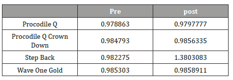
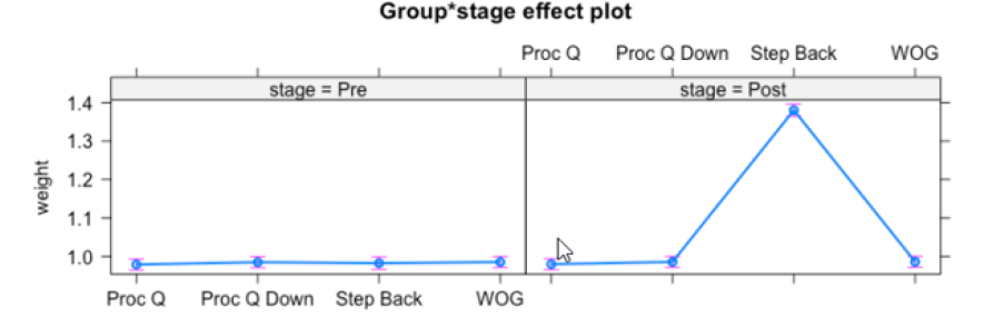
The dentinal debris extrusion was recorded for all four groups. There is statistical difference in between step back technique compared to Procodile Q (Crown Down) and WaveOne Gold systems (p<0.05). The highest mean extrusion value was produced by the Step Back technique. The mean value of pre and post extrusion by all the four groups are given in the following (chart/Table 1). All analyses were performed using R statistical software (R Core Team 2018). R Core Team (2018). R: A language and environment for statistical computing. R Foundation for Statistical Computing, Vienna, Austria. URL https://www.R-project.org/ [9] (Tables 1, 2).

Table 1: Pre and post weight of debris collection.

irispublishers-openaccess-dentistry-oral-health


Table 2: Pre-weight and post instrumentation weight of extruded debris.

irispublishers-openaccess-dentistry-oral-health


Observational findings of reciprocation systems

During the root canal instrumentation various important files features were observed.

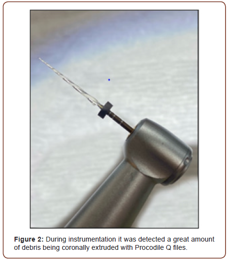
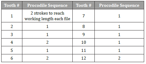
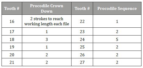
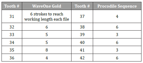
Procodile system was faster in reaching working length on both modalities when compared to WaveOne Gold. It was also observed more debris coronally extruded during instrumentation and good amount of debris attached to the file. As well, flexibility of Procodile file is by far, higher than the one observed in WaveOne Gold files (Tables 3-5) (Figures 2, 3).

irispublishers-openaccess-dentistry-oral-health

Table 2: Procodile sequence. Strokes necessary for each file to reach working length.

irispublishers-openaccess-dentistry-oral-health


Table 2: Procodile Crown Down. Strokes necessary for each file to reach working length.

irispublishers-openaccess-dentistry-oral-health


Table 2: WaveOne Gold. Strokes necessary for each file to reach working length.

irispublishers-openaccess-dentistry-oral-health


Discussion

Our results confirmed, the extrusion of debris through the apical foramen happens regardless of the instrumentation technique or system used for the debridement of the root canal space [5]. As expected, the Step back technique extruded more debris out of the four groups (p<0.05) and this finding is similar to other studies [10- 12]. The high R-Squared value for the Step back group, supports to the technique having a strong effect in the amount of debris extrusion. We attributed this outcome to the type of instrument, since in the initial phase of the Step back technique; the continuous .02 taper of the hand files and the close proximity of the flutes of the active portion of the file does not leave enough space to allow the debris to move coronally within the canal, additionally al- Omari, Dummer [6], concludes that the filling motion of the Step back technique pushes the debris apically. Even though the rest of the groups extruded debris through the apical foramen, it was of no statistical significance (p>0.05). There has been a change of WaveOne Gold technique since the last report done by Jaramillo (2015) [10]. WaveOne Gold has change its instrumentation technique by adding the peaking motion and constant irrigation, to help remove most of the produced debris and to prevent the pack down of dentin debris by a single direct apical pressure motion of the instrument. While the new Procodile version, Procodile Q design and motion, produces more abundant coronal extrusion of debris as the instruments advances faster apically, plus the peaking motion recommended by the manufacturer. The newer technology in instrument design increases the efficacy to prevent in larger amount, the debris extrusion during the instrumentation phase of the root canal treatment.

The study has some limitations like the inability to recreate the back pressure from the apical tissues being this a contributing factor for the extrusion of debris regardless of the technique used [5]. Moreover, the study was limited to samples with straight canals, thus the observed results should not be generalized to teeth with curvatures in the root. Additionally, the engine driven instruments used a different type of motion, WaveOne Gold uses a reciprocating motion, while Procodile Q operate with a continuous rotatory motion [13]. For future projects it would be advised to compare similar modes of motions [14-16].

Conclusion

The differences in the amount of debris extruded may be explained by the technique used to prepare the root canal space, the use of reciprocating motion or by the design of the files. Procodile Q, Procodile Q Crown-Down sequence, and WaveOne Gold produce minimal debris extrusion while Step back produced most of the debris extrusion.

To read more about this article...Open access Journal of Dentistry & Oral Health

Please follow the URL to access more information about this article
https://irispublishers.com/ojdoh/fulltext/comparison-of-dentin-debris-extrusion-after-the-use-of-waveone-gold-and-procodile-q-niti-file.ID.000626.php

To know more about our Journals...
Iris Publishers

To know about 
Open Access Publishers

Iris Publishers- Open access Journal of Agriculture and Soil Science | Current Application of Microencapsulation Technology in Bioremediation of Polluted Groundwater


Authored by Stalis Norma Ethica*,

Abstract

Major challenges in global groundwater supply mainly Come from population growth and climate change requiring innovative water technologies to keep steady supply of drinking water and decrease water pollution worldwide. In particular, agricultural activities impact groundwater pollution. Against this background, the adaptation of highly advanced bioremediation supported by microencapsulation technology to traditional process engineering offers new opportunities in technological developments for advanced groundwater and wastewater technology processes. In this study, benefits of microencapsulation technology for in situ bioremediation of polluted groundwater applications, particularly in bio-augmentation and bio-stimulation, are presented. In addition, an overview of recent advances in microencapsulation technology for contaminated groundwater treatment processes is provided, including the use of various micro-carrier materials are also summarized.

Keywords: Microencapsulation; Groundwater contamination; Micro-carrier; Bioremediation

Introduction

The long-term establishment of the global water supply and sustainability is closely related with the world population growth and global climate change. Steadfast growth of the world’s population forecasted to be almost multiplied by two from 3.4 to 6.3 billion between 2009 - 2050, is presented by a predicted required growth of 70% agriculture production by 2050 [1]. Hence, the need for fresh water is dramatically increasing, especially for food production. It is because 70% of the withdrawals of world’s freshwater are already adjudged for agricultural land irrigation. Today each year, 64 billion cubic meters of fresh water are consumed progressively by worlds’ population [2].

In developing countries, ground water contamination is a key issue, with high levels of pollutants being reported in various regions. Various contamination control and groundwater treatment technologies methods should be applied to overcome this problem [3]. Groundwater treatment technologies encompass physical, chemical, or biological treatment techniques. They could be divided as ex-situ or in-situ technologies.

Aside of world population growth and global climate change, in particular, agricultural activities have been known to give impact to groundwater pollution. For example, high nitrogen fertilizers application rates have been associated with the raise of groundwater pollution [4]. Groundwater has been found as vulnerable to pesticides used in agricultural land [5].

Public concern with polluted soil and groundwater encouraged the development of programs designed to control and remediate this contamination, as well as to prevent further contamination [1]. Bioremediation as an environmentally friendly, socially allowable and economically viable is among the best way to eliminate pollutants from the environment. In bioremediation, microorganisms with beneficial biological activity, including fungi, algae, bacteria, and yeast, could be utilized in their naturally occurring forms [6].

In situ bioremediation involving bio-stimulation and/ or bioaugmentation, being an economical and eco-friendly approach, has come out as the most beneficial soil and water clean-up technique for contaminated sites [7]. Systems involving degrading bacteria have been found helpful in supporting bioremediation option to treat the polluted groundwater [8]. Cells of degrading bacteria have been known as bioremediation agent [9]. Microencapsulation is among important strategy used in bio-augmentation and biostimulation improve the effectiveness of bioremediation processes [10].

Microencapsulation is among quality preservation techniques of vulnerable substances, such as enzymes, living bacterial cells, phytochemicals, and a method for generation of materials with novel precious characteristics. Microencapsulation is defined as a process of packing micron-sized particles in a polymeric shell. Various techniques are now available for the encapsulation of different entities. This mini review provides a literature review of different microencapsulation techniques applied in bioremediation of groundwater worldwide in the last ten years [1].

Discussion

Bioremediation for polluted groundwater

With latest advancements, bioremediation offers an environmentally friendly, socially acceptable and economically viable as well as choice option to deplete pollutants from the environment. There are three major ways of bioremediation including the use of microorganisms, plants and enzymes as remediation agent [11].

Bioremediation technology optimizes and exploits the natural role of microorganisms in the transformation and mineralization of these environmental pollutants. The range of contaminated environments may include surface and subsurface soils and surface and groundwater. Bioremediation for contaminated sites including groundwater containing heavy metals and/or organic pollutants usually involves bio-augmentation and/or bio-stimulation [10]. Bioaugmentation could be defined as addition of pre-grown microbial cultures to support the degradation of unwanted substances (contaminants), while bio-stimulation is the injection of nutrients and other supplementary substances to the indigenous microbial population to influence propagation at a stimulated rate [7].

As the concern towards environmental deterioration grows worldwide, new technological achievements become important for all countries. Among the technologies offering great potential of bioremediation is the microencapsulation of active material including living cells or microorganisms [12]. For bioremediation to be effective, microorganisms must enzymatically attack the pollutants and convert them to harmless products [11].

Role of microencapsulation in bioremediation

Microencapsulation is defined as a process of enclosing or encapsulating micron-sized particles of solids or small drops of liquids or gasses in an inert shell, which in turn protects and isolates them from the external environment [13]. Micro-particles are products obtained by microencapsulation. When the particles have diameter between 3–800mm, they are regarded as micro-particles, microspheres or microcapsules. Micro-capsules are distinguished from microspheres in terms of morphology and internal structure [14].

Microencapsulation is a technology developed to pack solids, liquids and gases in tiny, sealed capsules isolating and protecting them from harsh environmental factors, such as moisture, light, oxygen, and interaction with other substances. Such microcapsules could gradually release their contents under specific conditions at controlled rates. Those packs are spherical with a micrometer size; yet are highly affected by the structure of micro-carriers and the core components [15,16].

Degradation activities naturally mediated by microorganisms used as bioremediation agent could detoxify pollutants. It is also the goal bioremediation to develop reliable technology, which can accelerate this degradation process, to reduce health risks of the pollutants and to restore the affected site into its natural state. However, even though these organisms have high degradation performance, there are limitations in success including microbial inoculum distribution and handling, suppression by parasites and predators as well as nutrient limitation [17]. These factors highly affect microbial bioremediation agent to survive in the environment. To overcome the issues, possible strategies include improved delivery system of microbial inoculum on microencapsulation technology, which could provide protection through the making of micro-environments and allow controlled release of inoculum to the targeted site [18].

Bio-stimulation supported by microencapsulation

Bio-stimulation is a commonly used technique for bioremediation involving the addition of rate-limiting nutrients to speed up the biodegradation process. Bio-stimulation often includes the addition of oxygen and nutrients to aid indigenous microorganisms used as bioremediation agent. The nutrients are essential as the basic building blocks of life allowing the microorganisms to produce particular enzymes, which could degrade pollutants [11].

A number of studies have reported the use of controlled release of active materials as a way of bio-stimulation and providing the nutrients required or essential for the bioremediation process [12]. In this sense, bio-stimulation could be highly supported by microencapsulation. The use of microencapsulated microorganisms offers a great potential in degrading pollutants through bioremediation. Microencapsulation of living microbial cells in a semi-permeable gel or carrier materials bring more advantages over the free cell bio-augmentation. The microencapsulation could prevent microbial cells from bacteriophage infections and protozoa grazing. It supports both biological and physical stabilities, by decreasing risks such as brief and sudden variations of pH or temperature; covers from abiotic stresses coming from heavy metals or other toxic compounds [19]. In addition, microencapsulation using carboxymethyl-cellulose as microcarrier could form fine structures for nutrient release, producing bio-stimulation in biodegradation process [12]. Thus, in general, microencapsulation is beneficial in enhancing cell survival and high biomass concentration

Bio-augmentation supported by microencapsulation

Bioremediation of pollutants or contaminants by utilizing microorganisms is among the most important strategies to eliminate contaminants from groundwater. However, there are limitations of this approach since many contaminants are not efficiently removed [11]. To overcome these limitations, bio-augmentation also includes addition more specific and efficient pollutantbiodegrading microorganisms into a microbial community as a way to support the ability of this microbial community to biodegrade contaminants. In this aspect, microencapsulation of the pollutantbiodegrading microbial cells is relevant to allow steady supply of the bioremediation agent.

To date, the elimination of contaminants by bio-augmentation has been widely investigated in surface water, soil and groundwater [19]. However, although it has been practiced in agriculture and in wastewater treatment for years, bio-augmentation is still experimental. Many factors (e.g. predation, competition or sorption) conspire against it. However, a number of strategies have been explored to make bio-augmentation a beneficial technology in sites lacking significant populations of biodegrading microorganisms. The pollutant degradation rate under optimal local conditions, may increase upon addition of an inoculant to remediate a chemical spill; yet, the most successful examples of bio-augmentation occur in confined systems, such as bioreactors allowing controlled conditions to favor prolonged activity and survival of the exogenous microbial population [20].

Micro-carriers used in groundwater bioremediation applications

One of the vital steps in micro-coating is choosing the most suitable wall materials/ micro-carriers. Micro-carriers or coating materials usually are film-forming materials opted from various natural and synthetic polymers, or combination of both, depending on the inner component and the overall desired microcapsule characteristics [15,21]. Ideally, the wall or sphere material should be an emulsifier, so it could promote enough content release when reconstructed into the product, have a low viscosity due to high concentrations of solids, have good film-forming performance, and have high hygroscopicity.

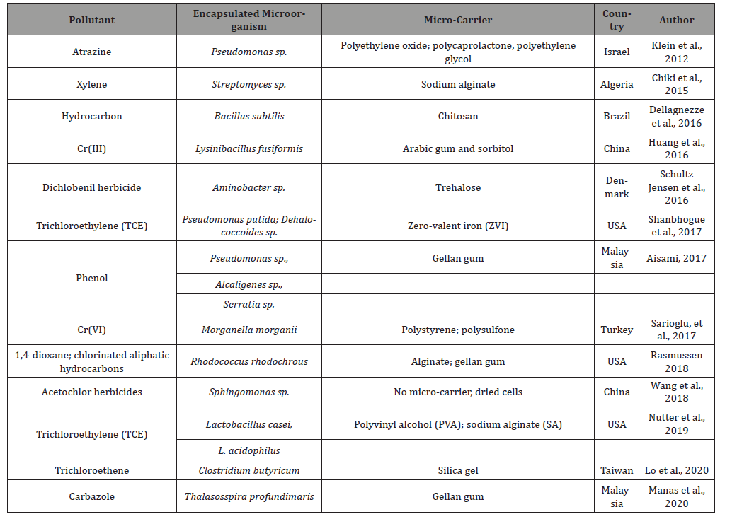
Over the past 10 years, the number of publications on the use of encapsulated microorganisms for the elimination of pollutants in contaminated groundwater has been increasing steadily. The following are among the most commonly used wall/ sphere materials in microencapsulation: carbohydrates (sucrose, starch, maltodextrins, and cyclodextrins), cellulose (carboxymethyl cellulose and its derivatives), gum (Arabic and agar), lipids (wax and fatty acids), and proteins (gelatin, gluten, and casein) [16,21]. Most of these materials have been used in the bioremediation of groundwater in the last decade as listed in Table 1.

Based on Table 1, in the last decade, microencapsulation technology has been widely applied in bioremediation of groundwater polluted by various substances including hydrocarbons, heavy metal, dioxin, herbicides, and plastics. Various micro-carriers grouped as alginates, gums, polymers have also been used as encapsulating materials providing protection as well as nutrition source in suitable environment allowing the release and growth of microbial cells. Interestingly, the microbial cell immobilization could be done by creation of dried cells, which means it does not necessarily need any micro-carrier [8].

Based on this literature review, microencapsulation technologies with various applied micro-carriers as single or combinations keep producing novel micro-engineered materials offering great potential for more innovations in the future. Such innovations are in particular very beneficial for the treatment for contaminated groundwater [22-29].

Table 1: Reported studies related with the application of microencapsulation technology for bioremediation of polluted groundwater in the last decade (2010-2020).

irispublishers-openaccess-agriculture-soil-science

Conclusion

There is a need for novel advanced groundwater bioremediation technologies, in particular to ensure a high quality of drinking water and to eliminate water pollutants using suitable treatment systems. Micro-engineered materials produced by microencapsulation technology offer the potential for novel water technologies that can be easily adapted to groundwater bioremediation applications. To date, microencapsulation with various micro-carriers keep producing novel micro-engineered materials offering great potential for more innovations in the coming decades, in particular for treating heavily degradable contaminants in groundwater.

To know about Open Access Publishers

Iris Publishers-Open access Journal of Hydrology & Meteorology | Influence of Community Resilience to Flood Risk and Coping Strategies in Bayelsa State, Southern Nigeria

  Authored by  Nwankwoala HO *, Abstract This study is aimed at assessing the influence of community resilience to flood risk and coping str...