Authored by Andrea Fischlowitz*,
Clinical Image
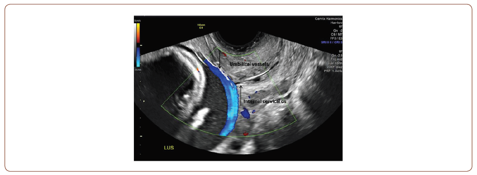
This image demonstrates the ultrasound diagnosis of vasa previa in a 37-year-old primigravid woman who presented for a routine detailed fetal anatomic survey at 21 weeks gestation. Obstetric management consisted of pelvic rest, serial fetal growth scans starting in the late second trimester, and antenatal corticosteroids for fetal lung maturity at 33 weeks. Due to the risk of rapid fetal exsanguination with rupture of the vasa previa, the patient was admitted to the antepartum service at 35 weeks for observation. The patient fortunately experienced an uncomplicated antepartum course with the delivery of a healthy infant via nonemergent cesarean section at 36w3d. The placental pathology was consistent with the prenatal diagnosis of a velamentous cord insertion leading to the vasa previa.

To read more about this article.....Open access Journal of Gynecology & Womens Health
Please follow the URL to access more information about this article
To know more about our Journals...Iris Publishers


No comments:
Post a Comment