Authored by Smail Kharoubi*,
Abstract
Rhinolithiasis is a rare condition often neglected or unknown. The most common symptom is a long-term unilateral purulent rhinorrhea and unilateral nasal obstruction. Nasal endoscopy and imaging are interesting to facilited diagnosis . Therapeutic management requires endonasal extraction of the rhinolith under local or general anesthesia.
Keywords: Endoscopy, Rhinolith, Computed tomography scan
Observation
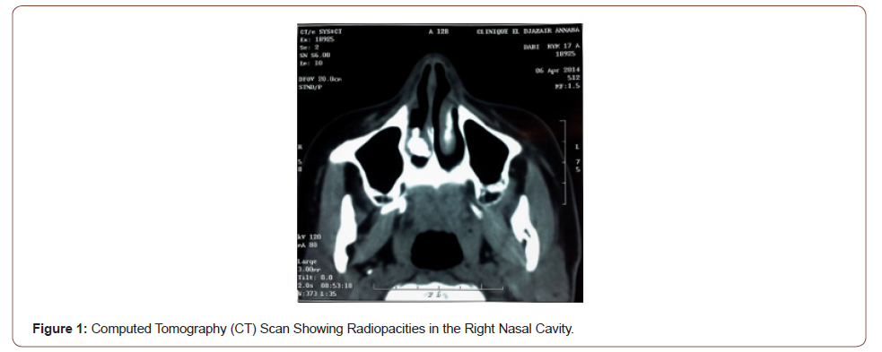
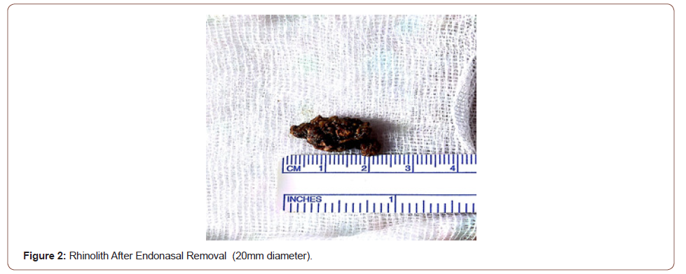
Patient D.A 17 year-old, consults for right nasal obstruction, rhinorhea end epistaxis during for 5 years. Endonasal exam showed purulent secretion and irregular gray’s mass hard palpation with sucing canula. Facial CT showed 25 mm long axis calcium density opacity between inferior turbinal and septal space. The diagnosis of rhinolithiasis is confirmed after imaging. Removal rhinolith with nasal endoscopy under general anesthesia was performed. Follow up was favorable after 14 months.
Discussion
Rhinolithiasis corresponds to a solid calcification by gradual deposition of calcareous salts around a central resorbable or non-resorbable foundation of varying shape and size [1]. They are rarely published and the first description was reported by BARTOLIN in 1654. In 1943 POLSON had collected 380 cases. We can estimate at 800 the number of cases published in the literature [1]. Rhinolithiasis can often occured in female patients with or without organic or non-organic forein body [2, 3]. Clinically the symptomatology is dominated by unilateral nasal obstruction, epistaxis, rhinorhea, headache, cacosmia, epiphora, halitosis and facial pain. rhinolithiasis me by found incidentally examination or imaging. Endoscopy showed hard gray’s mass between inferior turbinate and nasal septum with sensation of crackling during the button-styled exploration.
Imaging is very interesting exam and show calcic density opacity in posterior part of nasal cavity. sinusitis may to be associated and bone lysis is uncommon. Kodaka, et al. [4] analyzed the stone by electron microscopy and energy disperse X-ray analysis and found mainly phosphorus, magnesium, and calcium as a nucleus [4]. Rhinolith can induce septum perforation, nasooral fistula, dacryocystisis. It could be differentiated from other entities with similar presenting symptoms, such as sinusitis, nasal foreign body, osteoma, hemangioma, bone sequestration (syphilis, radiotherapy), osteosarcoma [5]. Rhinolithiasis is removed by forceps with endoscopic endonasal approach in local anesthesia. Big rhinolith, rhinolith associated with septal deviation, polyp, sinusitis pusillanimous patient, can requiring general anaesthesia. Ultrasound lithotripsy can be used to break up the stone. Reccurences are exceptionnel and outcomes generally favorable (Figures 1,2).


Conclusion
Rhinolithiasis is uncommun pathology often neglected that tends to disappear in developed countries. The most common symptom is unilateral purulent rhinorrhea and nasal obstruction. Nasal endoscopy and imaging data confirm the diagnosis. Therapeutic management requires endonasal extraction of the rhinolith generaly in local anesthesia.
To read more about this article....Open access Journal of Otolaryngology and Rhinology
Please follow the URL to access more information about this article
To know more about our Journals...Iris Publishers


No comments:
Post a Comment