Authored by Abdulbaset Naas*,
Abstract
Objective: To report complications related to the flap around cochlear implants and to discuss the possible causes and treatment options.
Method and Materials: This is a retrospective review of 264 of cochlear implant of pediatric and adult patients operated in our center during the period from April 2013 to April 2021, with review of complications related to flap around the implant, complications are divided into minor and major complications Minor complications include skin infection, wound dehiscence without exposure of the implant, wound seroma and hematoma. Major complications include flap necrosis and implant extrusion.
Results: Among 264 cochlear implant recipient 10 cases of seroma and/or hematoma, 9 of them treated with aspiration and one by surgical evacuation, compression and I/V antibiotics and they recovered completely, 7 cases of wound infection, delayed wound healing and wound dehiscence without exposure of the implant treated with daily dressing, debridement and intravenous antibiotics. 2 patients suffered with flap necrosis and extrusion of the implant, one of them presented after 3 years of surgery, treated with transposition of the implant, secured with a vascularized pericranial flap based upon temporalis muscle and rotation skin flap, the other patient presented 2 weeks after surgery and ended with explantation.
Conclusion: Complications related to flap in CI are considered uncommon with incidence of 7 %. In our study, only 0.75% of them are major complication. We consider the age, sex and preceding upper respiratory tract infection with acute otitis media and head trauma as possible related factors in these complications. Management of complications of CI should start with prevention and early detection and treatment of any wound infection, hematoma, and seroma.,/p>
Keywords: Flap; Cochlear; Implant; Complications
Introduction
Cochlear implant is now considered a relatively safe and effective surgery [1] in management of bilateral severe and profound sensori-neural hearing loss when there is no benefit from the maximum powerful hearing aid [2]. As there is increase in community awareness of deafness and neonate screening test of deafness, this result in marked increase in surgery of cochlear implant in last decades. The benefit of cochlear implant is great. In pediatric age group there is great access to sound, improvement in their auditory skills, speech understanding and oral linguistic development. In adults the restoration of hearing by cochlear implant prevents the humiliation, loneliness, social isolation, depression and loss of jobs.
Post- operative complications can occur like any other surgery, cochlear implant surgery is not exceptional, one of the most common complications after cochlear implant is related to the flap around the implant, these complications can be divided into two varieties [3]. Minor variety is referred to those complications which does not require admission to hospital and can be treated as outpatient like minor skin infections, delayed wound healing, hematoma and seroma. On the other hand, major complications include severe skin infection, dehiscence, device exposure and extrusion, such complications need admission to hospital, I/V antibiotics and further surgical intervention.
Management of scalp flap complications started with prevention by good preparation of patients before surgery, good flap design which should have adequate blood supply and venous drainage and very gentle handling of tissue.
Material and Methods
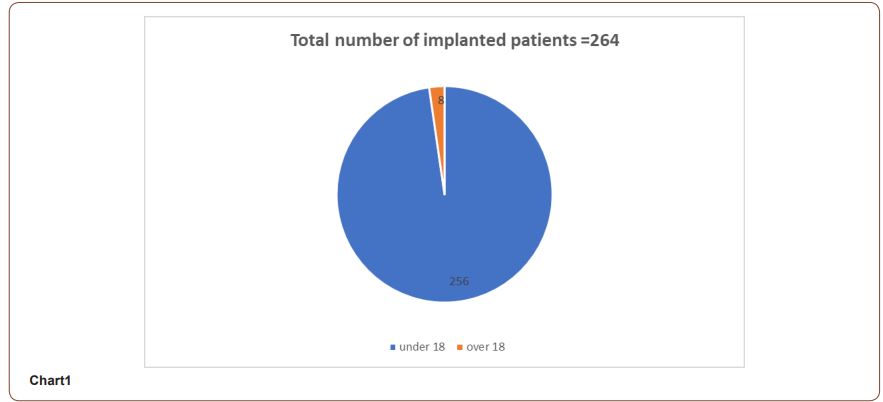
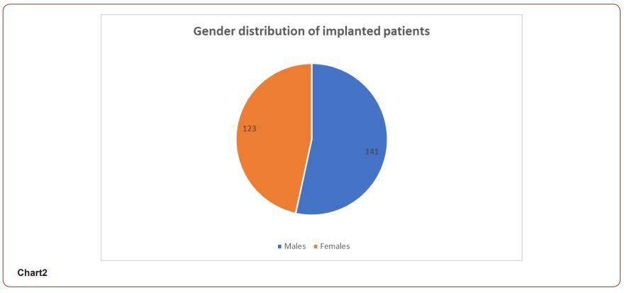
This is a retrospective study by reviewing the medical notes of 264 cochlear implant patients operated in our department during the period between April 2013-April 2021. From them 256 children less than 18 yrs. (117 females and 139 males) and 8 adults (6 females and 2 males), all of them were operated on by three senior surgeons using the same surgical protocol includes complete ENT examination, complete blood count and tympanometry one day before surgery. Shaving of operation site is done one hour before surgery. Our approach is short linear post-auricular incision, raising the flap, bony work starts by cortical mastoidectomy and preparing the bed for implant which is about 25mm from the mastoid cavity with depth of 4mm with anterior step then we thinning the posterior canal wall and we open facial recess and identify round window. The device is fixed in the bed and the electrode inserted in the round window. The wound is closed in layers using absorbable sutures and dressing with antiseptic solutions and then covered with compressive bandage. One dose of I/V antibiotic is given at the time of induction of on the 2nd post-operative day and every other day. If the condition is satisfactory, the patient is discharged from the hospital on the 3rd or the 4th post-operative day with follow up in OPD regularly (Charts 1, 2).
Complications and their management
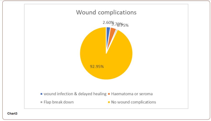
In our study there is 19 out of 264 cochlear implant recipients had complications related to the wound and/or the flap,17are minor and 2 major complications. The details of these complications as follows:
Minor complications
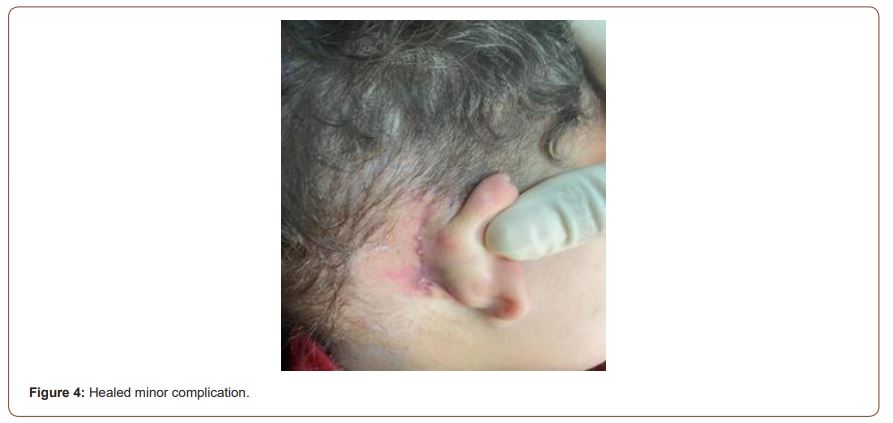
1. Wound infection, delayed wound healing and mild wound dehiscence without exposure of the device or the electrode, such complications occurred in 7 cases, and presented early during dressing with wet hyperemia and redness of the wound. All cases were treated with systemic antibiotics and daily dressing, and followed up until complete healing. All cases healed completely.
2. Hematoma around the device occurred in 4 cases, usually during the 1st or 2nd post-operative week. It presented as soft tender fluctuating swelling around the implant, microbiology study was negative, all cases treated with aspiration, compressive bandage and systemic antibiotics, all cases healed completely.
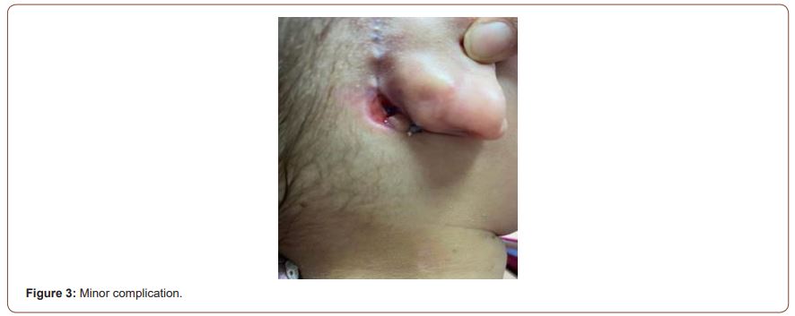
3. Seroma, which is a collection of serous fluid around the implant developed in 6 cases of our study several months or years after surgery, all cases kept on daily follow up and treated with aspiration which was done more than once. Overhead compressive bandage was applied and systemic antibiotics was given. 5 cases showed complete recovery and only one case had a recurrence of the seroma and treated with surgical evacuation through incision and compression.
Major complications
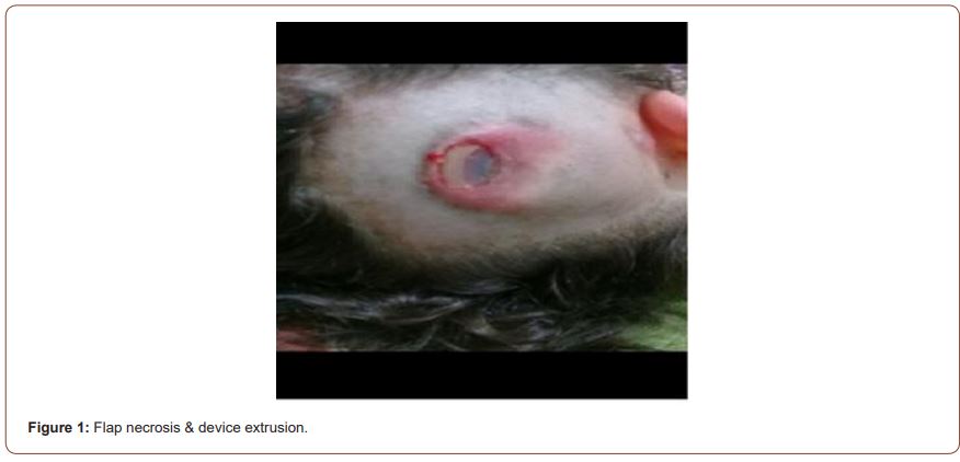
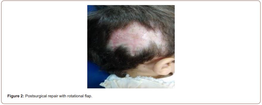
Break down and necrosis of the flap with exposure and extrusion of the implant. This complication occurred in 2 cases of our study. The first has occurred 3 years after the implantation. It has presented late with most of the implanted device exposed without any clear reason. The family denied any history of trauma. The child was admitted to hospital, microbiology swabs was taken for C/S but did not show any growth of bacteria. It was treated with antibiotic and dressing. Surgical intervention was carried out with debridement and preparation of another bed for the device, covered with per-cranial and temporalis muscle flaps and rotation skin flap. The healing was complete, and the child started using the external device after six weeks. In the other case the break down and necrosis occurred 2 weeks after surgery started with wound infection, followed by extrusion of the device. Bacteriological study showed growth of MRSA. Explantation of the device was carried out. The family has been counseled about implantation of the other ear but they refused any further surgical intervention (Chart 3) (Figures 1-4).
Discussion
Cochlear implant has made great progress in the past 30 years with the development in surgical equipment and techniques, generally post-operative complication is uncommon and the most common complications after cochlear implant are related to the surgical wound and the flap around the device [4]. In our study the rate of complications related to wound and the flap around implants was 7%, while rates of such complications reported in literature range from 0.06% to 10% [5]. All complications related to the flap around the implant takes place in children less than 5year. Male and female children had almost the same incidence of minor complications, but in major skin flap complications occurred in girls only. Wound infection and delayed wound healing occurred in our study in 7 cases out of 264 (2.6%) such complications occur even with the patients still on antibiotics, there is no clear explanation but it might be due to scratching with finger nail or allergy to dressing material or the plaster.
Collection of fluids around the implant after surgery is another minor complication [6]. This include hematoma and seroma, The incidence of such complications occurred in 10 patients (3.7%), the rate of such complications in literature range from (0.4% to 3.7%) [7], in our study hematoma takes place in 4 cases and presented early in the first days after surgery as soft swelling around the implant, aspiration show dark red blood with no bacterial growth, generally the condition might be caused by abnormal clotting mechanism or inadequate intraoperative hemostasis etc. In the other hand seroma occurred in 6 cases after months or years of surgery and presented with obvious swelling around the implant, 2 cases with postoperative seroma complains of altered function of the implant with pain in wearing the external part, in all cases aspiration of the serous fluid, they did not show any bacterial growth, all treated with aspiration , prophylactic antibiotics compressive bandage for one week and avoid wearing the external part for one month, all cases recovered completely except one patient where there is recurrence of the collection necessitate incision and evacuation and compressive bandage, the cause of post- operative seroma occurred months and years after surgery with apparently completely healed wound is unclear here we consider trauma although the patients and the family deny any H/O trauma.
The early identification and management of fluid collections around the implant is critical to prevent a serious complication including migration of the device, abscess formation, flap necrosis and break down and extrusion of the device, so we should keep the families aware of any changes occur around the flap for early detection and management.
Major skin flap complications (wound breakdown, flap necrosis, exposure and extrusion of the implant) occurred in 2 cases both of them are girls below the age of 5 years with incidence in our study 0.75% where is the rate of such complication in literature is ranged from 1.08% to 8.2% [8, 9], causes of major flap complications are not always clear but it may explained by three factors, first patient factors including the age of the patient; children more susceptible to trauma, thin skin, other factor is the sex of the patient as girls are more liable to flap complications where here skin is thinner, also girls usually have long hair, any associated comorbidity is another possible cause. Second is the surgical factors which include the design of the incision, handling of tissue, sterility, the way of closure of the surgical wound which should be in layers without undue tension. The third factor may be related to device like silicone allergy and Biofilms [10, 11].
Summary
Post cochlear implant complications are generally uncommon, those complications related to the flap are usually minor and can be managed in the outpatient clinic. On the other hand major flap complications are challenging to us, as they need prolonged stay in hospital and further surgical intervention. We can avoid such complications by good preparation of the patient, proper wound design, meticulous surgical techniques and regular postoperative follow up for early detection and management of any complications.
To read more about this article....Open access Journal of Otolaryngology and Rhinology
Please follow the URL to access more information about this article
To know more about our Journals...Iris Publishers


No comments:
Post a Comment