Monday, January 22, 2024

Iris Publishers-Open access Journal of Cardiology Research & Reports | Ultrasound-Guided Central Venous Access Insertion in Prone Decubitus Position in Severe Covid-19 Patients: Case Report


Authored by Felipe de Jesús Montelongo*,

Abstract

Objective: To describe the placement of ultrasound-guided central venous access in the prone position in patients with severe acute respiratory distress syndrome due to COVID-19.

Design: Transverse and prospective, with reports of cases of hospitalized patients.

Setting: Adult intensive care unit, of a second level teaching hospital in Ecatepec, State of Mexico.

Participants: Hospitalized patients diagnosed with severe PNEUMONIA due to COVID-19, who required prone position and placement of central venous access, were included from March to September 2020.

Primary outcome measures: Placement of central venous access in prone position guided by ultrasound.

Result: Three patients were included, with predominance of the male sex, with a grade II obesity in 66%, grade III obesity in 33%, with an average of 6 days in the prone position, two required the change and placement of catheter due to obstruction and one case for requiring the installation of a hemodialysis catheter due to the initiation of continuous slow therapy to replace renal function. The number of operators was one to two and the most common site of vascular insertion was the left jugular. The number of punctures was 1.6 times, with a puncture time to insertion of the metal guidewire of 45 to 55 seconds. None had complications.

Conclusion: The placement of central venous accesses in the prone position with ultrasound support is a procedure that facilitates insertion and is apparently fast and safe. However, it is a procedure of which there is little description within the international medical literature.

Background

COVID-19 is an emerging viral disease, which was detected in Wuhan, China in December 2019, with first cases being identified as pneumonia of unknown cause [1-3]. In February 2020, the World Health Organization described it as COVID-19 and the disease was declared an international emergency on January 30, 2020 [4]. Atypical pneumonia due to SARS-CoV-2 generally presents with severe acute respiratory distress syndrome (ARDS), producing an alteration in gas exchange. Patients with a < 150 PaO2/FiO2 ratio and a fraction of inspired oxygen requirements greater than 60% are considered as candidates for initiation of the prone position strategy, which seeks to improve the posterior lung recruitment to improve hypoxemia and the PaO2/FiO2 ratio, as well as the prognosis and survival of these patients. This is carried out for a 16-hour periods that can be prolonged for up to 72 hours (still considered as prone safe) when the withdrawal criteria are not met. After this time, the risk of complications increases [4,5].

Case Descriptions

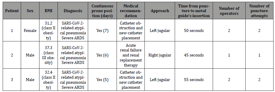
Herein, we present three cases of patients with critical pneumonia due to COVID-19 who developed severe ARDS and were admitted to the adult intensive care unit of the Hospital General de Ecatepec “Las Américas,” ISEM, State of Mexico, Mexico. These patients were intubated under mechanical ventilation in a prolonged prone position, with a 6-day mean, due to refractory hypoxemia. Two patients had to have the central venous catheter removed because of lumen obstruction and a 7 French tri-lumen catheter was placed. The third patient had a 12 French hemodialysis catheter installation as they developed acute renal failure and met criteria for initiation of renal replacement therapy (Table 1).

Table 1: Characteristics of the patients who underwent vascular access in the prone position. No patient presented complications to the post-procedure ultrasound scan.
irispublishers-openaccess-cardiovascular-research

Procedure and Technique

The purpose of the authors is to describe the performance of central venous access with the jugular approach in the prone position guided by ultrasound following personal protection measures for SARS-CoV-2. After obtaining the relatives’ consent for the procedure, the doctors proceeded to put on the personal protective equipment that consisted of a Tyvek suit, double gloves, surgical boots and cap, anti-fog goggles, and/or full-face mask. They also wore a sterile surgical gown and an extra pair of sterile gloves as barrier measures. It should be noted that these steps represent an additional degree of difficulty to the procedure.

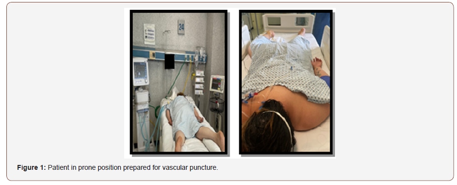
1. First, the patient in the prone position is placed in Trendelenburg position (Figure 1).
irispublishers-openaccess-cardiovascular-research

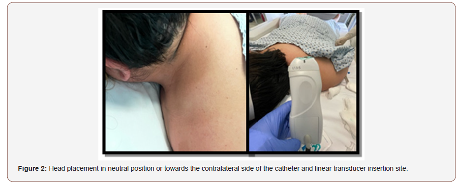
2. Then, the head is rotated to the opposite side of the chosen site to perform the jugular approach or, if there are head and face protections for the prone position, the head is left in a neutral position (facing the bed). During the procedure, both arms are placed close to the thorax, and are returned to a swimmer’s posture when the procedure is completed (Figure 2).
irispublishers-openaccess-cardiovascular-research

irispublishers-openaccess-cardiovascular-research

3. Site insolation should be performed before puncturing to ensure that the vessel exists and to confirm its position and/or possible anatomical abnormalities. Asepsis of the neck chosen for approach is performed and sterile drapes are placed on the back and head.

4. A 7-13 Hz linear transducer is used; sterile gel or sterile saline solution is placed on it using a sterile piping bag or pads to cover the transducer probe (Figure 2).

5. The traditional material for central venous access is prepared.

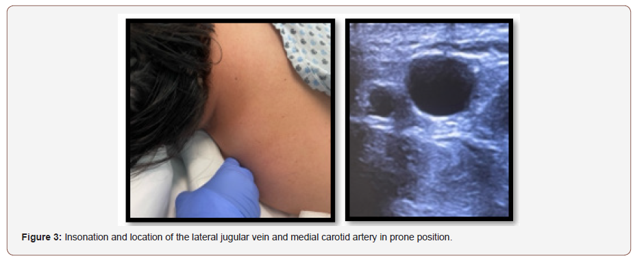
6. After identifying the area of the neck to be insolated, the linear transducer is positioned directing the mark towards the anterior region of the neck to identify the vessels in a transverse position since the external jugular vein is (often irregularly) completely compressible with the transducer and the pulsatile carotid artery has thick and usually round walls. Then, the mark is directed to a cephalic position to identify the longitudinal vessels (Figure 3). The subcutaneous cellular tissues and sternocleidomastoid muscle’s structures are identified with the vein in its longitudinal trajectory above the longitudinal trajectory of the carotid with a depth of about 1.5-to-3-cm. The site to be punctured is identified and local anesthesia is placed with 1% lidocaine. Afterwards, the puncture is carried out after visualizing the needle and the vein in the plane; the entry of the needle into the venous vessel is observed, thereby confirming the return of blood by aspiration and the insertion of the metal guide. The transducer is removed, and the dilator is inserted. The central venous catheter insertion is continued while the metallic guide is withdrawn. Once the venous catheter is inside the vessel, insolation is performed and its presence and venous return by lumens are confirmed. Lastly, the catheter is fixed, and the procedure is finished. The same technique is used for the hemodialysis catheter.

7. Finally, an ultrasound scan is performed on the vessel and puncture site, to evaluate the catheter position and rule out the presence of bruises, catheter knotting, etc. A pulmonary ultrasound is also done to evaluate the presence of pneumothorax, hemothorax or subcutaneous emphysema. This allows us to evaluate complications quickly and avoids the need to move the patient for a radiology study.

Patient and Public Involvement

Due to the nature of the pandemic and the severity of the disease in hospitalized cases, patients and the public were not involved in the development of the research, results, or in the design, recruitment, and conduct of the study.

Discussion

The severe acute respiratory distress syndrome due to COVID-19 requires handling strategies to improve the hypoxemia it generates. Among these strategies mentioned in the literature are the use of prone position, which recently has been used early and for a long time in patients with severe pneumonia in the intensive care unit. During this pandemic, it has been observed that patients cannot supinate because they do not meet the criteria or because when they are back in the supine position, they develop refractory hypoxemia. Therefore, being able to place a central venous access, which is very necessary in this type of patient, is problematic; so, the performance of the ultrasound-guided central venous access placement procedure in the prone position was considered as a solution.

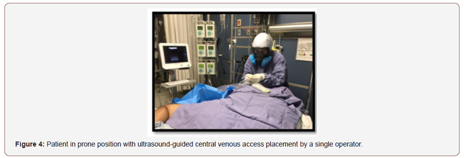
The benefits found in this technique are keeping the prone position, safety after ultrasound identification of the vessel to be punctured and, probably, less vascular puncture attempts and less time in the duration of the procedure. It is very important to take this last detail into account since personal protective equipment is worn, which increases technical difficulty, tiredness, and fatigue. The disadvantages found were the same as for the standard technique, such as the fact that, in obese patients or patients with a short neck, the adipose tissue makes placement difficult by reducing the space to maneuver the transducer and the puncture needle. In addition, in the case of personnel with little experience with ultrasound, it requires someone in charge of guiding the transducer and another person or operator performing the puncture and catheter placement (Figure 4). The use of personal protective equipment in this series of cases increased the technical difficulty, but the puncture time (50 seconds in average) and the presence of complications, such as local hematoma, pneumothorax, or subcutaneous emphysema, were the same.
irispublishers-openaccess-cardiovascular-research

Conclusion

The central venous access placement in prone position guided by ultrasound is a procedure that has not been widely described in the medical literature. Performing this approach by ultrasound facilitates placement with an approximate time of 0.5 to 1 minutes from puncture to insertion of the metal guide.

To read more about this article...Open access Journal of Cardiology Research & Reports

Please follow the URL to access more information about this article


To know more about our Journals...Iris Publishers

To know about Open Access Publishers

Iris Publishers- Open access Journal of Gastroenterology & Hepatology | Recurrence of Upper Gastrointestinal Hemorrhage in Sclerotherapy X Elastic Ligature: Systematic Review


Authored by Sander BQ*,

Introduction

Introduction: The first technique to be used as an intervention in this procedure was sclerotherapy, but with the passing of the years and improvement it was found in the elastic ligature technique the gold standard for the approach in these cases.

Objectives: To compare, through a systematic review, the recurrence of upper digestive hemorrhage in the techniques of sclerotherapy and elastic ligation.

Methods: The most relevant studies in the MedLine, Bireme and Scielo databases were analyzed. The search strategy used the following keyword combinations: (“High Digestive Hemorrhage, Sclerotherapy, Elastic Bandage”).

Results: There were 9 studies selected from the crossword of the keywords, and of these, only 6 were used in the studies after applying the Randomized Controlled Trial and Clinical Trials filters.

Conclusion: There is no definite consensus on which technique - sclerotherapy or elastic ligation - could be associated with a lower risk of recurrence in cases involving upper gastrointestinal bleeding. The literature shows advantages both in the technique of sclerotherapy and in the technique of elastic ligation, and in general, it is nowadays observed that the technique considered gold standard for these cases is the technique of elastic ligation, although many professionals still opt for sclerotherapy, especially in surgical intervention in pediatric patients.

Keywords: Upper gastrointestinal bleeding; Sclerotherapy; Elastic ligation

Introduction

Upper gastrointestinal bleeding is an important medical emergency, with a high rate of morbidity and lethality [1]. Over the past few years, a significant evolution in the methods and techniques available has been observed. Endoscopic therapy, whether with elastic ligation or sclerotherapy, is the most indicated for the management of upper gastrointestinal bleeding and for post-bleeding treatment [2]. However, both techniques have advantages and disadvantages, as well as different statistics about the likelihood of recurrences and rebleeds. This complication is conceptualized as gastrointestinal bleeding proximal to the Treitz angle, which has an increasing incidence due to the aging of the population and its comorbidities [3]. In addition, the causes of upper gastrointestinal bleeding may be due to non-varicose factors, or bleeding from esophageal varices, with a total of 80% of cases resulting from the first type mentioned [3].

Although the mortality rate from upper gastrointestinal bleeding has declined over the years, as a result of the implementation of several more effective treatment and care modalities, its mortality rates still reach the 20% range in up to 6 weeks [2]. The rate of a first episode of bleeding from varicose veins is 5-15% in 1 year and its risk is defined by the size of the varicose veins, presence of red signs, severity of liver disease and continuous alcohol intake. Among the prognostic factors, the size of the varicose vein is the most important, and the larger the caliber, the greater the risk of bleeding [2]. In view of these previous considerations, the objective of the present study was to compare, through a systematic review, the recurrence of upper digestive hemorrhage in the sclerotherapy and elastic ligation techniques.

Method

The most relevant studies originally published in the English language were analyzed, from 2003 to 2018, using the MEDLINE, Lilacs, SciELO databases as a reference. In order to select the studies with the greatest scientific evidence, only controlled and randomized controlled trials (ECCR) were identified, identified by previous reviews, and whose scope was updated and revised, in addition to systematic reviews with or without meta-analysis. The search strategy used the following keywords: sclerotherapy; elastic bandage and upper gastrointestinal bleeding. The inclusion and exclusion criteria were applied based on the types of studies, language, type of therapy and date of publication from the points raised in each item exposed (Chart 1). For the selection of studies, the inclusion and exclusion criteria presented in Table 1 were applied.

Table 1:Inclusion and exclusion criteria applied in the selection of studies.

irispublishers-openaccess-gastroenterology-hepatology

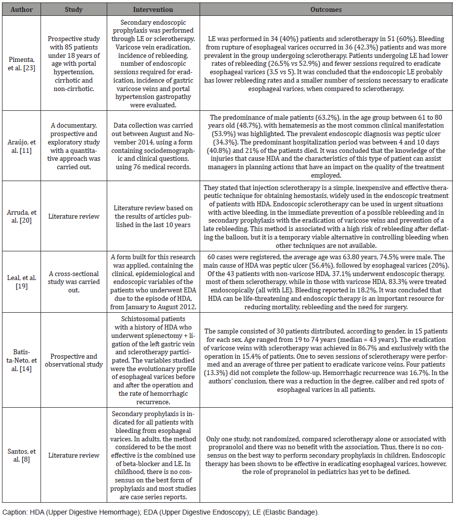
Table 2:Summary of studies and their main results.

irispublishers-openaccess-gastroenterology-hepatology

Result

By crossing the keywords sclerotherapy, elastic ligation and upper gastrointestinal bleeding, it was possible to obtain an initial sample of 9 articles. However, after applying the Randomized Controlled Trial and Clinical Trials filters and after reading the articles found, 6 articles were selected involving the theme and which were then part of the final sample. In Table 1 we will present a summary of the works selected and reviewed in the present study.

Discussion

Through this study, we sought to compare the recurrence of upper gastrointestinal bleeding in the sclerotherapy and elastic ligation techniques, pointing out the consensus that exists on this topic in national and international literature. What are the risks of each of these techniques? Which is more prone to recurrences and what are the advantages and limitations of each one in particular? Analyzing the results obtained through the bibliographic survey, it was possible to observe that both techniques have important advantages and are perfectly indicated. However, in short, the literature allows us to conclude that the technique considered the gold standard for these cases is the technique of elastic ligation, which is the one with the lowest risk of recurrences in comparison to the sclerotherapy technique, although many professionals still choose to use the technique of sclerotherapy, especially in surgical intervention in pediatric patients. This finding was evident in several of the most recent studies that highlighted the fact that sclerotherapy is a pioneering technique in this type of intervention, and although it is still widely used, its usefulness has fallen a lot since the appearance and improvement of the technique of elastic ligation. Sclerotherapy was the first option available, in terms of technique, to be used in these cases. However, over the years and with the evolution of available techniques and materials, the technique of elastic ligation started to gain its space and is currently recommended as being the gold standard in these cases [2, 3].

Guidoux, et al. [7] who added that, after the advent of elastic ligation, the preference for sclerotherapy has been revised, since, in the authors’ view, elastic ligation has lower rates of morbidity. Also, according to Guidoux, et al. [7] over the years, the elastic bandage has been replaced, representing, in the survey carried out in a hospital unit by these authors, a total of 76% of the cases in which surgical intervention related to upper gastrointestinal bleeding was required. However, in many situations, the use of sclerotherapy is still highly recommended, considering the advantages attributed by many to this technique. Specifically analyzing the issue of the occurrence of recurrences, it was possible to observe that most of the studies listed here presented data on this topic, a topic of great importance when talking about rehabilitation in these cases. Thus, the analyzed literature presented several studies comparing the occurrence of recurrences between both techniques. It was possible to observe that, in general, the results showed that the technique of elastic ligation is the most used and considered the one of choice when it comes to prevention of cases of variceal bleeding. However, there was no consensus on which technique can be considered the most effective when the theme is the prevention of the occurrence of recurrences.

In the study by Zanette, et al. [6], the authors stated that elastic ligation of esophageal varices is as effective as sclerotherapy itself, but also highlighted that the technique of elastic ligation is the one that most presented, according to the literature review, a higher frequency of recurrences. than the sclerotherapy technique. Among the studies that pointed to a higher risk of recurrence in the elastic ligation technique is Santos8, which obtained a rate of recurrence and greater complications in the elastic ligation technique compared to the cyanoacrylate injection technique. Also, in the study by Leal, et al, [19] in their study they observed that the rate of rebleeding after elastic ligation was common in 18.2%. It was also possible to observe that the patients who had recurrences were also the ones who had a high mortality rate. Otherwise, some authors have pointed out that there are no differences between the results obtained, in terms of recurrence, in the use of the technique of elastic ligation and sclerotherapy. This was evident in the study by Luz [9] who found similar results, which suggest that sclerosis and elastic ligation are equally effective in controlling acute variceal hemorrhage.

Now analyzing the results obtained with the sclerotherapy technique in terms of recurrences, it was possible to observe that some authors defended this technique as the safest and with the lowest risk of recurrences. This was observed in the study by Santos, et al. [13] who stated that this technique is the most used in children due to its safety and effectiveness, and that the risk of new episodes of upper gastrointestinal bleeding is low (0 to 11.9%), being the most cases from gastric varices or duodenal ulcer. Another advantage of this technique is the fact that it presents a low risk of serious complications, although mild reactions have been frequently reported. Also, in the study by Batista-Neto, et al. [14], sclerotherapy was performed on 30 patients, with a recurrence rate of 16.7%. To our understanding, analyzing the recurrence rates of the studies by Batista-Neto, et al. [14], who evaluated the relapse in sclerotherapy, and the study by Leal, et al. [19] who evaluated the rate in cases of elastic ligation, were very similar, reaching 16.7 % and 18.2% respectively. In the study by Pimenta, et al. [23], which was carried out with 85 patients, the authors observed that rebleeding was more frequent in patients undergoing sclerotherapy, reaching 42.3% in relation to the group submitted to elastic ligation, which reached 26.5%.

It is also noteworthy that the occurrence of upper gastrointestinal bleeding represents an important medical emergency, which requires immediate hospitalization and specialized intervention, in view of the risk of death. Initially analyzing the mortality rate of this type of pathology, according to the vision of Luz, et al. [2], this complication can present a rate of up to 20% of mortality, with a period of up to 6 weeks after the occurrence of the event. The occurrence of these cases has been associated with population aging in agreement with the comorbidities associated with it [3]. Studies have pointed out that the risks will depend on the severity and size of the varicose vein and the size of the varicose vein is still a factor considered essential in order to have a definition of the prognosis of each case [2]. In view of its risks, it is known that the therapeutic options in these cases are quite restricted. Therefore, in these situations, surgical intervention is recommended especially in those cases where treatment and endoscopic retreatment are not effective, according to Lourenço and Oliveira [5].

Therefore, in view of all the results obtained, we consider that it is not possible to present a definitive position as to which technique - sclerotherapy or elastic ligation - is the most effective in preventing recurrences in upper gastrointestinal bleeding. We also consisder that the evidence indicates that the sclerotherapy technique has been advocated as safer for pediatric patients, but that the technique of elastic ligation is considered the gold standard for cases of upper gastrointestinal bleeding in general, being preferred by a wide variety of professionals trained to carry out such intervention.

Conclusion

In view of the results found, there is no definitive consensus on which technique - sclerotherapy or elastic ligation - could be associated with a lower risk of recurrences in cases involving upper gastrointestinal bleeding. The literature points out advantages both in the sclerotherapy technique and in the technique of elastic ligation, and, in general, it is currently observed that the technique considered the gold standard for these cases is the technique of elastic ligation, although many professionals still opt for the use of sclerotherapy, especially in surgical intervention in pediatric patients.

To read more about this article...Open access Journal of Gastroenterology & Hepatology

Please follow the URL to access more information about this article

https://irispublishers.com/ajgh/fulltext/recurrence-of-upper-gastrointestinal-hemorrhage-in-sclerotherapy-x-elastic-ligature.ID.000547.php

Thursday, January 18, 2024

Iris Publishers-Open access Journal of Otolaryngology and Rhinology | A Rare Case of Eagle Syndrome Associated with Tonsil Hypertrophy: Case Report with Literature Review


Authored by Radhi M EL Hafed*,

Abstract

Eagle’s syndrome, or stylohyoid complex syndrome, is a rare facial pain syndrome characterised by latero-cervical pain radiating to the face, pharyngeal pain increased by swallowing, linked to an abnormal enlargement of the styloid or calcification of the stylo-hyoid ligament. The incidence of the presentation is about 4 to 8 per 10,000 individuals. The establishment of the diagnosis is a challenge in itself as we need to differentiate it from the neuralgias of cranial nerve such as glossopharyngeal neuralgia and superior laryngeal neuralgias. The surgical management of the Eagle syndrome consists in the shortening of the elongated styloid process, with the surgical access by intra oral or cervical. We report a case of a 28-yearold men admitted to our otorhinolaryngology department with sharp neck pain and a sensation of having a foreign body in the throat. Physical examination found bilateral tonsilar hypertrophia, with palpation of a solid immobile structure. CT scan with 3D reconstruction revealed elongated styloid processes with ossification of the left stylo-hyoid ligament. The patient was treated by tonsillectomy with excision of styloid processes by intra oral approach.

Keywords: Eagle syndrome; Tonsil hypertrophy; Intra oral surgery

Introduction

Eagle’s syndrome, or stylohyoid complex syndrome, is a rare facial pain syndrome characterised by latero-cervical pain radiating to the face, pharyngeal pain increased by swallowing, linked to an abnormal enlargement of the styloid or calcification of the stylo-hyoid ligament. Hyperplasia of these anatomical structures may develop as the result of trauma, genetic conditions, surgical procedures, or hormonal disorders [1,2]. Described more than a century ago [3], this condition remains an enigma and patients are usually found beingcross-referred from outpatient departments of neurology, dentistry, surgery and otorhinolaryngology. The average length of the styloid process has been determined to be 15.4-18.8mm in Asian population and 20-30mm in Caucasian population [4]. The incidence of the presentation is about 4 to 8 per 10,000 individuals.

The establishment of the diagnosis is a challenge in itself as we need to differentiate it from the neuralgias of cranial nerve such as glossopharyngeal neuralgia and superior laryngeal neuralgias. Furthermore, the decision of kind surgical approach; intra oral surgery or trans cervical is another dilemma in itself. We report a case of a 28-year-old men with classic Eagle’s syndrome associated with bilateral tonsil hypertrophy treated in our department by intra oral surgery. The patient remained asymptomatic until this day.

Case Presentation

28-year-old man presented to our otorhinolaryngology department with sharp neck pain and a sensation of having a foreign body in the throat aggravated on swallowing or in turning his head to the right side, symptoms that had developed during the nine past months. At times the pain extended to his right ear. There was no history of trauma or infection. On physical examination, no abnormality was present in the neck, thyroid, ear, sinuses or lymph nodes. Video laryngoscopy produced normal results as well. Thorough intraoral examination had been performed to rule out any source of dental infection and was unremarkable. In the region of the left tonsillar pillar, we found bilateral tonsilar hypertrophia, also a solid and immobile structure was palpated. The patient reported pain during palpation.

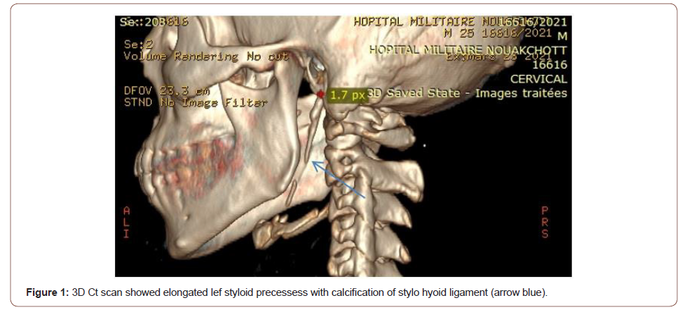
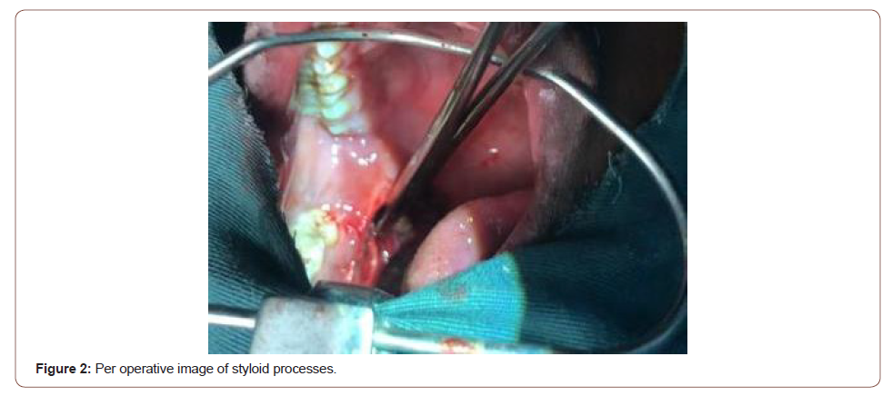
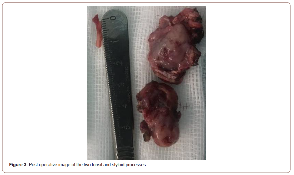
Computed tomography with 3D reconstruction revealed (Figure1) left elongated styloid processes measuring 3.9 cm with ossification of the stylo-hyoid ligament, the right styloid process was normal. A diagnosis of Eagle’s syndrome was made. The treatment option was surgical excision of the styloid processes with tonsillectomy. The chosen access was intraoral approach (Figures 2,3). The patient underwent general anaesthesia with nasotracheal intubation. With the patient’s mouth at the range of maximum opening, we first performing bilateral tonsillectomy, the region of the tonsillar pillar was palpated, indicating the location for incision with the electrocautery.

Tissue dissection was performed and the styloid processes were located. The tissue was dissected till the most proximal portion of the styloid processes. During surgery, an elongated styloid process was visualized and removed with a forceps and then incisions were closed with simple continuous 4-0 vicryl sutures. Symptoms resolved immediately after surgery. On follow-up 7 months after surgery, the patient remained asymptomatic and pain-free (Figures 1-3).

irispublishers-openaccess-textile-science-fashion

Discussion

Eagle first described two syndromes associated with elongation/ aberrant ossification of the stylo-hyoid apparatus. The first, classic Eagle syndrome was described as pain, dysphagia, and a foreign body sensation that presents immediately after tonsillectomy. Eagle also described stylocarotid sydrome in which an elongated styloid compresses the carotid artery and results in pain (parietal/ periorbital), visual disturbances, and syncope [5]. The diagnosis of Eagle’s syndrome is based on IHCD-3 [6] diagnostic criteria. Those criteria consist of any head, neck, pharyngeal and/or facial pain with at least two of the four following signs: Pain provoked or exacerbated by digital palpation of the stylohyoid ligament, pain provoked by head turning, pain improved by injection of local anaesthetic agent into the stylohyoid ligament, or by styloidectomy, or pain ipsilateral to the inflamed stylohyoid ligament. Also, there must be radiological evidence of a calcified or elongated stylohyoid ligament.

Eagle syndrome is diagnosed by radiography and physical examination. Pain during palpation of the tonsillar fossa could alert clinicians to this possible diagnosis Our case was a classic eagle syndrome associated with bilateral tonsil hypertrophy, treated several times previously as neuropathy pain by analgesic without significant result. There are many different etiologies that have been proposed to explain Eagle syndrome. three possible explanations was listed, which lead to abnormal stylohyoid complexes [7]. One of the theories relates to retained embryologic cartilage tissue from Reichert’s cartilage. The second theory is calcification of the stylomandibular ligament. The third explanation is expansion of osseous tissue at the origin of the stylomandibular ligament.

In our case, Eagle syndrome was the main diagnostic hypothesis after physical and CT scan result. The symptomatic management of Eagle syndrome with conservative medical treatment, such as antiinflammatory medications, anticonvulsants, antipsychotics, or other analgesics is usually reserved for patients unable to undergo surgical procedure [8]. Surgical treatment presents remission of symptoms without the need for continuous drug use [9] and it is indicated for all patients due to it being the definitive treatment for this condition. The option for the surgical treatment in this case was taken, considering the age of patient, worsening of the symptoms reported by the patient and the definitive character of the intervention.

The surgical management of the Eagle syndrome consists in the shortening of the elongated styloid process, with the surgical access remaining a matter of dispute. Intraoral access consists of the first stage involving the resection of the palatine tonsil (if no tonsillectomy had been performed before) followed by the second stage involving the incision of the tonsillar bed mucosa and partial resection of the process. Benefits of this technique consist in the possibilities to avoid a skin scar. However, the technique is associated with a high risk of complications due to the poor visibility of the operating field resulting in a likelihood of important anatomical structures within the parapharyngeal space being damaged in the course of the procedure. Another complication may consist in the infection of the parapharyngeal space and the neck [10,11].

Eagle syndrome has a large variety of clinical presentations as evidenced by the multitude of nonspecific symptoms. As a result, it is important to understand the diagnostic workup, relevant imaging, and ultimate treatment options. Intraoral resection of styloid process remains the preferred surgical management.

To read more about this article....Open access Journal of Otolaryngology and Rhinology

Please follow the URL to access more information about this article

To know more about our Journals...Iris Publishers

To know about Open Access Publishers

Iris Publishers-Open access Journal of Dentistry & Oral Health | Chief Complaint in A Dental and Maxillofacial Surgery Department in Abuja Nigeria


Authored by Nathan Ukuoghene Ikimi*,

Abstract

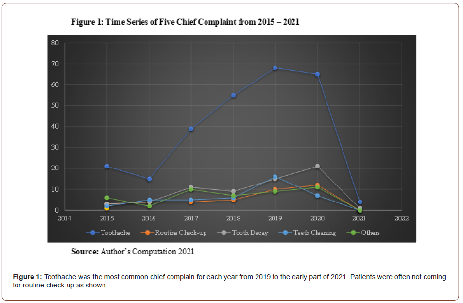
Chief complaints in the dental clinic are a series of complaints that a patient presents with in order of severity that needs urgent attention. The knowledge of chief complaints enables the clinician to focus appropriately on the area of dental challenge to treat his patient’s immediate complaint and plan preventive management. This is a retrospective study of cases that presented at the Dental and Maxillofacial Surgery department of State House Clinic from 2014 to 2021. Various Chief complaints that patients presented with in the clinic were prepared and used to review case files of patients. Statistical Package for social science (SPSS) version 21 was used to analyse the data collected; the relationship between chief complaints and age-group by sex of patients was examined. Toothaches was the most common chief complaints reported at 43.3% and this was more in females at 50.9%. Among the female sex, those within the age of 21-30 years reported more toothaches, while among the male sex those within 31-40 years of age reported more toothaches complaints and this was statistically significate.

The limitation of this study was that the sample size was small, and the study was conducted in one dental clinic only in Abuja. Thus, this might not be a complete reflection of pattern of chief complaints in Abuja, the Federal Capital Territory of Nigeria. Health planners, policy makers including non-governmental agencies should be interested in further studies since the result could be used in planning for dental needs of the populace.

Keywords: Chief complaints; Toothaches; State house clinic

Introduction

A chief complaint in dentistry could be defined as a subjective report provided by a patient in his own word that describes the most significant reason or serious symptoms of abnormality that caused him to seek an oral health care [1] and it serves as a source of information to the dentist which he uses to elicit signs in making a set of provisional diagnosis [2]. This is usually recorded in order of the most pressing dental challenges requiring urgent attention in the patient’s exact own words to know his awareness of the dental health challenges [1, 2]. Hence, recording accurately and identifying the patient’s chief complaints (CC) by asking appropriate question that would elicit a detailed history from the patient is considered as the foundation for developing a consistent treatment plan [3]. The severity of the chief complaint (CC) in dentistry reflects the urgency of the patient to seek dental care however, a patient’s CC is not designated to create a standard of care [4, 5] but rather it is the beginning or guide to taking an accurate dental history and is usually a short statement describing the symptom or problems that requires dental attention [1].

Studies carried out in Pakistan reported toothache as the CC of patients seeking dental treatment [2], and this is like the report of 31.5% patients, attending the University of Nairobi Dental hospital whose chief complain was pain, while 25.4% had orthodontic related challenges as their CC and finally, 19.7% gave dental tooth decay as their chief complaint [6]. A study in Pelotas, Brazil indicated that the CC of most patients visiting the dental hospital was dental pain originating from dental caries and these ones eventually underwent endodontic treatment [7]. Furthermore, it has been reported that in India, toothache was the chief complaint among dental patients, while oral hygiene and mal-occlusion were the next most common CC [8]. In addition, Akaji, et al. reported on 1663 patients with an average age of 33.218 years, attending the Dental Clinic of the University of Nigeria that almost half of the patients, 49.2% to be precise, recorded tooth dental pain as CC, 7.6% had swelling as CC and 5.7% were at the clinic for dental check-up [9].

In dental practice, it is important to give utmost attention to the problem that brought the patient to the dental clinic and treat appropriately so that the patient leaves the clinic satisfied and the time spent in the dental clinic is thus reduced [4]. The knowledge of the most common chief complain would enable the dentist to focus his attention on that speciality that manages that complaints and plan for more dental materials to handle such cases when patients with them turn up. Moreover, dental public health campaigns would be geared towards enlightening the populace on preventive measures that could be done to reduce incidence of the chief complaints, while making them to appreciate the importance of visiting the dental clinic for dental check-ups early so that serious dental challenges are detected on time and treated to prevent them from developing into more serious challenges in the future [10].

However, literature on CC of patients visiting the dental clinic in Nigeria seems to be scanty from our search and there was none to the best of our knowledge on study carried out in the northern region of Nigeria. Thus, this study aims to provide an update on the chief complaint of patients residing in Abuja, the Northcentral region of Nigeria so that adequate training and emphasis would be focused on the area of oral health needs of patients in this region and preventive strategies can be developed and implemented to reduce the man-hour spent in dental clinics by patients.

Methodology

This is a five-year retrospective study of chief complaints (CC) of patients attending the Dental and Maxillofacial Surgery Department of State House Clinic, Abuja. The State House Clinic was initially restricted to the President, the Vice-President, and their families, including a few senior government officials especial since it was not created to generate funds. However, with the creation of the National Health Insurance Scheme (NHIS) in June 2005 [11], the clinic was opened to employees of the State House and those patients who choose the clinic as their NHIS accredited Hospital. The NHIS has resulted in an increase in number of patients accessing the State House Clinic and this is similar to the effect of NHIS reported in Lagos state [12]. Ethical clearance for this study was obtained from the Federal Capital Health research ethics committee, Abuja.

Information such as age, sex, marital status excluding their names was collected from the case files of the patients in the Medical Records Unit of the clinic. A table of various CC was prepared with CC commonly heard from patients such as “My tooth is paining me”, “I want to remove my tooth”, “I want to wash my teeth”, “I want to do dental check-up”, “I do not like the arrangement of my child’s teeth” and two trained investigators were required to tick on the table, the most appropriate CC found in the case file.

Chi-square test was conducted to find if there were any relationship between CC and age-group by gender of patients. Statistical Package for social science (SPSS) used is version 21.

Results

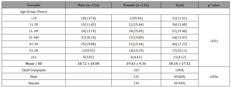
Table 1: Sex Distribution of Respondents and Chief Complaint.

irispublishers-openaccess-dentistry-oral-health

ʆ in the period under review, toothache complain was higher in females than in males, however this was not statistically significant.

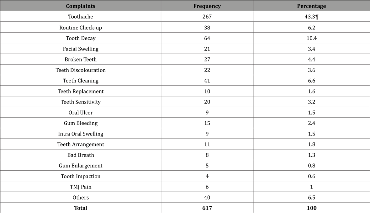
Table 2: Chief Complaints of Respondents.

irispublishers-openaccess-dentistry-oral-health

¶ Toothache is the most common complaints by patients.
§ More females than males under the age-group of 21-30 complained of toothaches.
Β Toothache complain was highest among males of 31-40 agegroups and this correlation is statistically significant at the 0.01 level (1-tailed).

irispublishers-openaccess-dentistry-oral-health

Table 3: Age Group by Sex Distribution of Toothache.

irispublishers-openaccess-dentistry-oral-health

Discussion

This is a retrospective study of the chief complaints of patients who attended the dental and oral maxillofacial surgery department of the State House Clinic Abuja, over a period of 5 years, for the management of oral health challenges. The sample size of this study is a limitation that may affect its interpretation and it may not be a general reflection of chief complaints pattern in Abuja, Federal Capital Territory (FCT), Nigeria. Moreover, the study was done at a clinic that does not have a good mix of the general population of the federal capital territory, Abuja. Access to the State House Clinic (SHC) is limited to a certain category of government employees and those who chose SHC as their Health Provider in the National Health Insurance Scheme (NHIS) [11, 13]. In addition, SHC was originally created to operate as a clinic but became a Medical Centre as the facility was expanded during President Obasanjo period as the President of Nigeria (1999-2007). However, due to insufficient funding plus the fact that the medical centre was not originally established to generate funds, it was changed back from a Medical Centre to a Clinic. With these change, new restrictions were introduced which affected the number of patients that could access healthcare at the clinic as this number dropped drastically [14]. A reduced attendance in SHC by extension, also affected dental and maxillofacial surgery department, hence the low number of cases that were recruited for this study. Therefore, it is the authors believe that a better reflection of chief complaints in Abuja would be provided by a result from a retrospective study of Government Dental Centres where patients from diverse educational and social background have unrestricted access.

Nevertheless, in SHC the percentage of females that reported with toothache as Chief Complaints (CC) was 50.9%, and this was higher than males who recorded 49.1%. This result agreed with a similar study conducted in Benin city Edo state, and some parts of the world where a higher female percentage with toothache as CC were reported [15-17]. While this study did not investigate the reason for the higher female percentages, possible reason could be that the females were more concern about the state of their teeth than the males which led to a higher percentage of females attending the dental clinic more than their male counterparts. Maheswaran et al study in Malaysia agrees with that assertion because he concluded that females’ attendance pattern to the dental clinic was more preventive oriented than that of males [8]. Additionally, it is also possible that the females were not employed and were more at home which gave them more flexible time schedule to attend dental care facilities more than the males.

In our study, the total number of patients, male and female that presented with toothaches as chief complaints was 43.3% of the 267 patients whose dental records were reviewed. This result is in consonance with the study conducted by Ali in Iraqi among 407 patients, in which 47.02% were at the dental clinic for toothaches [18]. Additionally, Warnakulasuriya in Sri Lanka reported that dental caries was the chief complaints among the patients that were investigated [19]. This may not be surprising because toothaches resulting from dental caries has been described as “the most prevalent chronic diseases of people worldwide” [20].

In our study, toothache complain was highest among males of 31-40 age-groups and this correlation is statistically significant. Agreeing with this report is an Australia study that reported prevalence of toothaches being common among young people below the age group of 45-61 [21] and a Brazilian report of a prevalence of 34.8% which was among middle aged adults of 35-44 years old [22]. This age group is the economically active age group, therefore if this number keep increasing and more males keep spending time at the dental clinic, then the negative outcome would be a lower number of males reporting for work at their place of employment and this would have a ripple effect on the country’s economy with much funds being lost [22].

Conclusion

This study has investigated the chief complaints of patients attending the Dental and Maxillofacial Surgery Department of State House Clinic. The result indicates that toothache is the highest on the list of patients’ complaints. It was also observed in the study that young adults in their prime both in males and females had more toothaches. This population represents the active workforce of any Country. Adequate planning on preventive dentistry should be the focus so the man-hour spent in the dental clinic would be reduced.

To read more about this article...Open access Journal of Dentistry & Oral Health

Please follow the URL to access more information about this article
https://irispublishers.com/ojdoh/fulltext/chief-complaint-in-a-dental-and-maxillofacial-surgery-department-in-abuja-nigeria.ID.000613.php

To know more about our Journals...
Iris Publishers

To know about 
Open Access Publishers

Iris Publishers-Open access Journal of Hydrology & Meteorology | Influence of Community Resilience to Flood Risk and Coping Strategies in Bayelsa State, Southern Nigeria

  Authored by  Nwankwoala HO *, Abstract This study is aimed at assessing the influence of community resilience to flood risk and coping str...